Review
. 2011 Jun;259(3):641-57.
doi: 10.1148/radiol.11081489.

Transcatheter intraarterial therapies: rationale and overview

Affiliations
Review

Transcatheter intraarterial therapies: rationale and overview

Robert J Lewandowski et al. Radiology. 2011 Jun.

Abstract

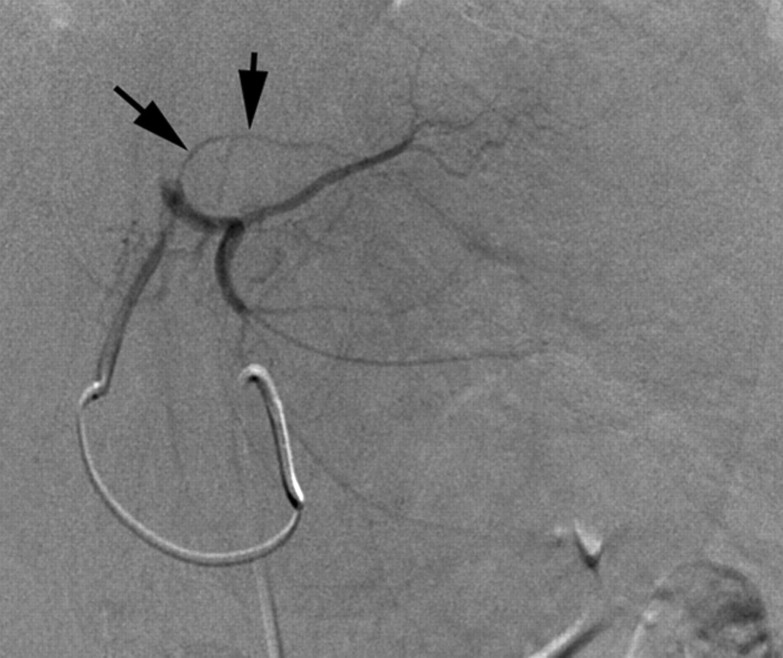
Transcatheter intraarterial therapies have proved valuable in the battle against primary and secondary hepatic malignancies. The unique aspects of all such therapies are their reduced toxicity profiles and highly effective tumor responses. These unique characteristics coupled with their minimally invasive nature provide an attractive therapeutic option in patients who may have previously had few alternatives. The concept of all catheter-based intraarterial therapies is to selectively deliver anticancer treatment to tumor(s). These therapies, which include transarterial embolization, intraarterial chemoinfusion, transarterial chemoembolization with or without drug-eluting beads, and radioembolization with use of yttrium 90, inflict lethal insult to tumors while preserving normal hepatic parenchyma. This is possible because hepatic neoplasms preferentially derive their blood supply from an arterial source while the majority of noncancerous liver is supplied by the portal vein. As part of the interventional oncology review series, in this article we describe the rationale behind each of these transcatheter therapies and provide a review of the existing medical literature.


Figures

Figure 1a:
Figure 1a:
Common extrahepatic arteries that need to be recognized and potentially embolized prior to transcatheter intraarterial therapy. (a) Celiac angiogram reveals right gastric artery (arrows) originating from the proximal left hepatic artery. (b) Right hepatic angiogram reveals cystic artery (arrow) originating from the proximal right hepatic artery. (c) Celiac angiogram in a patient with a replaced right hepatic artery from the superior mesenteric artery. Arrows delineate a faint linear vessel coursing from the segment IV branch of the left hepatic artery. (d) Selective left hepatic angiogram better demonstrates the falciform artery (black arrows). The tumor blush is now visible (white arrows). (e) Left hepatic angiogram reveals an accessory left gastric artery (arrows). (f) Delayed phase selective angiogram of the accessory left gastric artery shows the draining coronary vein (arrow), confirming gastric perfusion. (g) Celiac angiogram reveals arterial-portal fistula in the right hepatic lobe. Off the left hepatic artery, there is a vessel coursing under the left hemidiaphragm (arrows). (h) Left hepatic angiogram better demonstrates this vessel (arrows) as the left inferior phrenic artery.
Figure 1b:
Figure 1b:
Common extrahepatic arteries that need to be recognized and potentially embolized prior to transcatheter intraarterial therapy. (a) Celiac angiogram reveals right gastric artery (arrows) originating from the proximal left hepatic artery. (b) Right hepatic angiogram reveals cystic artery (arrow) originating from the proximal right hepatic artery. (c) Celiac angiogram in a patient with a replaced right hepatic artery from the superior mesenteric artery. Arrows delineate a faint linear vessel coursing from the segment IV branch of the left hepatic artery. (d) Selective left hepatic angiogram better demonstrates the falciform artery (black arrows). The tumor blush is now visible (white arrows). (e) Left hepatic angiogram reveals an accessory left gastric artery (arrows). (f) Delayed phase selective angiogram of the accessory left gastric artery shows the draining coronary vein (arrow), confirming gastric perfusion. (g) Celiac angiogram reveals arterial-portal fistula in the right hepatic lobe. Off the left hepatic artery, there is a vessel coursing under the left hemidiaphragm (arrows). (h) Left hepatic angiogram better demonstrates this vessel (arrows) as the left inferior phrenic artery.
Figure 1c:
Figure 1c:
Common extrahepatic arteries that need to be recognized and potentially embolized prior to transcatheter intraarterial therapy. (a) Celiac angiogram reveals right gastric artery (arrows) originating from the proximal left hepatic artery. (b) Right hepatic angiogram reveals cystic artery (arrow) originating from the proximal right hepatic artery. (c) Celiac angiogram in a patient with a replaced right hepatic artery from the superior mesenteric artery. Arrows delineate a faint linear vessel coursing from the segment IV branch of the left hepatic artery. (d) Selective left hepatic angiogram better demonstrates the falciform artery (black arrows). The tumor blush is now visible (white arrows). (e) Left hepatic angiogram reveals an accessory left gastric artery (arrows). (f) Delayed phase selective angiogram of the accessory left gastric artery shows the draining coronary vein (arrow), confirming gastric perfusion. (g) Celiac angiogram reveals arterial-portal fistula in the right hepatic lobe. Off the left hepatic artery, there is a vessel coursing under the left hemidiaphragm (arrows). (h) Left hepatic angiogram better demonstrates this vessel (arrows) as the left inferior phrenic artery.
Figure 1d:
Figure 1d:
Common extrahepatic arteries that need to be recognized and potentially embolized prior to transcatheter intraarterial therapy. (a) Celiac angiogram reveals right gastric artery (arrows) originating from the proximal left hepatic artery. (b) Right hepatic angiogram reveals cystic artery (arrow) originating from the proximal right hepatic artery. (c) Celiac angiogram in a patient with a replaced right hepatic artery from the superior mesenteric artery. Arrows delineate a faint linear vessel coursing from the segment IV branch of the left hepatic artery. (d) Selective left hepatic angiogram better demonstrates the falciform artery (black arrows). The tumor blush is now visible (white arrows). (e) Left hepatic angiogram reveals an accessory left gastric artery (arrows). (f) Delayed phase selective angiogram of the accessory left gastric artery shows the draining coronary vein (arrow), confirming gastric perfusion. (g) Celiac angiogram reveals arterial-portal fistula in the right hepatic lobe. Off the left hepatic artery, there is a vessel coursing under the left hemidiaphragm (arrows). (h) Left hepatic angiogram better demonstrates this vessel (arrows) as the left inferior phrenic artery.
Figure 1e:
Figure 1e:
Common extrahepatic arteries that need to be recognized and potentially embolized prior to transcatheter intraarterial therapy. (a) Celiac angiogram reveals right gastric artery (arrows) originating from the proximal left hepatic artery. (b) Right hepatic angiogram reveals cystic artery (arrow) originating from the proximal right hepatic artery. (c) Celiac angiogram in a patient with a replaced right hepatic artery from the superior mesenteric artery. Arrows delineate a faint linear vessel coursing from the segment IV branch of the left hepatic artery. (d) Selective left hepatic angiogram better demonstrates the falciform artery (black arrows). The tumor blush is now visible (white arrows). (e) Left hepatic angiogram reveals an accessory left gastric artery (arrows). (f) Delayed phase selective angiogram of the accessory left gastric artery shows the draining coronary vein (arrow), confirming gastric perfusion. (g) Celiac angiogram reveals arterial-portal fistula in the right hepatic lobe. Off the left hepatic artery, there is a vessel coursing under the left hemidiaphragm (arrows). (h) Left hepatic angiogram better demonstrates this vessel (arrows) as the left inferior phrenic artery.
Figure 1f:
Figure 1f:
Common extrahepatic arteries that need to be recognized and potentially embolized prior to transcatheter intraarterial therapy. (a) Celiac angiogram reveals right gastric artery (arrows) originating from the proximal left hepatic artery. (b) Right hepatic angiogram reveals cystic artery (arrow) originating from the proximal right hepatic artery. (c) Celiac angiogram in a patient with a replaced right hepatic artery from the superior mesenteric artery. Arrows delineate a faint linear vessel coursing from the segment IV branch of the left hepatic artery. (d) Selective left hepatic angiogram better demonstrates the falciform artery (black arrows). The tumor blush is now visible (white arrows). (e) Left hepatic angiogram reveals an accessory left gastric artery (arrows). (f) Delayed phase selective angiogram of the accessory left gastric artery shows the draining coronary vein (arrow), confirming gastric perfusion. (g) Celiac angiogram reveals arterial-portal fistula in the right hepatic lobe. Off the left hepatic artery, there is a vessel coursing under the left hemidiaphragm (arrows). (h) Left hepatic angiogram better demonstrates this vessel (arrows) as the left inferior phrenic artery.
Figure 1g:
Figure 1g:
Common extrahepatic arteries that need to be recognized and potentially embolized prior to transcatheter intraarterial therapy. (a) Celiac angiogram reveals right gastric artery (arrows) originating from the proximal left hepatic artery. (b) Right hepatic angiogram reveals cystic artery (arrow) originating from the proximal right hepatic artery. (c) Celiac angiogram in a patient with a replaced right hepatic artery from the superior mesenteric artery. Arrows delineate a faint linear vessel coursing from the segment IV branch of the left hepatic artery. (d) Selective left hepatic angiogram better demonstrates the falciform artery (black arrows). The tumor blush is now visible (white arrows). (e) Left hepatic angiogram reveals an accessory left gastric artery (arrows). (f) Delayed phase selective angiogram of the accessory left gastric artery shows the draining coronary vein (arrow), confirming gastric perfusion. (g) Celiac angiogram reveals arterial-portal fistula in the right hepatic lobe. Off the left hepatic artery, there is a vessel coursing under the left hemidiaphragm (arrows). (h) Left hepatic angiogram better demonstrates this vessel (arrows) as the left inferior phrenic artery.
Figure 1h:
Figure 1h:
Common extrahepatic arteries that need to be recognized and potentially embolized prior to transcatheter intraarterial therapy. (a) Celiac angiogram reveals right gastric artery (arrows) originating from the proximal left hepatic artery. (b) Right hepatic angiogram reveals cystic artery (arrow) originating from the proximal right hepatic artery. (c) Celiac angiogram in a patient with a replaced right hepatic artery from the superior mesenteric artery. Arrows delineate a faint linear vessel coursing from the segment IV branch of the left hepatic artery. (d) Selective left hepatic angiogram better demonstrates the falciform artery (black arrows). The tumor blush is now visible (white arrows). (e) Left hepatic angiogram reveals an accessory left gastric artery (arrows). (f) Delayed phase selective angiogram of the accessory left gastric artery shows the draining coronary vein (arrow), confirming gastric perfusion. (g) Celiac angiogram reveals arterial-portal fistula in the right hepatic lobe. Off the left hepatic artery, there is a vessel coursing under the left hemidiaphragm (arrows). (h) Left hepatic angiogram better demonstrates this vessel (arrows) as the left inferior phrenic artery.
Figure 2a:
Figure 2a:
Example of combination therapy with transcatheter chemoembolization and radiofrequency ablation. The patient has a history of HCC and underwent prior right hepatic lobe resection. Surveillance imaging revealed subcentimeter focus of tumor recurrence in the left hepatic lobe. A multidiscipline conference recommended percutaneous ablation, but the tumor could not be seen at US. (a) Pretreatment gadolinium-enhanced magnetic resonance (MR) image reveals a small arterial enhancing focus (arrow) in the left hepatic lobe. This tumor washed out at the delayed phases, consistent with recurrent HCC. (b) Left hepatic angiogram depicts a small hypervascular tumor (arrow) correlating with the preprocedure MR imaging. Transcatheter chemoembolization was performed to permit appropriate tumor targeting for percutaneous radiofrequency ablation. (c) Nonenhanced CT scan immediately after transcatheter chemoembolization reveals dense accumulation of ethiodol within the tumor (arrow). (d) Nonenhanced CT scan obtained during radiofrequency ablation depicts the electrode placed through the ethiodol-stained tumor (arrow).
Figure 2b:
Figure 2b:
Example of combination therapy with transcatheter chemoembolization and radiofrequency ablation. The patient has a history of HCC and underwent prior right hepatic lobe resection. Surveillance imaging revealed subcentimeter focus of tumor recurrence in the left hepatic lobe. A multidiscipline conference recommended percutaneous ablation, but the tumor could not be seen at US. (a) Pretreatment gadolinium-enhanced magnetic resonance (MR) image reveals a small arterial enhancing focus (arrow) in the left hepatic lobe. This tumor washed out at the delayed phases, consistent with recurrent HCC. (b) Left hepatic angiogram depicts a small hypervascular tumor (arrow) correlating with the preprocedure MR imaging. Transcatheter chemoembolization was performed to permit appropriate tumor targeting for percutaneous radiofrequency ablation. (c) Nonenhanced CT scan immediately after transcatheter chemoembolization reveals dense accumulation of ethiodol within the tumor (arrow). (d) Nonenhanced CT scan obtained during radiofrequency ablation depicts the electrode placed through the ethiodol-stained tumor (arrow).
Figure 2c:
Figure 2c:
Example of combination therapy with transcatheter chemoembolization and radiofrequency ablation. The patient has a history of HCC and underwent prior right hepatic lobe resection. Surveillance imaging revealed subcentimeter focus of tumor recurrence in the left hepatic lobe. A multidiscipline conference recommended percutaneous ablation, but the tumor could not be seen at US. (a) Pretreatment gadolinium-enhanced magnetic resonance (MR) image reveals a small arterial enhancing focus (arrow) in the left hepatic lobe. This tumor washed out at the delayed phases, consistent with recurrent HCC. (b) Left hepatic angiogram depicts a small hypervascular tumor (arrow) correlating with the preprocedure MR imaging. Transcatheter chemoembolization was performed to permit appropriate tumor targeting for percutaneous radiofrequency ablation. (c) Nonenhanced CT scan immediately after transcatheter chemoembolization reveals dense accumulation of ethiodol within the tumor (arrow). (d) Nonenhanced CT scan obtained during radiofrequency ablation depicts the electrode placed through the ethiodol-stained tumor (arrow).
Figure 2d:
Figure 2d:
Example of combination therapy with transcatheter chemoembolization and radiofrequency ablation. The patient has a history of HCC and underwent prior right hepatic lobe resection. Surveillance imaging revealed subcentimeter focus of tumor recurrence in the left hepatic lobe. A multidiscipline conference recommended percutaneous ablation, but the tumor could not be seen at US. (a) Pretreatment gadolinium-enhanced magnetic resonance (MR) image reveals a small arterial enhancing focus (arrow) in the left hepatic lobe. This tumor washed out at the delayed phases, consistent with recurrent HCC. (b) Left hepatic angiogram depicts a small hypervascular tumor (arrow) correlating with the preprocedure MR imaging. Transcatheter chemoembolization was performed to permit appropriate tumor targeting for percutaneous radiofrequency ablation. (c) Nonenhanced CT scan immediately after transcatheter chemoembolization reveals dense accumulation of ethiodol within the tumor (arrow). (d) Nonenhanced CT scan obtained during radiofrequency ablation depicts the electrode placed through the ethiodol-stained tumor (arrow).
Figure 3a:
Figure 3a:
Images in a patient with unresectable HCC. (a) Gadolinium-enhanced MR image reveals a hypervascular tumor (arrow) in the right hepatic lobe. (b) C-arm CT image with the catheter selectively positioned in a tumor-feeding branch reveals marked tumor perfusion (arrow) and minimal perfusion to the adjacent parenchyma, enabling the uninvolved parenchyma to be spared exposure to toxic therapeutics.
Figure 3b:
Figure 3b:
Images in a patient with unresectable HCC. (a) Gadolinium-enhanced MR image reveals a hypervascular tumor (arrow) in the right hepatic lobe. (b) C-arm CT image with the catheter selectively positioned in a tumor-feeding branch reveals marked tumor perfusion (arrow) and minimal perfusion to the adjacent parenchyma, enabling the uninvolved parenchyma to be spared exposure to toxic therapeutics.
Figure 4a:
Figure 4a:
(a) Contrast-enhanced CT scan reveals infiltrating right hepatic lobe tumor invading the portal vein (arrows). (Image courtesy of Karen Brown, MD, Memorial Sloan-Kettering Cancer Center.) (b) After transarterial embolization, contrast-enhanced CT scan reveals marked necrosis and clear demarcation of this previously infiltrating tumor (arrows). Small gas bubbles within the tumor are an indication of necrosis. (Image courtesy of Karen Brown, MD, Memorial Sloan-Kettering Cancer Center.)
Figure 4b:
Figure 4b:
(a) Contrast-enhanced CT scan reveals infiltrating right hepatic lobe tumor invading the portal vein (arrows). (Image courtesy of Karen Brown, MD, Memorial Sloan-Kettering Cancer Center.) (b) After transarterial embolization, contrast-enhanced CT scan reveals marked necrosis and clear demarcation of this previously infiltrating tumor (arrows). Small gas bubbles within the tumor are an indication of necrosis. (Image courtesy of Karen Brown, MD, Memorial Sloan-Kettering Cancer Center.)
Figure 5a:
Figure 5a:
(a) Contrast-enhanced CT scan reveals multifocal 2–3-cm bilobar hepatic metastases from colorectal cancer. (Image courtesy of Karen Brown, MD, Memorial Sloan-Kettering Cancer Center.) (b) Following chemoinfusion therapy with FUDR, contrast-enhanced CT scan reveals disappearance of these tumors, which is consistent with a complete response. (Image courtesy of Karen Brown, MD, Memorial Sloan-Kettering Cancer Center.)
Figure 5b:
Figure 5b:
(a) Contrast-enhanced CT scan reveals multifocal 2–3-cm bilobar hepatic metastases from colorectal cancer. (Image courtesy of Karen Brown, MD, Memorial Sloan-Kettering Cancer Center.) (b) Following chemoinfusion therapy with FUDR, contrast-enhanced CT scan reveals disappearance of these tumors, which is consistent with a complete response. (Image courtesy of Karen Brown, MD, Memorial Sloan-Kettering Cancer Center.)
Figure 6a:
Figure 6a:
(a) Gadolinium-enhanced MR image reveals a 3.2-cm hypervascular HCC (arrow) in the right hepatic lobe. (b) Nonenhanced CT scan immediately after transarterial chemoembolization demonstrates dense ethiodol staining with the tumor (arrow). (c) Gadolinium-enhanced MR image after transcatheter chemoembolization, reveals necrosis and tumor size reduction to 2.3 cm (arrow), which is consistent with a favorable response to therapy.
Figure 6b:
Figure 6b:
(a) Gadolinium-enhanced MR image reveals a 3.2-cm hypervascular HCC (arrow) in the right hepatic lobe. (b) Nonenhanced CT scan immediately after transarterial chemoembolization demonstrates dense ethiodol staining with the tumor (arrow). (c) Gadolinium-enhanced MR image after transcatheter chemoembolization, reveals necrosis and tumor size reduction to 2.3 cm (arrow), which is consistent with a favorable response to therapy.
Figure 6c:
Figure 6c:
(a) Gadolinium-enhanced MR image reveals a 3.2-cm hypervascular HCC (arrow) in the right hepatic lobe. (b) Nonenhanced CT scan immediately after transarterial chemoembolization demonstrates dense ethiodol staining with the tumor (arrow). (c) Gadolinium-enhanced MR image after transcatheter chemoembolization, reveals necrosis and tumor size reduction to 2.3 cm (arrow), which is consistent with a favorable response to therapy.
Figure 7a:
Figure 7a:
(a) Gadolinium-enhanced MR image reveals a 6.5-cm hypervascular HCC (arrow) in the right hepatic lobe. (b) Following chemoembolization with drug-eluting beads, gadolinium-enhanced MR image reveals complete necrosis (arrow), which is consistent with a favorable response to therapy. The tumor has only minimally decreased in size (6.1 cm).
Figure 7b:
Figure 7b:
(a) Gadolinium-enhanced MR image reveals a 6.5-cm hypervascular HCC (arrow) in the right hepatic lobe. (b) Following chemoembolization with drug-eluting beads, gadolinium-enhanced MR image reveals complete necrosis (arrow), which is consistent with a favorable response to therapy. The tumor has only minimally decreased in size (6.1 cm).
Figure 8a:
Figure 8a:
(a) Gadolinium-enhanced MR image reveals a 5.8-cm hypervascular HCC (arrow) in the right hepatic lobe. (b) Following radioembolization, MR image reveals necrosis and decreased tumor size (4.6 cm) (arrow), which is consistent with a favorable response to therapy. The pattern of necrosis is in a segmental distribution. This has been termed radiation segmentectomy.
Figure 8b:
Figure 8b:
(a) Gadolinium-enhanced MR image reveals a 5.8-cm hypervascular HCC (arrow) in the right hepatic lobe. (b) Following radioembolization, MR image reveals necrosis and decreased tumor size (4.6 cm) (arrow), which is consistent with a favorable response to therapy. The pattern of necrosis is in a segmental distribution. This has been termed radiation segmentectomy.

References

    1. Lammer J, Malagari K, Vogl T, et al. Prospective randomized study of doxorubicin-eluting-bead embolization in the treatment of hepatocellular carcinoma: results of the PRECISION V study. Cardiovasc Intervent Radiol 2010;33(1):41–52 - PMC - PubMed
    1. Varela M, Real MI, Burrel M, et al. Chemoembolization of hepatocellular carcinoma with drug eluting beads: efficacy and doxorubicin pharmacokinetics. J Hepatol 2007;46(3):474–481 - PubMed
    1. Lewandowski RJ, Mulcahy MF, Kulik LM, et al. Chemoembolization for hepatocellular carcinoma: comprehensive imaging and survival analysis in a 172-patient cohort. Radiology 2010;255(3):955–965 - PMC - PubMed
    1. Salem R, Lewandowski RJ, Kulik L, et al. Radioembolization results in longer time-to-progression and reduced toxicity compared with chemoembolization in patients with hepatocellular carcinoma. Gastroenterology 2011;140(2):497-507 - PMC - PubMed
    1. Sato K, Lewandowski RJ, Bui JT, et al. Treatment of unresectable primary and metastatic liver cancer with yttrium-90 microspheres (TheraSphere): assessment of hepatic arterial embolization. Cardiovasc Intervent Radiol 2006;29(4):522–529 - PubMed

Publication types

MeSH terms