. 2011 Jul 21;118(3):576-85.
doi: 10.1182/blood-2011-01-333641. Epub 2011 May 20.

SALL4 is a robust stimulator for the expansion of hematopoietic stem cells

Affiliations

SALL4 is a robust stimulator for the expansion of hematopoietic stem cells

Jerell R Aguila et al. Blood. .

Abstract

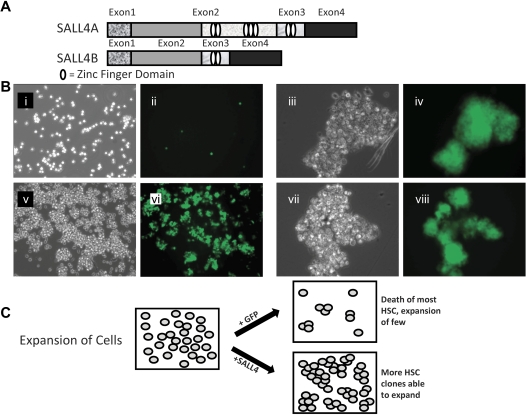
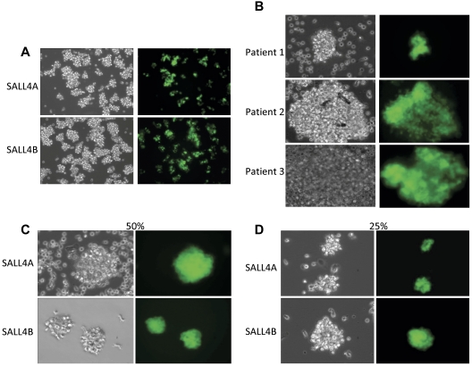
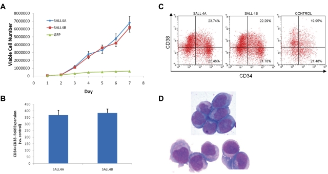
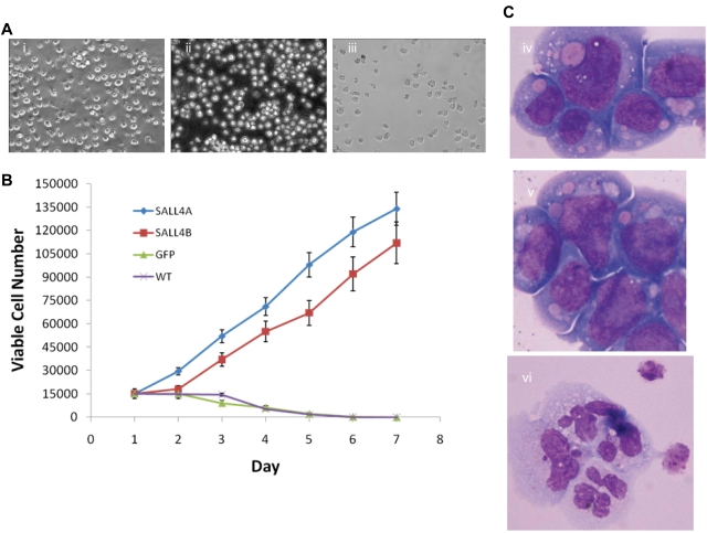
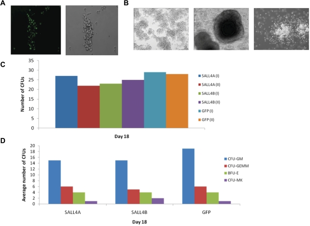
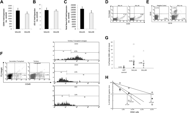
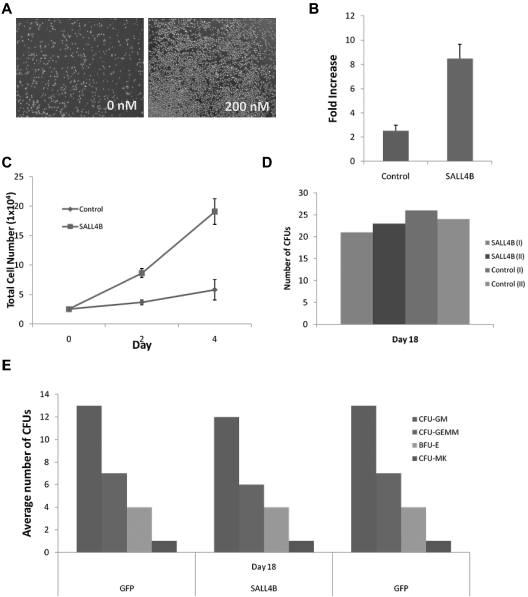
HSCs are rare cells that have the unique ability to self-renew and differentiate into cells of all hematopoietic lineages. The lack of donors and current inability to rapidly and efficiently expand HSCs are roadblocks in the development of successful cell therapies. Thus, the challenge of ex vivo human HSC expansion remains a fertile and critically important area of investigation. Here, we show that either SALL4A- or SALL4B-transduced human HSCs obtained from the mobilized peripheral blood are capable of rapid and efficient expansion ex vivo by >10 000-fold for both CD34(+)/CD38(-) and CD34(+)/CD38(+) cells in the presence of appropriate cytokines. We found that these cells retained hematopoietic precursor cell immunophenotypes and morphology as well as normal in vitro or vivo potential for differentiation. The SALL4-mediated expansion was associated with enhanced stem cell engraftment and long-term repopulation capacity in vivo. Also, we demonstrated that constitutive expression of SALL4 inhibited granulocytic differentiation and permitted expansion of undifferentiated cells in 32D myeloid progenitors. Furthermore, a TAT-SALL4B fusion rapidly expanded CD34(+) cells, and it is thus feasible to translate this study into the clinical setting. Our findings provide a new avenue for investigating mechanisms of stem cell self-renewal and achieving clinically significant expansion of human HSCs.


Figures

Figure 1
Figure 1
SALL4 isoforms and SALL4-transduced HSC expansion. (A) Schematic diagram of the SALL4A and SALL4B isoforms demonstrating the variable number of zinc-finger domains possessed by each. The HSCs were transduced with either the SALL4A or SALL4B gene using a lentiviral transfection system. (B) Bright field and fluorescent images of human bone marrow CD34+ cells transduced with GFP (i-ii, 10×) or representative SALL4 isoform B (v-vi, 10×) 9 days after infection. Initially, 50 000 CD34+/CD38 cells were plated. High magnification of SALL4B-transduced HSC clusters (iii,iv,vii,viii, 40×). The GFP cell clusters signified positive overexpression of SALL4B. With SALL4B overexpression, HSC cell clusters are able to survive and are rapidly expanding at 9 days after infection. (C) Model of SALL4-mediated ex vivo HSC expansion. The primary culture was divided and transduced with a SALL4 or GFP control. The viable HSCs without SALL4 overexpression decreased in number because of differentiation or death leading to a net HSC decline. In contrast, HSCs in which SALL4 was overexpressed, many clones were able to survive and expand in the culture. A net HSC expansion was exhibited with numerous expanding clusters throughout the culture.
Figure 2
Figure 2
Expansion of SALL4-transduced HSCs. (A) HSCs transduced with SALL4A and SALL4B are able to survive and expand rapidly 7 days after lentiviral infection (10×). (B) CD34+ cells isolated from peripheral blood stem cells of 3 different patients. CD34+ cells were isolated from the stem cell pool using magnetic anti-CD34+ human microbeads. The CD34+-enriched cells were transduced with SALL4A and imaged under bright field and fluorescent microscopy (40×). All 3 samples from the various patients were successfully transduced with SALL4A and expanded rapidly in culture. In addition, SALL4-induced HSCs are able to expand when growth factor concentrations are decreased. When SALL4-transduced HSCs were cultured in growth media containing 50% less cytokines (C), they were still able to survive and expand 6 days after lentiviral infection (40×). Furthermore, when growth factor concentrations were decreased to 25% of original values (D), the SALL4-transduced HSCs continued to proliferate (40×). In contrast, control cells had undergone cell death by day 6.
Figure 3
Figure 3
Characterization of SALL4-transduced HSC cells. (A) Growth curves of CD34+ cells transduced with SALL4A, SALL4B, or GFP and cultured in media containing 75% less cytokines. After transduction, 50 000 cells of each group were cultured in stringent conditions in which normal cytokine concentrations were decreased by 75%. HSCs transduced with SALL4A or SALL4B continued to survive and expand over 7 days, whereas control cells growth halted at day 5. (B) Fold expansion of CD34+/CD38 cells 14 days after infection of lenti-SALL4A or -SALL4B versus control. Cells transduced with SALL4A demonstrated a 368-fold increase of CD34+/CD38 cells over control, whereas those transduced with SALL4B showed a 384-fold increase. Values are means + SD. (C) Phenotypic analysis of SALL4-induced hematopoietic stem cells 31 days after lentiviral infection. Human-specific antibodies CD34-PE and CD38-APC were used to compare SALL4-transduced HSCs versus 3-day control cells. Thirty-one days after lentiviral infection, the aged SALL4-induced cells continued to demonstrate similar phenotypic ratios compared with control cells for CD34+/C38. FLOW analysis was carried out on 3 separate samples. Therefore, many of these aged cells still attained progenitor characteristics and had the ability to differentiate into various cells lines. (D) Thirty-one-day old SALL4-induced HSCs attain blastlike morphology. Aged 31-day-old SALL4-induced HSCs were Wright-Giemsa–stained. Many cells showed blastlike morphology including large nuclei and scant cytoplasm (100×). These cells represented a population of undifferentiated cells still visible 31 days after SALL4-lentiviral infection and expansion.
Figure 4
Figure 4
Overexpression of SALL4-immortalized 32D cells and blocks G-CSF–dependent differentiation. (A) SALL4-induced expansion of 32D cells proliferate after the removal of IL-3 and addition of G-CSF. Three days after the removal of IL-3 and addition of G-CSF to the growth media of the cells, the SALL4A- and SALL4B-induced cells continue to expand, whereas the GFP-induced cells exhibit a decrease in cell number. At 7 days, the SALL4A- (i, 20×) and SALL4B-induced (ii, 20×) cells continue to proliferate, whereas the control cells (iii, 20×) have undergone cell death. (B) Growth curves of SALL4-induced 32D cells cultured only with G-CSF. 32D cells were transduced with SALL4A, SALL4B, or GFP lentivirus and then cultured for 3 days in growth media containing IL-3. On the day 4, 15 000 cells were aliquoted from each group and placed in new growth media with G-CSF and without IL-3. Cell growth was monitored daily, and the viable number of cells in each group was recorded. In cells that were transduced with SALL4A, an 8-fold increase in the number of cells was observed from day 1 to day 7. Cells that were transduced with SALL4B exhibited a 7-fold expansion of cells. In contrast, cells that were only transduced with GFP and wild type (WT, no lentiviral infection) demonstrated a decrease in the number of cells over the same period with almost all the cells undergoing cell death by day 5. (C) Wright-Giemsa staining of 32D cells (100×). Morphology of 32D cells with IL-3 alone (iv), transduced with SALL4A with G-CSF (v), and with G-CSF alone (vi). Cells given IL-3 or transduced with SALL4A continue to demonstrate blastlike morphology (iv and v), whereas the cells not transduced with SALL4A and given G-CSF exhibit neutrophil morphology (vi).
Figure 5
Figure 5
CFU assays of SALL4-transduced cells. (A) CFU progenitors are GFP-positive. HSC cells selected for CFU assays were GFP-positive, which verified that the cells were successfully transduced and overexpressing SALL4 (10×). (B) Various CFU colonies are able to differentiate from SALL4-induced HSCs. Aged SALL4-induced HSCs were plated in MethoCult and observed for CFU colonies. Numerous lineages were observed in CFU assays using the SALL4-induced HSCs, including CFU-GEMM, BFU-E, and CFU-GM colonies (10×). These data demonstrated that the aged HSCs transduced with SALL4 were capable of differentiating into different blood cell lineages. (C) Number of CFU colonies formed from SALL4-induced hematopoietic stem cells. The number of CFU colonies was counted 13-18 days after SALL4-induced or GFP-induced cells were cultured in CFU MethoCult media. The representative data from day 18 are shown. (D) Types of CFU colonies formed at day 18.
Figure 6
Figure 6
Relative amplification of total stem cells and human cell engraftment into NOD/SCID mice. (A) After 1 month of cell culture, CD34+ cells transduced with SALL4A or SALL4B had 1780- and 1463-fold increases, respectively, relative to control cells. Values are means + SD. (B) Furthermore, SALL4-transduced cells showed 9.32-fold increases for SALL4A and 8.88-fold increases for SALL4B versus controls for the total number of LTC-ICs after 1 month. Values are means + SD. (C) Overall, SALL4A-transduced cells had a total fold CD34+/CD38 stem cell expansion 16 776 over control, whereas SALL4B-transduced cells showed 13 320-fold increases. Values are means + SD. (D) Representative flow cytometry analysis 4 weeks after injection for CD45+ human leukocytes from peripheral blood of NOD/SCID recipients transplanted with SALL4A- or SALL4B-transduced HSCs. (E) Representative flow cytometry profile 4 weeks after injection of a mouse exhibiting multilineage repopulation of human cells by engrafted cells. Although the negative control animal showed no engraftment of human cells, the experimental animal showed both CD15+ myeloid and CD19+ lymphoid human cell engraftment. (F) Flow analysis of secondary and tertiary bone marrow transplant NOD/SCID mice. The animals were positive for CD45+ cells in both secondary (2.74%) and tertiary (3.29%) transplants (left panel). The threshold bars for positive and negative populations were set according to independent staining controls. When the CD45+ population in the tertiary transplant was analyzed further for specific lineages, CD33 myeloid and CD19/CD3 lymphoid cells were positively measured (right panel). (G) Amount of human engraftment in the peripheral blood of NOD/SCID mice transplanted with 20 000 (SALL4A, ■; SALL4B, ▴, or GFP, ♦) or 40 000 (SALL4A, □; SALL4B, ▵; or GFP, ◊) initial human CD34+ cells. (H) Limiting dilution analysis of CD34+ bone marrow cells injected into NOD/SCID mice (n = 72) after lentiviral transfection with SALL4A (□), SALL4B (▴), or GFP (○).
Figure 7
Figure 7
Human bone marrow CD34+ cells expand at a higher rate when treated with TAT-SALL4B protein. (A) Bright field images of CD34+ cells after 3 days of protein treatment (10×). (B) Fold increase and total cell number (C) of TAT-SALL4B–treated bone marrow cells versus control cells treated solely with BSA. (D) Number of CFU colonies formed from hematopoietic stem cells treated with TAT-SALL4B protein compared with unmanipulated CD34+ cells. (E) Types of CFU colonies formed after treatment with TAT-SALL4B protein.

References

    1. Delaney C, Heimfeld S, Brashem-Stein C, Voorhies H, Manger RL, Bernstein ID. Notch-mediated expansion of human cord blood progenitor cells capable of rapid myeloid reconstitution. Nat Med. 2010;16(2):232–236. - PMC - PubMed
    1. Kelly SS, Sola CB, de Lima M, Shpall E. Ex vivo expansion of cord blood. Bone Marrow Transplant. 2009;44(10):673–681. - PMC - PubMed
    1. Astori G, Adami V, Mambrini G, et al. Evaluation of ex vivo expansion and engraftment in NOD-SCID mice of umbilical cord blood CD34+ cells using the DIDECO “Pluricell System”. Bone Marrow Transplant. 2005;35(11):1101–1106. - PubMed
    1. Boitano AE, Wang J, Romeo R, et al. Aryl hydrocarbon receptor antagonists promote the expansion of human hematopoietic stem cells. Science. 2010;329(5997):1345–1348. - PMC - PubMed
    1. Domashenko AD, Danet-Desnoyers G, Aron A, Carroll MP, Emerson SG. TAT-mediated transduction of NF-Ya peptide induces the ex vivo proliferation and engraftment potential of human hematopoietic progenitor cells. Blood. 2010;116(15):2676–83. - PMC - PubMed

Publication types

MeSH terms