. 2011 Jul;115(1):189-204.
doi: 10.1097/ALN.0b013e31821b1ac5.

Antagonism of nerve growth factor-TrkA signaling and the relief of pain

Affiliations

Antagonism of nerve growth factor-TrkA signaling and the relief of pain

Patrick W Mantyh et al. Anesthesiology. 2011 Jul.

Abstract

Nerve growth factor (NGF) was originally discovered as a neurotrophic factor essential for the survival of sensory and sympathetic neurons during development. However, in the adult NGF has been found to play an important role in nociceptor sensitization after tissue injury. The authors outline mechanisms by which NGF activation of its cognate receptor, tropomyosin-related kinase A receptor, regulates a host of ion channels, receptors, and signaling molecules to enhance acute and chronic pain. The authors also document that peripherally restricted antagonism of NGF-tropomyosin-related kinase A receptor signaling is effective for controlling human pain while appearing to maintain normal nociceptor function. Understanding whether there are any unexpected adverse events and how humans may change their behavior and use of the injured/degenerating tissue after significant pain relief without sedation will be required to fully appreciate the patient populations that may benefit from these therapies targeting NGF.


Conflict of interest statement

Conflict of interest: Karen Burrow and Aideen Young wrote the first rough draft of the manuscript in September 2009. Patrick Mantyh (PM), Martin Koltzenburg (MK), Lorne Mendell (LM), David Shelton (DS) and Leslie Tive (LT) then extensively edited and rewrote the manuscript in 20 separate drafts during the next 12 months. PM served as the lead author in rewriting, deleting, and adding new material and sections to the manuscript. PM drew the rough outlines of the figures which were then redrawn in professional format by Annemarie Johnson at Wake Forest University. PM, MK and LM are responsible for the views and opinions of this manuscript and did not receive any fees eror compensation for the writing of this review.

Figures

Fig. 1
Fig. 1
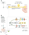
Schematic showing the neurotransmitters, receptors and ion channels that are modulated and up-regulated by NGF binding to TrkA+ primary afferent sensory nerve fibers. (A) Tropomyosin-related kinase A receptor (TrkA)+ primary afferent have their cell body in the dorsal root ganglia (DRG) and transmit sensory information from the periphery to the spinal cord and brain. During inflammation, injury or certain diseases, inflammatory/immune/Schwann cells release nerve growth factor (NGF) that binds to TrkA, which in turns directly activates and/or sensitizes nociceptors. NGF and its cognate receptor TrkA are retrogradely transported to the DRG, resulting in increased synthesis of neuropeptides (e.g.: substance P (SP), brain-derived neurotrophic factor (BDNF)), receptors, ion channels, and anterograde transport of certain neurotransmitters, receptors and ion channels from the DRG to the periphery tissue and spinal cord. (B) NGF is released during inflammatory injury, principally from mast cells, but also from other recruited cells. Binding of NGF to TrkA on mast cells causes release of inflammatory mediators, such as histamine, serotonin (5HT), and protons (H+) as well as NGF. Binding of NGF to TrkA on the peptidergic (TrkA+) fiber terminal activates intracellular signaling pathways (represented by arrows), which results in either increased expression (bold) or modulation (↑ or ↓) at the membrane surface of a number of receptors, including, bradykinin (BK) receptors (B2R), ion channels, including transient receptor potential vanilloid 1 (TRPV1), acid-sensing ion channels (ASIC) 2/3, voltage-gated sodium (Nav) or calcium (Cav) ion channels, delayed rectifier potassium (K+) currents and putative mechanotransducers. These rapid changes (taking from minutes to hours) in the afferent terminal modify the sensory fiber’s response to sensory stimuli, and the propagation of sensory impulses to the dorsal horn. CGRP = calcitonin gene-related peptide
Fig. 2
Fig. 2
Changes at the dorsal horn synapse following activation of a TrkA+ sensory nerve fiber. Longer-term (days) post-translational effects of nerve growth factor (NGF)-Tropomyosin-related kinase A receptor (TrkA) binding and transport to the dorsal root ganglia (DRG), include an increase (shown as ↑) in the concentration of peptides (e.g., substance P [SP], calcitonin gene-related peptide [CGRP], and brain-derived neurotrophic factor [BDNF]) in dorsal horn terminals of peptidergic (TrkA+) primary afferent neurons. Release of these peptides, in addition to glutamate acting on α-amino-3-hydroxy-5-methyl-4-isoxazolepropionic acid (AMPA) receptors, on subsequent stimulation of peptidergic (TrkA+) primary afferent neurons, and binding to their respective receptors (SP to NK-1; CGRP to CGRP-R, BDNF to TrkB) may cause strong depolarization of the post-synaptic second order projection neuron, changes in transcriptional activity in the second order projection neuron (e.g., increased expression of c-fos), and ultimately removal of the magnesium (Mg2+) block of the glutamatergic N-Methyl-D-Aspartate (NMDA) receptor. BDNF acts specifically as a central modulator, via binding to post-synaptic TrkB receptors, whereupon the BDNF-TrkB complex switches on intracellular protein kinases leading to phosphorylation of NMDA receptors and facilitated opening. This increases the probability of central sensitization and facilitated transmission through the dorsal horn synapse and via third-order neurons to the sensory cortex in the brain.
Fig. 3
Fig. 3
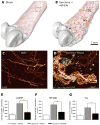
Nerve growth factor (NGF) induces sprouting and neuroma formation by sensory and sympathetic nerve fibers in a model of skeletal pain. Confocal images of periosteum of bone were acquired from whole mount preparations, tiled and overlaid (to scale) on a three-dimensional micro-computed tomography rendering of a sham femur (A) or sarcoma + vehicle femur (B), respectively, using Amira software. Note that the tumor-injected femur (B) has significant cortical bone deterioration and a pathological reorganization of calcitonin gene-related peptide (CGRP) nerve fibers (in red) compared to the sham bone (A). The boxed areas in (A) and (B) correspond to the confocal images in (C) and (D), respectively. High power confocal images of non-decalcified whole mount preparations of the femoral periosteum from sham + vehicle (C) or sarcoma + vehicle mice (D) showing CGRP+ nerve fibers and green fluorescent protein (GFP)+ sarcoma cancer cells (green). When GFP+ tumor cells invade the periosteum, they induce ectopic sprouting of CGRP+ sensory fibers (D, arrow) and the formation of neuroma-like structures. Administration of NGF sequestering therapy (10 mg/kg; intraperitoneal, given at days 6, 12, and 18 post cell injection) reduces sarcoma-induced nerve sprouting of CGRP+ (E), 200kd neurofilament (NF200)+ (F), and tyrosine hydroxylase (TH)+ (G) nerve fibers at day 20 post-cancer cell injection. Nerve fiber density was determined by measuring the total length of nerve fibers per unit volume in the periosteum. *p<0.05. Bars represent the mean ± SEM. Reproduced and modified with permission from Mantyh et al. 2010.
Fig. 4
Fig. 4
Therapies that sequester nerve growth factor (NGF) or inhibit tropomyosin-related kinase A receptor (TrkA) demonstrate significant analgesic efficacy in mouse and a human model of non-malignant skeletal pain. In a mouse model of bone fracture, pain-related behaviors (the time spent guarding of the fractured limb over a 2-minute observation) were significantly reduced by (A) anti-NGF therapy (10 mg/kg, i.p., administered at day 1, day 6 and day 11 post-fracture) and the pan-Trk antagonist ARRY-470 (30 mg/kg, p.o., administered twice daily beginning on day 1 post fracture). Note that anti-NGF therapy (A) and the pan-Trk inhibitor (B) both reduced non-malignant fracture pain-related behaviors by approximately 50%. (C) Anti-NGF therapy reduced walking pain in human patients with moderate to severe osteoarthritis pain. The patient’s assessment of knee pain while walking in response to therapy were obtained at baseline and at the indicated times with the use of a visual-analogue scale that ranged from 0 to 100. In the case of knee pain, a decrease in the score indicates improvement (i.e., less pain). Changes are reported as least-squares means ±SE. P<0.001 for the comparisons of all doses of anti-NGF (tanezumab) with placebo in the assessment of knee pain, except for the comparison of 10 μg of tanezumab per kilogram of body weight with placebo in the patient’s global assessment, for which P = 0.001. Reproduced with permission from Koewler et al. 2007, Ghilardi et al., 2011, and Lane et al. 2010.
Fig. 5
Fig. 5
There are differences in the percentages of tropomyosin-related kinase A receptor (TrkA)+ sensory nerve fibers that innervate the bone vs skin. The skin is innervated by thickly myelinated A-beta fibers (TrkA), thinly myelinated A delta fibers (both TkA and TrkA+), unmyelinated peptide-rich C fibers (TrkA+) and unmyelinated peptide-poor C-fibers (TrkA). In contrast, the bone appears to be predominantly innervated by thinly myelinated A-delta fibers (TrkA but mostly TrkA+) and peptide-rich C-fibers (mostly TrkA+ and a small proportion TrkA). The percentages and types of sensory nerve fibers innervating the skin ,,, and bone,,, were estimated from previous studies. As greater than 80% of all sensory nerve fibers that innervate the bone are TrkA+ whereas only 30% of the sensory nerve fibers that innervate skin are TrkA+, these data might help explain why blocking nerve growth factor or TrkA is highly efficacious in attenuating skeletal pain. Modified from. Castaneda-Hernandez et al. 2011.

References

    1. Hardt J, Jacobsen C, Goldberg J, Nickel R, Buchwald D. Prevalence of chronic pain in a representative sample in the United States. Pain Med. 2008;9:803–12. - PubMed
    1. Breivik H, Collett B, Ventafridda V, Cohen R, Gallacher D. Survey of chronic pain in Europe: Prevalence, impact on daily life, and treatment. Eur J Pain. 2006;10:287–333. - PubMed
    1. Fishman SM, Teichera D. Challenges and choices in drug therapy for chronic pain. Cleve Clin J Med. 2003;70:119–38. - PubMed
    1. Katz WA, Barkin RL. Dilemmas in chronic/persistent pain management. Dis Mon. 2010;56:233–50. - PubMed
    1. Benyamin R, Trescot AM, Datta S, Buenaventura R, Adlaka R, Sehgal N, Glaser SE, Vallejo R. Opioid complications and side effects. Pain Physician. 2008;11:S105–20. - PubMed

Publication types

MeSH terms