. 2011 Jun;13(6):700-7.
doi: 10.1038/ncb2259. Epub 2011 May 22.

Subcellular spatial regulation of canonical Wnt signalling at the primary cilium

Affiliations

Subcellular spatial regulation of canonical Wnt signalling at the primary cilium

Madeline A Lancaster et al. Nat Cell Biol. 2011 Jun.

Abstract

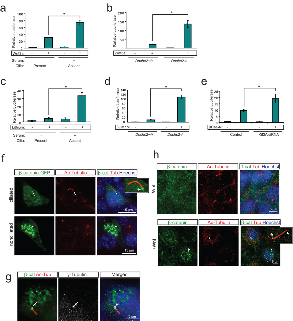
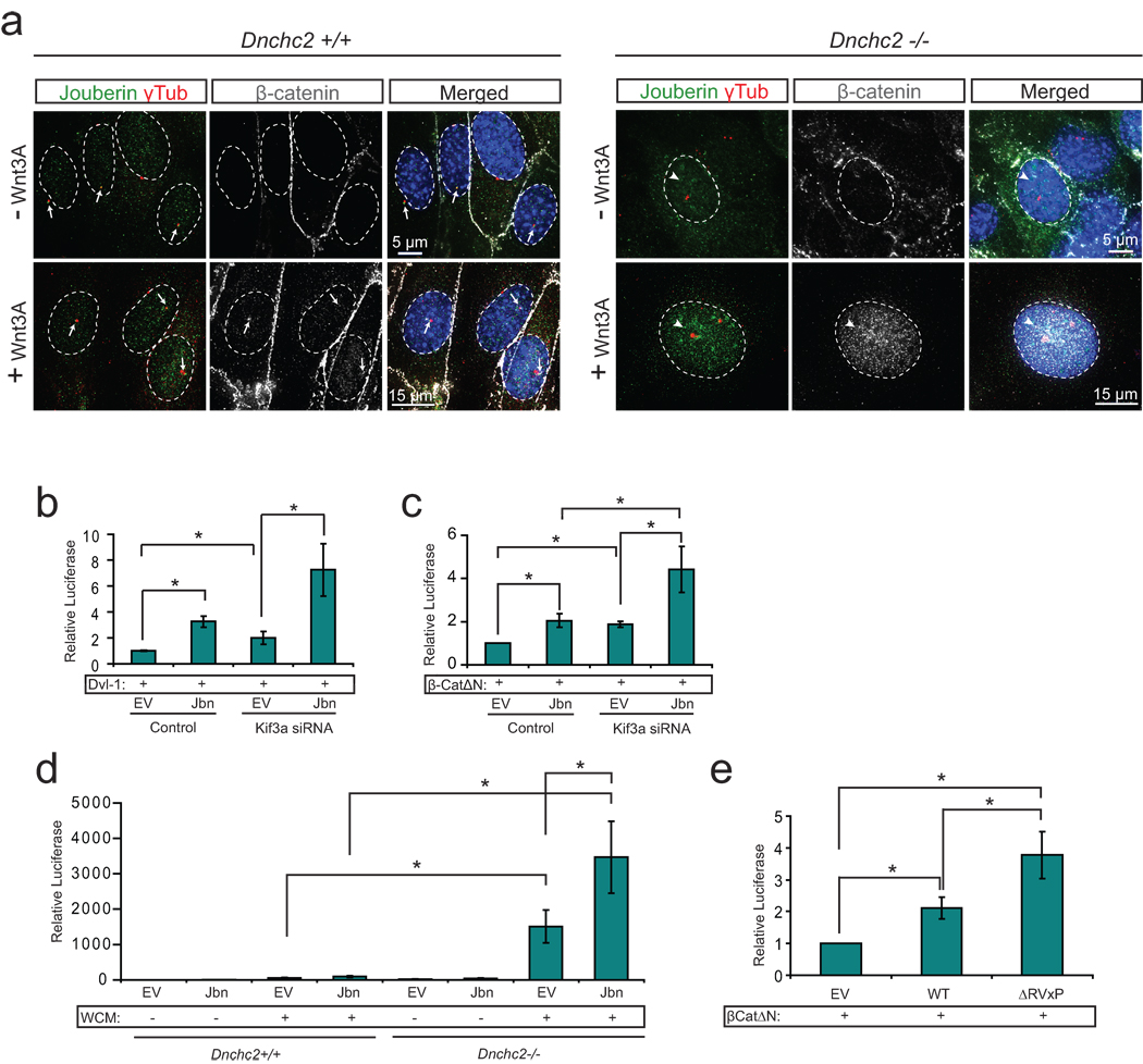
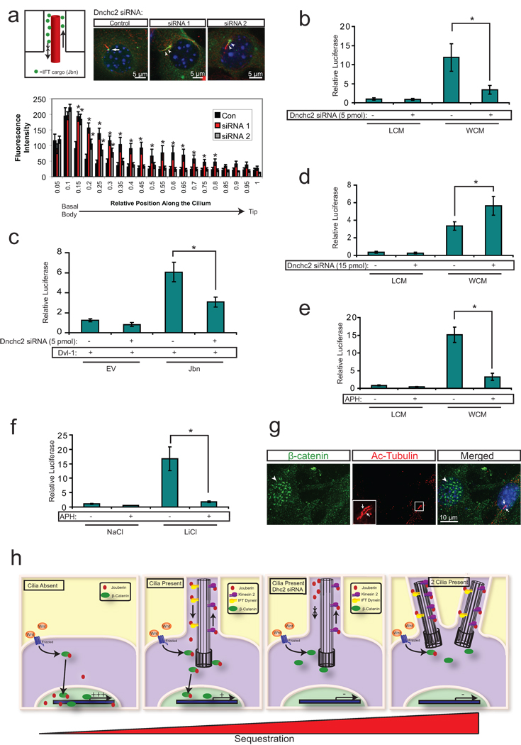
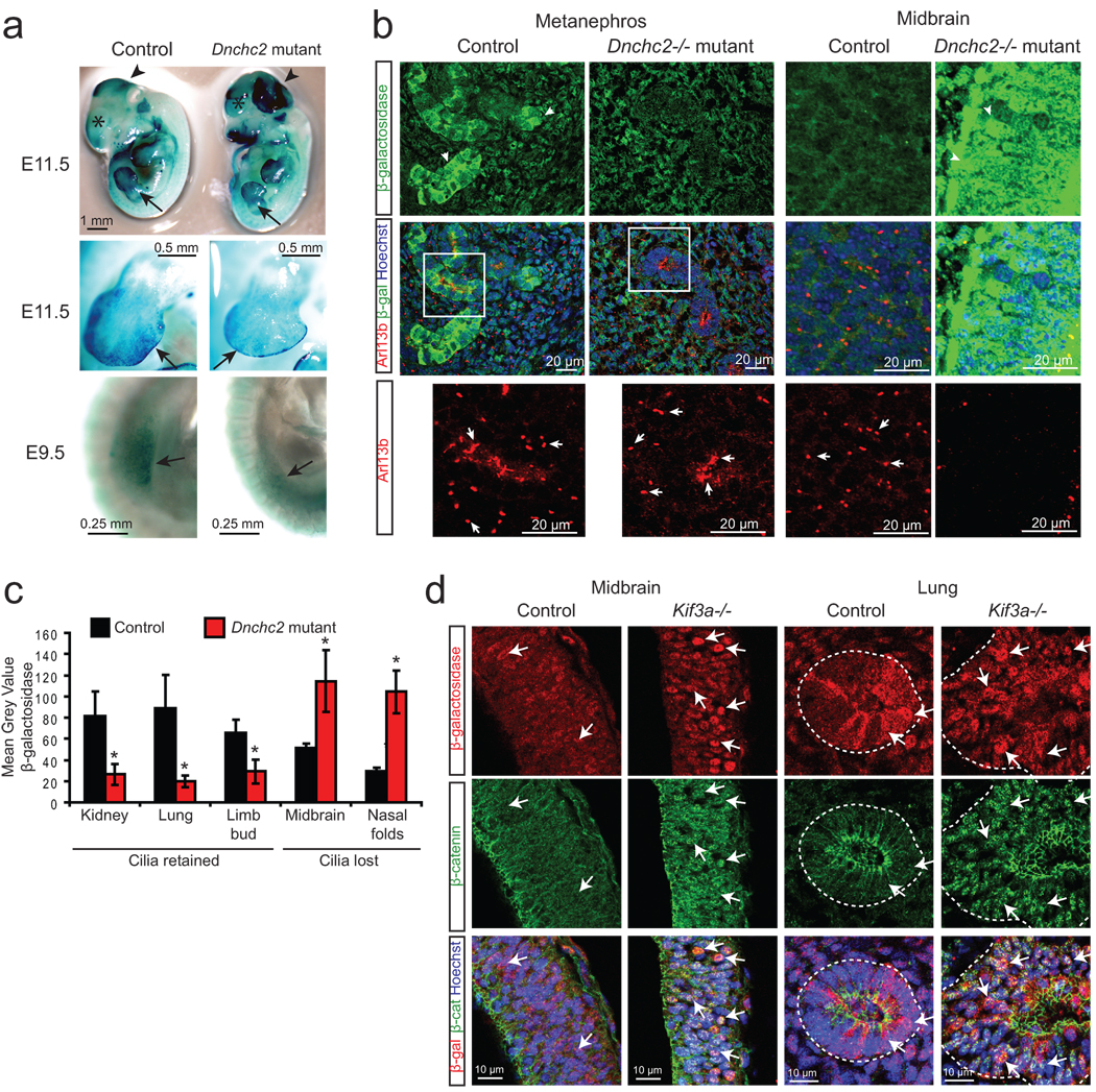
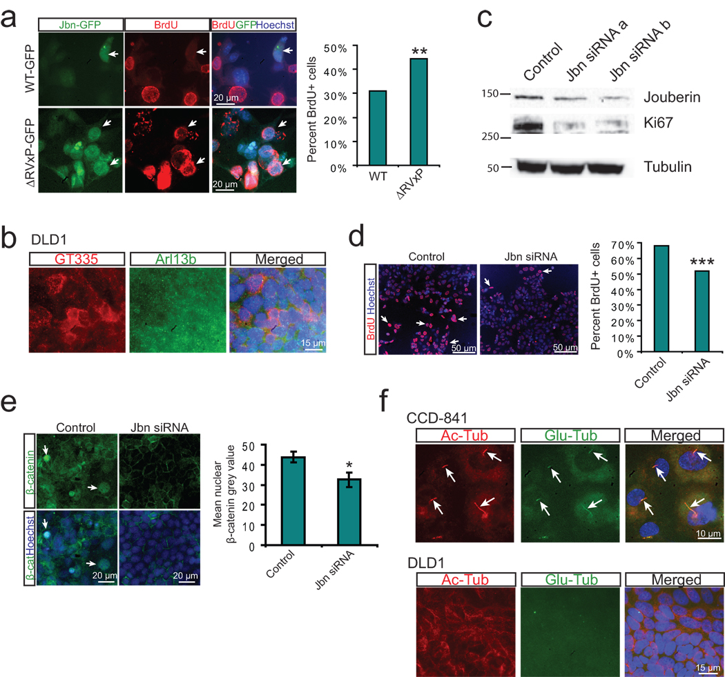
Mechanisms of signal transduction regulation remain a fundamental question in a variety of biological processes and diseases. Previous evidence indicates that the primary cilium can act as a signalling hub, but its exact role in many of the described pathways has remained elusive. Here, we investigate the mechanism of cilia-mediated regulation of the canonical Wnt pathway. We found that primary cilia dampen canonical Wnt signalling through a spatial mechanism involving compartmentalization of signalling components. The cilium, through regulated intraflagellar transport, diverts Jouberin (Jbn), a ciliopathy protein and context-specific Wnt pathway regulator, away from the nucleus and limits β-catenin nuclear entry. This repressive regulation does not silence the pathway, but instead maintains a discrete range of Wnt responsiveness; cells without cilia have potentiated Wnt responses, whereas cells with multiple cilia have inhibited responses. Furthermore, we show that this regulation occurs during embryonic development and is disrupted in cancer cell proliferation. Together these data explain a spatial mechanism of Wnt signalling regulation that may provide insight into ciliary regulation of other signalling pathways.


Figures

Figure 1
Figure 1. The primary cilium dampens Wnt activity by regulating β-catenin
a. Luciferase response to Wnt3a conditioned media in a wild-type MEF cell line following serum starvation and re-addition. *P<0.05, n=3 independent experiments, Student’s t-test. b. Luciferase assay in Dnchc2 mutant MEFs and littermate control MEFs reveals increased Wnt response to Wnt3a conditioned media in Dnchc2−/− cilium mutant MEFs. *P<0.05, n=3 independent experiments, Student’s t-test. c. Luciferase response to LiCl in wild-type MEFs following serum starvation and re-addition. *P<0.05, n=3 independent experiments, Student’s t-test. d. Luciferase activity in Dnchc2 MEFs with activation of the pathway with transfected β-catΔN. *P<0.05, n=3 independent experiments, Student’s t-test. e. Luciferase activity in MEFs transfected with Kif3a siRNA and activation of the pathway with cotransfected β-catΔN. *P<0.05, n=3 independent experiments, Student’s t-test. Values for luciferase assays were normalized across measurements for each experiment and for transfection efficiency relative to β-gal. Error bars represent S.E.M. f. Localization of β-catenin-GFP (green) in wild-type MEFs treated with Wnt3a conditioned media and stained with acetylated tubulin (red) to mark cilia and Hoechst (blue) as a nuclear marker. Arrows point to basal body localization of β-catenin while arrowheads indicate nuclear localization in a nearby nonciliated cell. g. Colocalization of β-catenin-GFP (green) with the basal body marker γ-tubulin (grey, arrow) and cilia (acetylated tubulin, red). Hoechst (blue) labels the nucleus. h. Localization of endogenous β-catenin (green) in wild-type MEFs in the presence and absence of Wnt3a conditioned media. Cilia are labeled with acetylated-tubulin (red) and nuclei are labeled with Hoechst (blue). Arrow marks ciliary localization of β-catenin while the arrowhead indicates nuclear localization in a neighboring nonciliated cell when treated with Wnt3a.
Figure 2
Figure 2. The primary cilium inhibits Jouberin mediated Wnt pathway regulation
a. Localization of endogenous β-catenin (grey) and Jbn (green) in Dnchc2 MEFs. Control MEFs (top) exhibit basal body localization (γ-tubulin, red) of β-catenin and Jbn (arrows) when treated with Wnt3A conditioned media, whereas Dnchc2−/− MEFs exhibit increased nuclear (Hoechst, blue) staining (arrowheads) of both β-catenin and Jbn. b. Luciferase activity in 293T cells transfected with Kif3a siRNA and overexpressed Jbn or empty vector (EV) with activation of the pathway by cotransfection of Dvl-1. *P<0.05, n=3 independent experiments, Student’s t-test. c. Luciferase activity in 293Ts transfected with Kif3a siRNA and overexpressed Jbn or EV with activation of the pathway with cotransfected β-catΔN. *P<0.05, n=3 independent experiments, Student’s t-test. d. Luciferase activity in Dnchc2 MEFs transfected with Jbn or EV with activation of the pathway with Wnt3a conditioned media (WCM). *P<0.05, n=3 independent experiments, Student’s t-test. e. Luciferase activity in MEFs with overexpression of EV, wild-type Jbn or ΔRVxP mutant construct and cotransfected β-catΔN. *P<0.05, n=3 independent experiments, Student’s t-test. Values for luciferase assays were normalized across measurements for each experiment and for transfection efficiency relative to β-gal. Error bars represent S.E.M.
Figure 3
Figure 3. The primary cilium sequesters Jouberin and β-catenin away from the nucleus
a. 3T3s transfected with two siRNAs to Dnchc2 exhibit accumulation of Jbn-GFP (green) along the length of the cilium (labeled with antibody to acetylated tubulin, red). The nucleus is labeled with Hoechst (blue). Schematic represents the experimental approach and interpretation of these results. Quantification of the green fluorescence along the cilium reveals a shifted localization along the ciliary axoneme in siRNA transfected cells with significantly increased levels of GFP beyond the basal body. Average fluorescence intensities of cells transfected with negative control siRNA (n=7), Dnchc2 siRNA #1 (n=10), or Dnchc2 siRNA #2 (n=11) were normalized and displayed in arbitrary grey value units. Position along the cilium was normalized for total cilia length and results were binned into 20 equal segments. b. Luciferase activity in MEFs transfected with 5 pmol of Dnchc2 siRNA and treated with Wnt3a conditioned media (WCM) or control L-cell conditioned media (LCM). *P<0.05, n=7 from 6 independent experiments, Student’s t-test. c. Wnt activity as reported by luciferase in 293T cells with overexpression of wild-type Jbn or EV and cotransfection of 5 pmol negative control siRNA or Dnchc2 siRNA. Cells were cotransfected with Dvl-1 to activate the Wnt pathway. *P<0.05, n=6 from three independent experiments, Student’s t-test. d. Luciferase activity in MEFs transfected with 3-fold increased Dnchc2 siRNA dose and treated with WCM or LCM. *P<0.05, n=7 from 6 independent experiments, Student’s t-test. e. Luciferase activity in MEFs treated with 1 µg/ml aphidicolin (APH) following serum starvation and treated with WCM or LCM. *P<0.05, n=6 from 5 independent experiments, Student’s t-test. f. Luciferase activity in MEFs treated with 1 µg/ml APH following serum starvation and treated with LiCl or NaCl. *P<0.05, n=3 independent experiments, Student’s t-test. Values for luciferase assays were normalized across measurements for each experiment and for transfection efficiency relative to β-gal. Error bars represent S.E.M. g. Localization of endogenous β-catenin (green) in MEFs treated with APH. Arrows point to two cilia (red, acetylated tubulin) within one cell while arrowheads point to nuclear β-catenin in a neighboring nonciliated cell. Hoechst (blue) labels nuclei. h. Model of the proposed mechanism of ciliary inhibition of canonical Wnt signaling. Jbn is proposed to interact with β-catenin upon Wnt activation and facilitate its translocation into the nucleus. In the absence of the cilium, this translocation is uninhibited compared to ciliated cells where nuclear translocation is highly regulated. This regulation is dependent upon intact retrograde IFT of Jbn down the cilium to the basal body where it can then interact with β-catenin and facilitate its nuclear accumulation. Furthermore, the presence of two cilia more strictly regulates the nuclear import of β-catenin, further dampening the pathway.
Figure 4
Figure 4. Wnt is regulated by the primary cilium during embryonic development
a. X-gal staining (blue) of Dnchc2 mutants at E11.5 and E9.5. Arrows point to developing limb bud, arrowheads demarcate the mid-hindbrain region, and asterisks label the developing telencephalon. b. Representative E11.5 embryonic sections of the developing kidney (metanephros) and midbrain stained for cilia (Arl13b, red) and Wnt activity (β-galactosidase, green). Hoechst labels nuclei (blue). Arrows point to cilia in control and mutant tissue, which are shown as magnified insets for the metanephros. Images for control and mutant tissues were acquired and analyzed identically. c. Quantification of average staining intensity for β-galactosidase in representative tissues from three E11.5 littermate control and Dnchc2 mutant embryos. Tissues that are ciliated (kidney, lung, limb bud) and nonciliated (midbrain, nasal folds) in the mutant are shown for comparison of the effect of loss of cilia compared with specific retrograde IFT defect. *P<0.05, Student’s t-test. Error bars represent S.E.M. d. β-galactosidase staining (red) and β-catenin staining (green) in E9.5 midbrain and lung from Kif3a mutant and littermate control embryos. Arrows point to β-galactosidase reporter activity and β-catenin nuclear staining, both of which are increased in Kif3a mutant tissues.
Figure 5
Figure 5. Regulation of cancer cell proliferation by cilia-localized Jouberin
a. BrdU staining (red) in MEFs transfected with GFP tagged wild-type or ΔRVxP mutant construct (green). Hoechst labels nuclei (blue). Arrows point to GFP positive transfected cells. Quantification of the percent of 200 counted GFP positive cells which stained positive for BrdU is shown at right. **P<0.0005, Chi-square test. b. Staining for cilia in DLD1 cells reveals a lack of cilia. Cilia markers include GT335 (red), Arl13b (green), while Hoechst labels nuclei. c. Western blot analysis of Jbn and Ki67 expression from whole cell lysates of DLD1 cells transfected with Jbn siRNAs. Alpha-tubulin is shown as the loading control. d. BrdU staining (red) of DLD1 cells transfected with Jbn siRNA and treated for 16 hr with BrdU. Hoechst labels nuclei (blue). Quantification of 3000 cells from images acquired and analyzed identically is shown at the right. ***P<10−5, Chi-square test. e. β-catenin staining (green) in DLD1 cells transfected with Jbn siRNA. Hoechst labels nuclei (blue). Arrows point to nuclear β-catenin staining. Quantification of average nuclear staining intensity for β-catenin from over 100 cells in three regions of the well is shown at the bottom. *P<0.05, Student’s t-test. Error bars represent S.E.M. f. Cilia staining using two markers: acetylated tubulin (red) and glutamylated tubulin (green) in human colon CCD-841 cells which exhibited cilia in 75% of cells (over 200 cells examined) whereas DLD1 cells completely lacked cilia. Hoechst (blue) labels nuclei.

Comment in

References

    1. Berbari NF, O'Connor AK, Haycraft CJ, Yoder BK. The primary cilium as a complex signaling center. Curr Biol. 2009;19:R526–R535. - PMC - PubMed
    1. Lancaster MA, et al. Impaired Wnt-beta-catenin signaling disrupts adult renal homeostasis and leads to cystic kidney ciliopathy. Nat Med. 2009;15:1046–1054. - PMC - PubMed
    1. MacDonald BT, Tamai K, He X. Wnt/beta-catenin signaling: components, mechanisms, and diseases. Dev Cell. 2009;17:9–26. - PMC - PubMed
    1. Lai SL, Chien AJ, Moon RT. Wnt/Fz signaling and the cytoskeleton: potential roles in tumorigenesis. Cell Res. 2009;19:532–545. - PubMed
    1. Sharma N, Berbari NF, Yoder BK. Ciliary dysfunction in developmental abnormalities and diseases. Curr Top Dev Biol. 2008;85:371–427. - PubMed

Publication types

Substances