. 2011 Jun;17(6):684-91.
doi: 10.1038/nm.2388. Epub 2011 May 22.

Lrp5 functions in bone to regulate bone mass

Affiliations

Lrp5 functions in bone to regulate bone mass

Yajun Cui et al. Nat Med. 2011 Jun.

Abstract

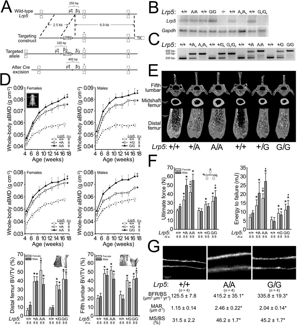
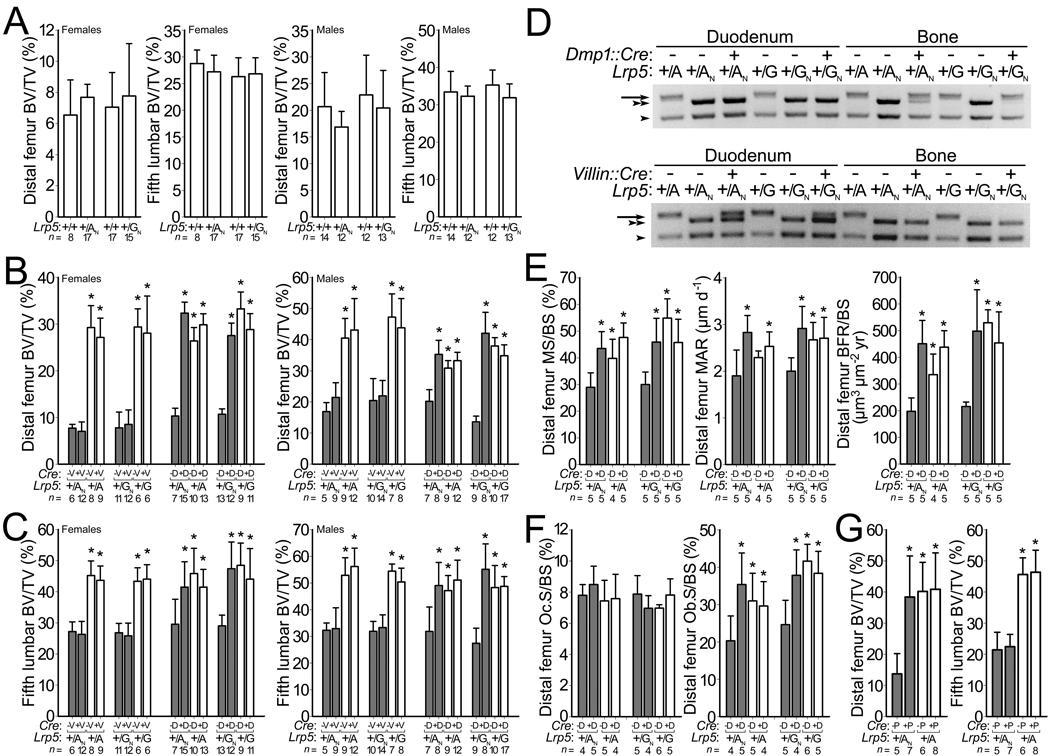
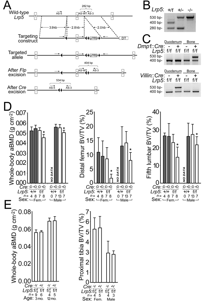
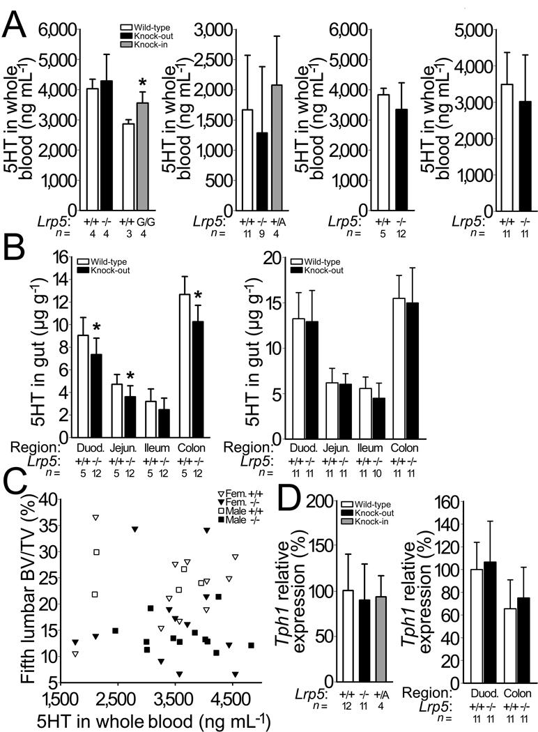
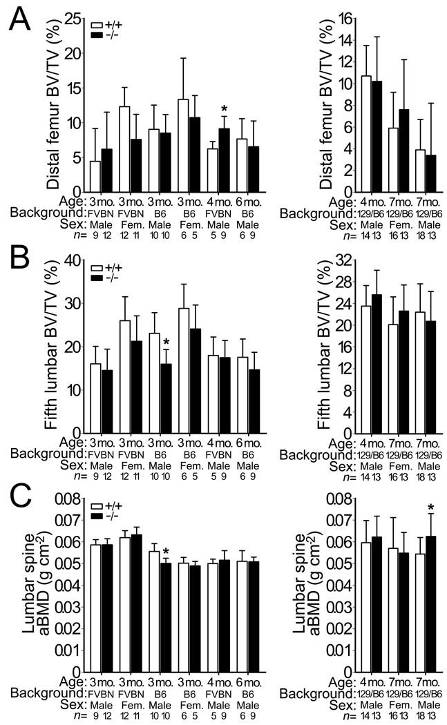
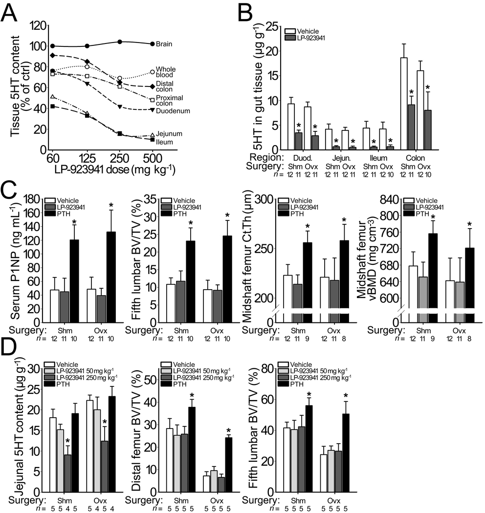
The human skeleton is affected by mutations in low-density lipoprotein receptor-related protein 5 (LRP5). To understand how LRP5 influences bone properties, we generated mice with osteocyte-specific expression of inducible Lrp5 mutations that cause high and low bone mass phenotypes in humans. We found that bone properties in these mice were comparable to bone properties in mice with inherited mutations. We also induced an Lrp5 mutation in cells that form the appendicular skeleton but not in cells that form the axial skeleton; we observed that bone properties were altered in the limb but not in the spine. These data indicate that Lrp5 signaling functions locally, and they suggest that increasing LRP5 signaling in mature bone cells may be a strategy for treating human disorders associated with low bone mass, such as osteoporosis.


Figures

Figure 1
Figure 1
Generation and characterization of HBM Lrp5 knockin mice. (a) Schematic depicting the targeting and genotyping strategies for HBM Lrp5 alleles. The 5’ targeting arm contains a neomycin-resistance cassette (NeoR) flanked by LoxP sites (arrowheads). The 3’ targeting arm begins in intron 2 and extends into intron 4, and is followed by a thymidine kinase (TK) cassette. Site-directed mutagenesis altered specific amino acid residues encoded by Lrp5 exon 3 (asterisk). The relative locations and orientation of primers (P1, P2, and P3) used for PCR genotyping and their expected amplimer sizes are noted. (b) Autoradiographs of a northern blot containing whole bone total RNA from mice with different Lrp5 genotypes initially hybridized with a radioactive Lrp5 cDNA probe (upper) and subsequently with a Gapdh cDNA probe which serves as a loading control (lower). (c) Photograph of an agarose gel containing PCR amplimers derived from genomic DNA of mice with different Lrp5 genotypes. (d) Graphs depicting the areal bone mineral density (aBMD) measured by DEXA in mice with different Lrp5 genotypes followed to 16.5-weeks-old (top and middle rows). Graphs depicting the percent trabecular bone volume in the total volume (BV/TV) of the distal femurs and 5th lumbar vertebrae of 16.5-wk-old male and female mice with different Lrp5 genotypes (bottom row). (e) Representative µCT scan images obtained from 16.5-wk-old mice with different Lrp5 genotypes. (f) Graphs depicting biomechanical properties of whole femurs in a 3-point bending assay from 16.5-wk-old mice with different Lrp5 genotypes. (g) Representative images of new bone formation assessed by double calcein labeling of mice with different Lrp5 genotypes. Bone formation rates/bone surface area (BFR/BS), mineral apposition rates (MAR), and mineralizing surface/bone surface (MS/BS). The numbers of mice studied (n =) are indicated, as are error bars equal to 1 s.d. Asterisks (*) indicate a significant difference (p < 0.05) when compared to WT mice, whereas # indicates a significant difference (p < 0.05) when compared to heterozygous HBM Lrp5 mice.
Figure 2
Figure 2
Effect of conditionally activating NeoR-containing HBM Lrp5 alleles. Graphs depicting (a) femoral and vertebral trabecular BV/TV in WT mice and in mice with NeoR-containing HBM Lrp5 alleles. (b) femoral trabecular BV/TV and (c) vertebral trabecular BV/TV in mice with (shaded bars) and without (unshaded bars) inherited NeoR-containing HBM Lrp5 alleles (+/AN or +/GN, and +/A or +G, respectively), and with and without Villin::Cre (+V and −V, respectively) or Dmp1::Cre (+D and −D, respectively) transgenes. (d) Photographs of agarose gels containing PCR amplimers derived from mouse genomic DNA extracted from either duodenum or femur cortex of mice with different Lrp5 and Cre-transgene genotypes. PCR amplimers correspond to the sizes depicted in Fig. 1a. (top) amplimers from the Dmp1::Cre cross. (bottom) amplimers from the Villin::Cre cross. WT allele (arrowhead), AN or GN allele (double arrowhead), A or G allele (arrow). (e) Graphs depicting fluorochrome-derived bone formation parameters in the distal femur from 9-wk-old female mice administered double calcein labeling. X-axis group notations follow those described for panel b. (f) Graphs depicting the proportion of distal femur trabecular bone surface covered by osteoclasts (left) and osteoblasts (right). (g) Graphs depicting femur and vertebra trabecular BV/TV in 12-wk-old HBM Lrp5 mice with (shaded bars) and without (unshaded bars) inherited NeoR-containing alleles (+/AN and +/A), respectively, and with and without the Prx1::Cre transgene (+P and −P, respectively). MS/BS, MAR, BFR/BS are as defined in Fig. 1. The numbers of mice studied (n =) are indicated, as are error bars equal to 1 s.d. Asterisks (*) indicate a significant difference (p < 0.05) when compared to NeoR-containing littermates that did not inherit a Cre-transgene.
Figure 3
Figure 3
Generation and characterization of mice with a conditional knockout allele of Lrp5. (a) Schematic depicting the creation of the Lrp5 floxed allele. LoxP sites (arrowheads), the neomycin-resistance cassette (NeoR), FRT sites (diamonds), and the diptheria toxin (DT) cassette are shown. The relative locations and orientation of the 3 primers (arrows) used for PCR genotyping and their expected amplimer sizes are noted. (b) Photograph of agarose gel depicting PCR amplimers for WT (+), floxed (f), and knockout (−) Lrp5 alleles from genomic DNA of mice with different Lrp5 genotypes. (c) Photographs of agarose gels containing PCR amplimers derived from mouse genomic DNA extracted from either duodenum or femur cortex of floxed Lrp5 mice, with or without the Dmp1::Cre transgene (top) and with or without the Villin::Cre transgene (bottom). (d) Graphs depicting whole-body aBMD (left), femoral trabecular BV/TV (middle), and vertebral trabecular BV/TV (right) in 16-wk-old mice homozygous for WT or floxed Lrp5 alleles, with or without the Dmp1::Cre transgene. (e) Graphs depicting whole-body aBMD and tibial trabecular BV/TV in 3-mo-old and 12-mo-old mice heterozygous or homozygous for floxed Lrp5 alleles with or without the Villin::Cre transgene. The numbers of mice studied (n =) are indicated, as are error bars equal to 1 s.d. Asterisks (*) indicate a significant difference (p < 0.05) when compared to floxed Lrp5 littermates that did not inherit the Cre transgene.
Figure 4
Figure 4
Effect of Lrp5 genotype on serotonin (5HT) levels and on Tph1 expression. (a) Graphs depicting whole blood 5HT measured by HPLC in 6-mo-old Lrp5 WT and knockout mice that had been backcrossed to C57BL6/J, and in Lrp5 WT and HBM knockin (G/G) mice on a mixed 129Sv/C57BL/6J background (far left), in 3-mo-old Lrp5 WT, knockout and HBM knockin (+/A) mice on a mixed 129Sv/C57BL/6J genetic background (middle left), in 3-mo-old male (middle right) and 13-mo-old female (far right) WT and knockout littermates on a mixed 129SvEvBrd/ C57BL/6J-Tyrc-Brd background. (b) Graphs depicting the quantity of 5HT extracted from several regions of the intestine, beginning in the duodenum and proceeding through the jejunum, ileum, and proximal colon in 3-mo-old male (left) and in 13-mo-old female (right) Lrp5 WT and knockout littermates on a mixed 129SvEvBrd/ C57BL/6J-Tyrc-Brd background. (c) Scattergram depicting vertebral trabecular BV/TV and whole blood 5HT measurements in individual Lrp5 WT (open symbols) and knockout (filled symbols) littermates. Correlations between BV/TV and whole blood serotonin were r2=0.13 (p = 0.16) for male mice, and r2=0.02 (p = 0.53) for female mice. (d) Graphs depicting normalized Tph1 transcript levels in duodenum RNA extracts from Lrp5 WT, knockout, and HBM knockin (+/A) mice on a mixed 129Sv/C57BL/6J genetic background (left) and duodenum and colon RNA extracts from Lrp5 WT and knockout mice on a 129SvEvBrd/C57BL/6J-Tyrc-Brd background (right) with Gapdh serving as the internal control. The mean Tph1 expression level for Lrp5 WT duodenum is set as 100%. The numbers of mice studied (n =) are indicated, as are error bars equal to 1 s.d. Asterisks (*) indicate a significant difference (p < 0.05) compared to WT mice.
Figure 5
Figure 5
Bone mass in WT and Tph1−/− mice. (a) Graphs depicting femoral trabecular BV/TV in WT and Tph1−/− mice on either FVB/N or C57BL/6 backgrounds (left) or on a mixed 129SvEvBrd/C57BL/6J-Tyrc-Brd background (right). (b) Graphs depicting the vertebral trabecular BV/TV of the 5th lumbar vertebra in the same mice described in panel a. (c) Graphs depicting lumbar spine aBMD, as measured by DEXA, in the same mice described in panel a. The numbers of mice studied (n =) are indicated, as are 1 s.d. error bars. Asterisks (*) indicate a significant difference (p < 0.05) compared to WT mice using an unpaired t-test; none of these differences remain significant after correcting for multiple testing.
Figure 6
Figure 6
Bone mass following pharmacologic inhibition of Tph1 activity. (a) Graph depicting dose dependent changes in 5HT content, compared to vehicle-treated controls, in 9-wk-old WT female C57BL/6 mice after receiving daily doses of LP-923941 for 7 days. A daily dose of 250 mg/kg lowered 5HT levels in whole blood and in intestine, but not in brain. (b) Graph depicting changes in the intestinal 5HT content of sham-operated (SHM) and ovariectomized (OVX) mice that received vehicle or LP-923941 (250 mg/kg/day) for 6 wks. OVX alone reduced 5HT content in the duodenum and colon by ~12% (p < 0.05) compared to SHM mice. Treatment with LP-923941 significantly reduced serotonin content equally in all regions of the intestine in SHM and in OVX mice. (c) Effect of treatment with LP-923941 (250 mg/kg/day) or teriparatide (80 µg/kg/day), the 1–34 residue amino-terminal fragment of human parathyroid hormone (PTH), on serum PINP levels, a marker of bone formation (left), vertebral trabecular BV/TV (middle left), midshaft femur cortical thickness (middle right), and midshaft femoral volumetric BMD (right) in SHM and in OVX mice. (d) Effect of treating SHM and OVX rats with LP-923941 (50 or 250 mg/kg/day) or teriparatide (80 µg/kg/day) for 6 weeks on jejunal 5HT content (left), femoral trabecular BV/TV (middle), and vertebral trabecular BV/TV (right). The numbers of mice studied (n =) are indicated, as are error bars equal to 1 s.d. Asterisks (*) indicate a significant difference (p < 0.05) compared to vehicle treated mice.

Comment in

References

    1. Gong Y, et al. LDL receptor-related protein 5 (LRP5) affects bone accrual and eye development. Cell. 2001;107:513–523. - PubMed
    1. Little RD, et al. A mutation in the LDL receptor-related protein 5 gene results in the autosomal dominant high-bone-mass trait. Am J Hum Genet. 2002;70:11–19. - PMC - PubMed
    1. Boyden LM, et al. High bone density due to a mutation in LDL-receptor-related protein 5. N Engl J Med. 2002;346:1513–1521. - PubMed
    1. Balemans W, et al. Increased bone density in sclerosteosis is due to the deficiency of a novel secreted protein (SOST) Hum Mol Genet. 2001;10:537–543. - PubMed
    1. Brunkow ME, et al. Bone dysplasia sclerosteosis results from loss of the SOST gene product, a novel cystine knot-containing protein. Am J Hum Genet. 2001;68:577–589. - PMC - PubMed

Publication types

MeSH terms