. 2011 May 22;12(7):607-15.
doi: 10.1038/ni.2043.

Tumor necrosis factor induces GSK3 kinase-mediated cross-tolerance to endotoxin in macrophages

Affiliations

Tumor necrosis factor induces GSK3 kinase-mediated cross-tolerance to endotoxin in macrophages

Sung Ho Park et al. Nat Immunol. .

Abstract

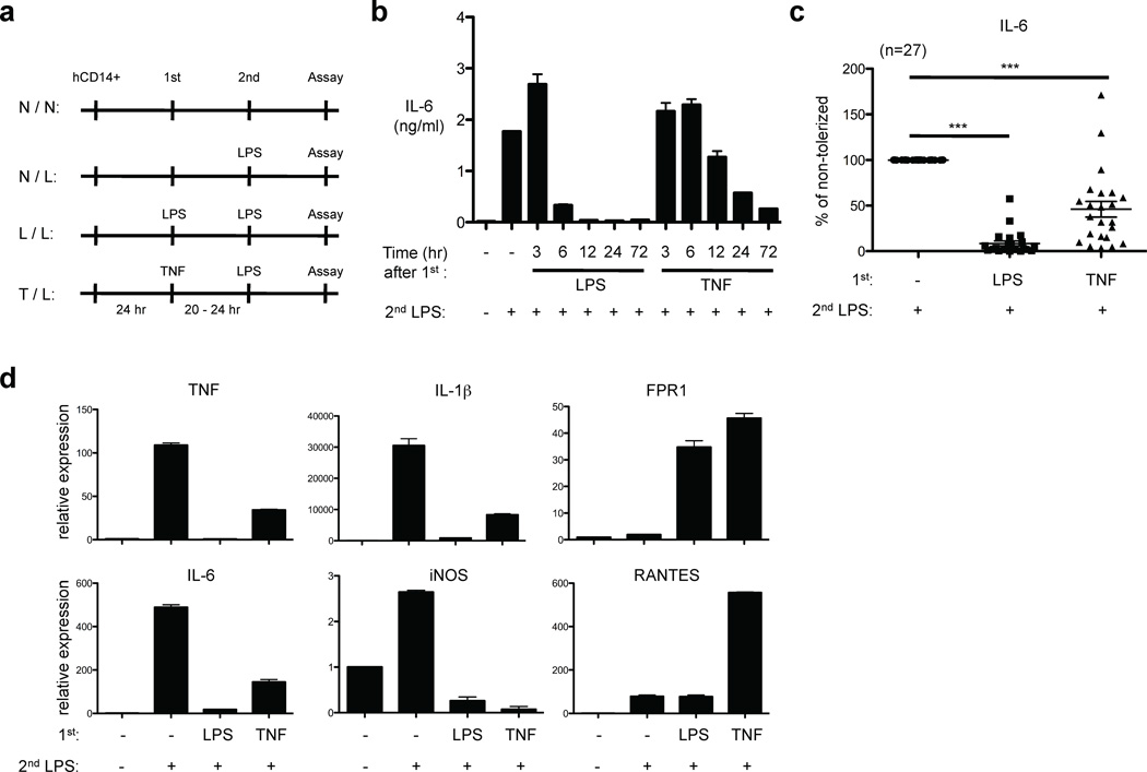
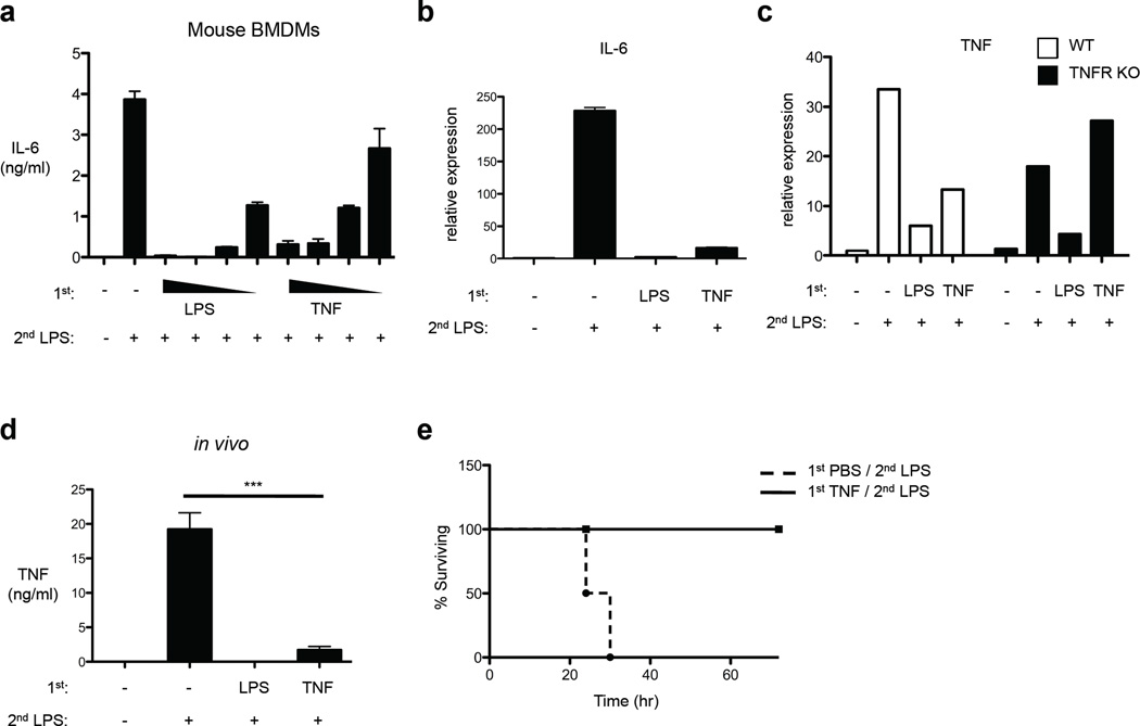
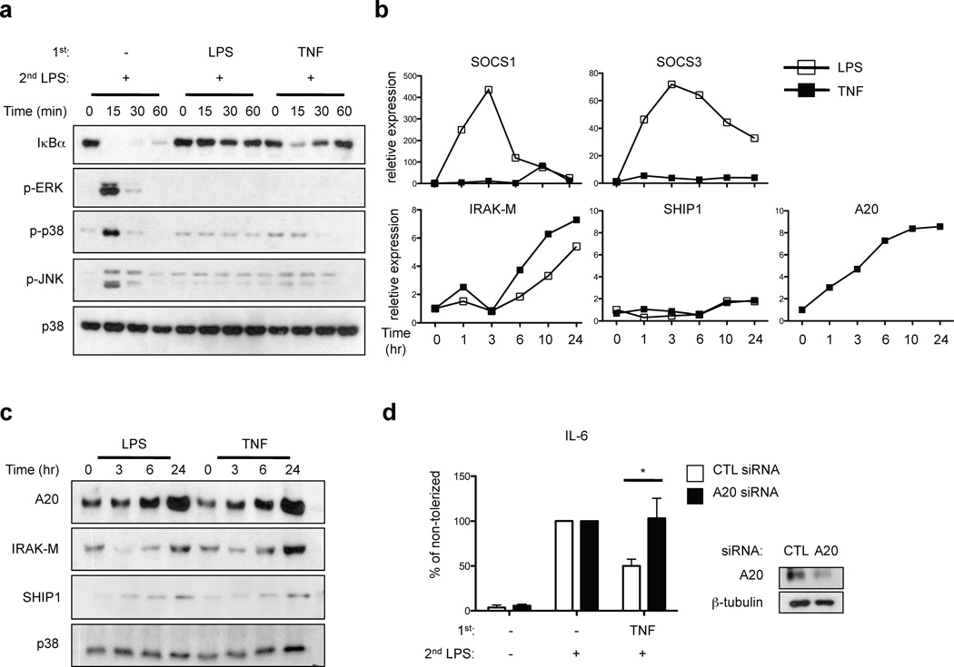
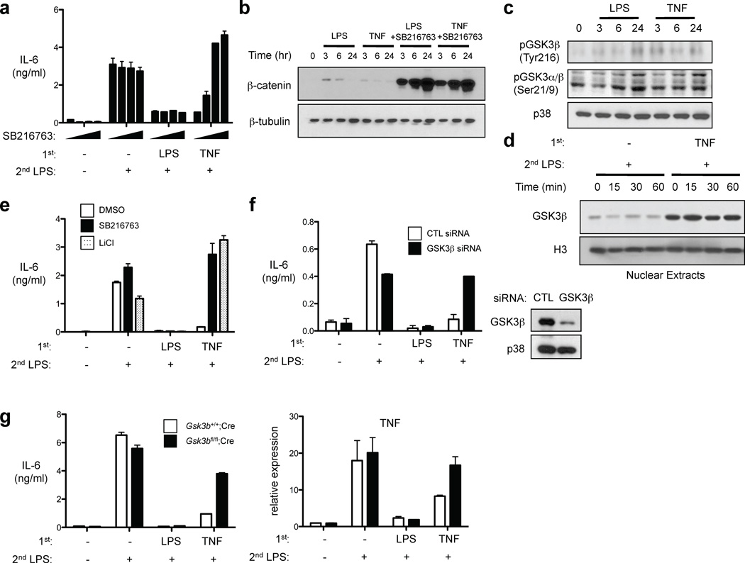
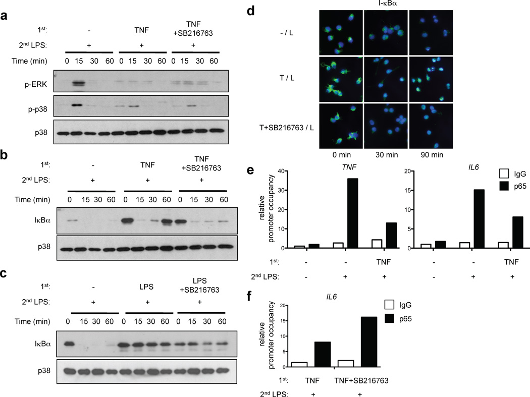
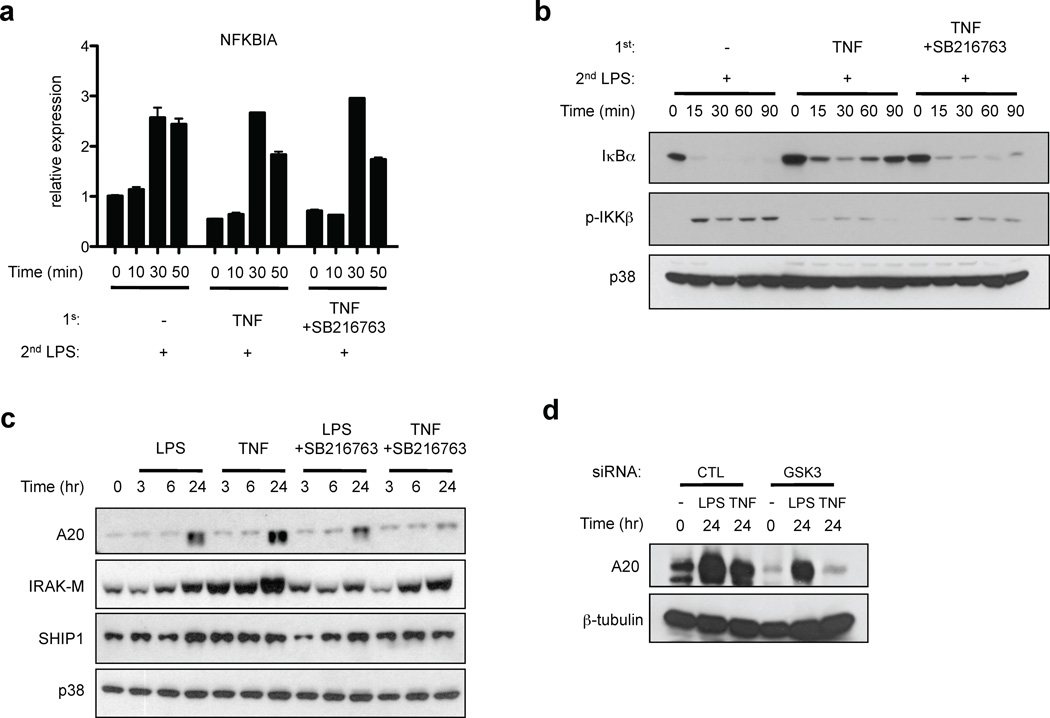
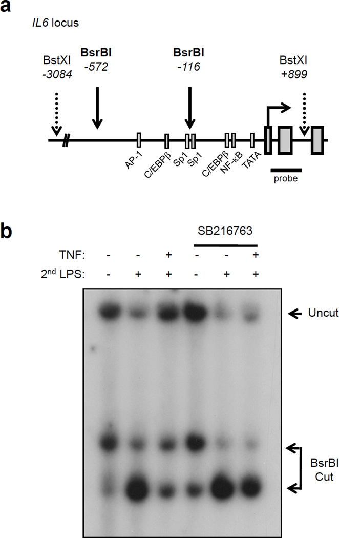
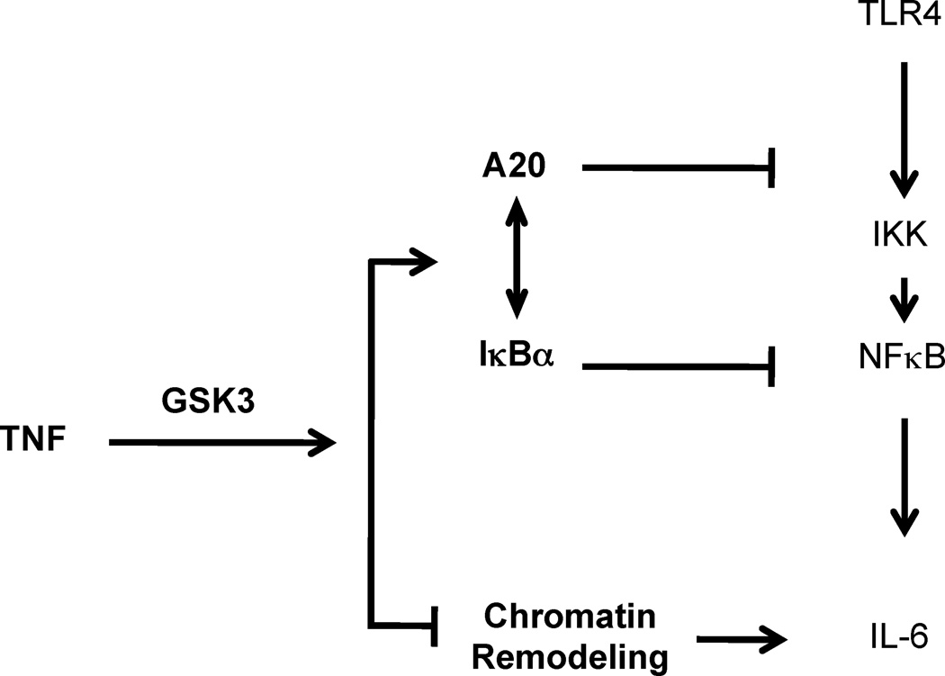
Endotoxin tolerance, a key mechanism for suppressing excessive inflammatory cytokine production, is induced by prior exposure of macrophages to Toll-like receptor (TLR) ligands. Induction of cross-tolerance to endotoxin by endogenous cytokines has not been investigated. Here we show that prior exposure to tumor necrosis factor (TNF) induced a tolerant state in macrophages, with less cytokine production after challenge with lipopolysaccharide (LPS) and protection from LPS-induced death. TNF-induced cross-tolerization was mediated by suppression of LPS-induced signaling and chromatin remodeling. TNF-induced cross-tolerance was dependent on the kinase GSK3, which suppressed chromatin accessibility and promoted rapid termination of signaling via the transcription factor NF-κB by augmenting negative feedback by the signaling inhibitors A20 and IκBα. Our results demonstrate an unexpected homeostatic function for TNF and a GSK3-mediated mechanism for the prevention of prolonged and excessive inflammation.


Figures

Figure 1
Figure 1
TNF pretreatment suppresses induction of pro-inflammatory cytokines on secondary challenge by LPS. (a) Experimental design. (b) Human primary macrophages were stimulated with LPS (10 ng/ml) or TNF (10 ng/ml) for the indicated times and challenged with 10 ng/ml LPS for 24 hr. IL-6 in culture supernatants was measured by ELISA. (c) Percent tolerization of IL-6 was calculated by dividing IL-6 production of tolerized (LPS- or TNF-pretreated) cells by that of non-tolerized cells. Data are a summary of 27 independent donors. P values were calculated by paired Student’s t-test. ***, P < 0.0001 (d) Human primary macrophages were stimulated with LPS (10 ng/ml) or TNF (10 ng/ml) for 24 hr and challenged with 10 ng/ml LPS for 1 hr (TNF, IL-1β, iNOS and RANTES) or 3 hr (IL-6 and FPR1). Real-time PCR was used to measure mRNA levels. Data are representative of four independent donors (mean ± s.d. of triplicate determinations).
Figure 2
Figure 2
TNF suppresses cytokine production in vivo and protects mice from LPS-induced lethality. (a) Mouse BMDMs were stimulated with increasing doses of LPS (1–100 ng/ml) or TNF (1–80 ng/ml) for 24 hr and then challenged with 10 ng/ml LPS. IL-6 in culture supernatants was measured by ELISA. (b) LPS or TNF-pretreated BMDMs as in (a) were stimulated with 10 ng/ml LPS for 1 hr. Real-time PCR was used to measure IL-6 mRNA levels (mean ± s.d. of triplicate determinations). (c) BMDMs from mice lacking TNFR1 and TNFR2 (TNFR KO) or genetically matched controls (WT) were stimulated with LPS (100 ng/ml) or TNF (40 ng/ml) for 24 hr and then challenged with 10 ng/ml LPS for 1 hr. TNF mRNA levels were determined by real-time PCR. Data are representative of three independent experiments. (d) Age- and sex-matched C57/BL6J mice received intraperitoneal (IP) or intravenous (IV) injection of LPS (100 µg) or of TNF (2 µg), respectively. After 24 hr, secondary LPS challenge (200 µg) was given by IP injection and after 90 min serum TNF was determined by ELISA (n=4 mice per group). P value was calculated by unpaired Student’s t-test. ***, P = 0.0004. Similar results were obtained in an additional 2 experiments using different TNF dosing regimens. (e) Mice were injected intravenously with TNF (2 µg), followed by injection of 500 µg of LPS after 24 hr. Survival rates were scored every 6 h for 4 days (n=4 mice per group). Similar results were obtained in an additional 3 experiments using different TNF pretreatment doses.
Figure 3
Figure 3
TNF suppresses TLR4 signaling and induces A20 expression. (a) Human primary macrophages were stimulated with LPS (10 ng/ml) or TNF (10 ng/ml) for 24 hr and challenged with 10 ng/ml LPS for the indicated times. I-κBα amounts and ERK, p38 and JNK phosphorylation were assessed by immunoblotting. (b, c) Human primary macrophages were stimulated with LPS or TNF for the indicated times. SOCS1, SOCS3, IRAK-M, SHIP1 and A20 expression was measured by (b) real-time PCR and (c) immunoblotting. Data are representative of four to twelve independent experiments. (d) Human macrophages transfected with control or A20-specific siRNA were treated with LPS or TNF for 24 hr and then challenged with 10 ng/ml LPS. IL-6 in culture supernatants was measured by ELISA. Percent tolerization of IL-6 was calculated by dividing IL-6 production of LPS- or TNF-pretreated cells by that of non-tolerized cells. Data are a summary of 3 independent experiments. P value was calculated by unpaired Student’s t-test. *, P = 0.038.
Figure 4
Figure 4
TNF-induced tolerance is mediated by GSK3. (a) Human macrophages treated with or without SB216763 (1–20 µM) were cultured with LPS or TNF for 24 hr and then challenged with 10 ng/ml LPS. Culture supernatants were harvested 24 hr later and IL-6 was measured by ELISA. (b, c) Human macrophages treated with or without SB216763 (10 µM) were stimulated with LPS or TNF for the indicated times. Whole cell extracts were analyzed by immunoblotting. (d) Human macrophages treated with or without SB216763 (10 µM) were stimulated with LPS or TNF for 24 hr and then challenged with 10 ng/ml LPS for the indicated times. Nuclear extracts were analyzed by immunoblotting. (e) Human macrophages treated with or without SB216763 (10 µM) or LiCl (20 mM) were cultured with LPS or TNF for 24 hr and then challenged with 10 ng/ml LPS and IL-6 was measured by ELISA. (f) Human macrophages transfected with control or GSK3β-specific short interfering RNA duplexes (siRNA) were treated with LPS or TNF for 24 hr and then challenged with 10 ng/ml LPS. IL-6 in culture supernatants was measured by ELISA. (g) BMDMs from mice lacking myeloid GSK3β or genetically matched controls were stimulated with LPS (100 ng/ml) or TNF (40 ng/ml) for 24 hr and then challenged with 10 ng/ml LPS for 1 hr (right panel) or 24 hr (left panel). TNF mRNA and IL-6 protein were assessed by real-time PCR and ELISA, respectively. Data are representative of at least three experiments.
Figure 5
Figure 5
GSK3 regulates kinetics of I-κBα expression and postinduction repression of NF-κB signaling in LPS-stimulated TNF-tolerized macrophages. (a – c) Human macrophages were pretreated for 24 hr with LPS or TNF with or without SB216763 (10 µM) and then challenged with 10 ng/ml LPS for the indicated times. IκBα amounts and ERK and p38 phosphorylation were assessed by immunoblotting of whole cell lysates. (d) Human macrophages were pretreated with LPS or TNF with or without SB216763 (10 µM) and then challenged with 10 ng/ml LPS for the indicated times. Leptomycin B (10 µM) was added together with LPS to inhibit CRM1/exportin1-mediated nuclear export. Cells were stained with anti-I-κBα (green) and DAPI (blue). (e, f) Human macrophages were pretreated for 24 hr with LPS or TNF with or without SB216763 (10 µM) and then stimulated with 10 ng/ml LPS for 1 hr or 3 hr, for analysis of TNF and IL6, respectively. NF-κB p65 recruitment to TNF and IL6 promoters was assessed by ChIP. Data are representative of three to eight independent experiments.
Figure 6
Figure 6
TNF-induced A20 expression is mediated by GSK3. (a – c) Human macrophages were pretreated for 24 hr with LPS or TNF with or without SB216763 (10 µM) and then challenged with 10 ng/ml LPS for the indicated times. (a) I-κBα mRNA and (b, c) I-κBα, A20, IRAK-M and SHIP1 amounts and IKKβ phosphorylation were assessed by real-time PCR and immunoblotting, respectively. (d) Human macrophages transfected with control or GSK3-specific RNAi were treated with LPS or TNF for 24 hr. A20 expression was determined by immunoblotting. Data are representative of at least three independent experiments.
Figure 7
Figure 7
TNF and GSK3 regulate chromatin accessibility at the IL6 promoter. (a) Schematic representation of the proximal IL6 promoter. (b) Human macrophages were pretreated for 24 hr with LPS or TNF with or without SB216763 (10 µM) and then challenged with 10 ng/ml LPS for 3 hr. Nuclei were digested with 50 U of BsrBI for 20 min at 37°C. Purified genomic DNA was analyzed by Southern blot. Data shown are representative of three independent experiments.
Figure 8
Figure 8
Model for regulation of TNF-induced tolerance by GSK3. TNF induces endotoxin tolerance by two complementary GSK3-dependent inhibitory mechanisms. 1. Pretreatment with TNF attenuates proximal TLR4-induced signaling by GSK3-mediated increases in A20 and I-κBα expression. 2. TNF pretreatment suppresses chromatin remodeling in a GSK3-mediated manner. These two inhibitory mechanisms can work in series to fine tune the amplitude and specificity of TLR4-induced gene expression.

References

    1. Kawai T, Akira S. The role of pattern-recognition receptors in innate immunity: update on Toll-like receptors. Nat Immunol. 2010;11:373–384. - PubMed
    1. Beutler B. Microbe sensing, positive feedback loops, and the pathogenesis of inflammatory diseases. Immunol Rev. 2009;227:248–263. - PMC - PubMed
    1. Liew FY, Xu D, Brint EK, O'Neill LAJ. Negative regulation of toll-like receptor-mediated immune responses. Nat Rev Immunol. 2005;5:446–458. - PubMed
    1. Biswas SK, Lopez-Collazo E. Endotoxin tolerance: new mechanisms, molecules and clinical significance. Trends Immunol. 2009;30:475–487. - PubMed
    1. Foster SL, Hargreaves DC, Medzhitov R. Gene-specific control of inflammation by TLR-induced chromatin modifications. Nature. 2007;447:972–978. - PubMed

Publication types

MeSH terms

LinkOut - more resources