. 2011 Jun;14(6):744-9.
doi: 10.1038/nn.2832. Epub 2011 May 22.

The SK2-long isoform directs synaptic localization and function of SK2-containing channels

Affiliations

The SK2-long isoform directs synaptic localization and function of SK2-containing channels

Duane Allen et al. Nat Neurosci. 2011 Jun.

Abstract

SK2-containing channels are expressed in the postsynaptic density (PSD) of dendritic spines on mouse hippocampal area CA1 pyramidal neurons and influence synaptic responses, plasticity and learning. The Sk2 gene (also known as Kcnn2) encodes two isoforms that differ only in the length of their N-terminal domains. SK2-long (SK2-L) and SK2-short (SK2-S) are coexpressed in CA1 pyramidal neurons and likely form heteromeric channels. In mice lacking SK2-L (SK2-S only mice), SK2-S-containing channels were expressed in the extrasynaptic membrane, but were excluded from the PSD. The SK channel contribution to excitatory postsynaptic potentials was absent in SK2-S only mice and was restored by SK2-L re-expression. Blocking SK channels increased the amount of long-term potentiation induced in area CA1 in slices from wild-type mice but had no effect in slices from SK2-S only mice. Furthermore, SK2-S only mice outperformed wild-type mice in the novel object recognition task. These results indicate that SK2-L directs synaptic SK2-containing channel expression and is important for normal synaptic signaling, plasticity and learning.


Figures

Figure 1
Figure 1. Subcellular localization of SK2-L in dendritic spines of wild type CA1 pyramidal neurons
(a) Using the pre-embedding technique, immunoparticles for SK2-L were detected along the extrasynaptic plasma membrane (arrows) of dendritic spines (s). (b) Using the post-embedding technique, immunoparticles for SK2-L were detected along the PSD (arrows) of dendritic spines (s). (c) Using the SDS-FRL technique, the synaptic membrane was identified by immunoparticles for PSD-95 (arrows). SK2-L immunoparticles were detected in dendritic spines intermingled with immunoparticles for PSD-95. at: axon terminal; scale bars: 200 nm.
Figure 2
Figure 2. SK2 gene locus and Western blot
(a) The 5’ region of the mouse SK2 gene. Boxes represent exons. The positions of the translational initiator methionine (Met) codons for SK2-L and SK2-S are shown as dotted lines. The 5’ end of the longest SK2-L cDNA resides within exon A, while the CAP site for the SK2-S mRNA resides in exon 1. The shaded areas indicate positions of the promoters that drive SK2-L and SK2-S expression. The lines above the exon-intron mosaic indicate the transcripts for SK2-L and SK2-S. The triangles show the positions of the LoxP sites in the floxed SK2 allele. (b) The mouse SK2 T allele. The position of the tet gene switch is indicated in the SK2-S 5’ untranslated region, 5’ of the SK2-S initiator Met. The SK2-L transcript is terminated within the inserted tet cassette, abolishing SK2-L expression. The SK2-S promoter drives expression of the tetracycline transactivator (tTA) mRNA. The tTA protein binds to Tet operator sequences at the 3’ end of the tet cassette and enables SK2-S over-expression from the minimal CMV promoter. (c) Western blots of hippocampal proteins probed for SK2 in wild type, SK2-Sonly, and SK2−/− mice using the SK2-L antibody (top blot) and the pan-SK2 antibody (bottom blot).
Figure 3
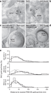
Figure 3. Subcellular localization of SK2 in wild type and SK2-Sonly CA1 pyramidal neurons
(a) Using the post-embedding technique in wild type, immunoparticles for SK2 were detected along the PSD (arrows) of dendritic spines. (b) Using the post-embedding technique in SK2-Sonly, immunoparticles for SK2 were detected along the extrasynaptic membrane (arrows) of dendritic spines but not along the PSD. (c) Using the SDS-FRL technique in wild type, SK2 immunoparticles were detected in dendritic spines intermingled with immunoparticles for PSD-95. (d) Using the SDS-FRL technique in SK2-Sonly, and double immunolabeling for SK2 and PSD-95, SK2 immunoparticles were detected in dendritic spines but segregated from immunoparticles for PSD-95. Scale bars: 200 nm. (e) For SDS-FRL immunolabeling, the percentage of SK2 immunoparticles was plotted as a function of distance from the nearest PSD-95 immunoparticle, and fit by Gaussians for the indicated groups. Dashed line in middle and bottom panels is the fit from wild type in upper panel.
Figure 4
Figure 4. SK2-containing channels are expressed in the plasma membrane of SK2-Sonly CA1 pyramidal neurons
(a) top: Voltage protocol used to evoke tail currents. Cells were voltage clamped at −55 mV, and tail currents were elicited after 200 ms depolarizing steps to 20 mV; bottom: representative tail currents from a wild type CA1 neuron before (black trace) and after (grey trace) apamin application. The inset shows the subtracted apamin-sensitive current. (b) Section through hippocampus from an SK2-Sonly mouse injected with rAAV directing separate expression of GFP and SK2-L. (c) top: Representative apamin-sensitive currents from wild type (left), SK2-Sonly inj. ctrl. (middle), and SK2-Sonly inj. green (left) CA1 pyramidal neurons; bottom: Summary plot shows that the amplitude of apamin-sensitive currents (ISK) for the three groups of neurons. Error bars indicate s.e.m. *P < 0.05 for wild type compared to SK2-Sonly inj. ctrl. or SK2-Sonly inj. green.
Figure 5
Figure 5. SK2-L re-expression re-instates apamin sensitivity to synaptically evoked glutamatergic EPSPs
(a) Time course of the normalized EPSP amplitude (mean ± s.e.m.) before and after apamin application for wild type (left), SK2-Sonly inj. ctrl. (middle), and SK2-Sonly inj. green (right) CA1 pyramidal neurons. Insets show representative average of 20 EPSPs, mean ± s.e.m. (shaded area), taken from the indicated shaded time points in control condition before (black line; left) and after (red line; right) apamin application. (b) Bar graph of the increase in EPSP following apamin application. Error bars indicate s.e.m. *P < 0.05 for SK2-Sonly inj. ctrl. and SK2-Sonly inj. green compared to wild type.
Figure 6
Figure 6. SK channel activity affects LTP in wild type but not SK2-Sonly mice
Field potentials were recorded from the CA1 region of hippocampal slices. (a) In wild type stimulation of Schaffer collateral axons induced more LTP in the presence of apamin (filled circles) than in control bath solution (open circles); (b): LTP was not different with (filled circles) or without (open circles) apamin application in SK2-Sonly slices. LTP was measured 50–60 min after stimulation (black bars). (c) Summary plot of LTP from the indicated groups. Error bars indicate s.e.m. *P < 0.05.
Figure 7
Figure 7. Spatial memory is impaired in SK2-Sonly mice
(a) The mean latency to escape on to the hidden platform was significantly different between wild type (open circles) and SK2-Sonly mice (filled circles). There was no difference between genotypes in mean latency on Trial 1 indicating that both groups exhibited nearly equivalent performance at the start of training. (b) The mean % dwell in the correct quadrant of the pool during the probe test of spatial memory retention imposed after the 4th (P1), 12th (P2) and the 20th training trial (P3). The dashed line indicates chance performance. The SK2-Sonly mice (filled circles), but not wild type (open circles) failed to exhibit a significant preference for searching in the correct quadrant of the pool. (c) Representative paths taken by a wild type mouse (left trace) and two SK2-Sonly mice (center and right traces) during the P3 probe test. The path of the WT mouse is direct to training quadrant and then characterized by repetitive passes through the precise location of the pool where the platform was placed during training (gray open circle). Note that the paths of the SK2-Sonly mice are much less accurate and more circuitous than that of the wild type mice.

References

    1. Bredt DS, Nicoll RA. AMPA receptor trafficking at excitatory synapses. Neuron. 2003;40:361–379. - PubMed
    1. Petralia RS, Wenthold RJ. Light and electron immunocytochemical localization of AMPA-selective glutamate receptors in the rat brain. J Comp Neurol. 1992;318:329–354. - PubMed
    1. Carroll RC, Zukin RS. NMDA-receptor trafficking and targeting: implications for synaptic transmission and plasticity. Trends Neurosci. 2002;25:571–577. - PubMed
    1. Adesnik H, Nicoll RA, England PM. Photoinactivation of native AMPA receptors reveals their real-time trafficking. Neuron. 2005;48:977–985. - PubMed
    1. Harris AZ, Pettit DL. Recruiting extrasynaptic NMDA receptors augments synaptic signaling. J Neurophysiol. 2008;99:524–533. - PubMed

Publication types

MeSH terms

Substances

LinkOut - more resources