. 2010 Dec 1;5(6):867-897.
doi: 10.2217/clp.10.67.

Mammalian diseases of phosphatidylinositol transfer proteins and their homologs

Affiliations

Mammalian diseases of phosphatidylinositol transfer proteins and their homologs

Aaron H Nile et al. Clin Lipidol. .

Abstract

Inositol and phosphoinositide signaling pathways represent major regulatory systems in eukaryotes. The physiological importance of these pathways is amply demonstrated by the variety of diseases that involve derangements in individual steps in inositide and phosphoinositide production and degradation. These diseases include numerous cancers, lipodystrophies and neurological syndromes. Phosphatidylinositol transfer proteins (PITPs) are emerging as fascinating regulators of phosphoinositide metabolism. Recent advances identify PITPs (and PITP-like proteins) to be coincidence detectors, which spatially and temporally coordinate the activities of diverse aspects of the cellular lipid metabolome with phosphoinositide signaling. These insights are providing new ideas regarding mechanisms of inherited mammalian diseases associated with derangements in the activities of PITPs and PITP-like proteins.


Figures

Figure 1
Figure 1. Transfer versus nanoreactor models for phosphatidylinositol transfer protein function
(A) Lipid transfer models invoke a vectorial carrier function for PITPs where PtdIns is transported from membranes of high PtdIns concentration (endoplasmic reticulum) to relatively PtdIns-poor membranes of the distal compartments of the secretory pathway which house PtdIns-4-OH kinase activity (TGN/endosomes or plasma membrane). These models describe productive transfer as involving one heterotypic exchange reaction per donor and acceptor membrane (i.e., two such exchanges per cycle). (B) The ‘nanoreactor’ model predicts that PITPs stimulate PI4-OH kinase activity by executing multiple rounds of phospholipid-exchange at a single membrane site. Only heterotypic exchange reactions generate PtdIns configurations suitable for effective PtdIns presentation. PI4P: Phosphatidylinositol-4-phosphate; PITP: Phosphatidylinositol transfer protein; PO4: Phosphate; PtdCho: Phosphatidylcholine; PtdIns: Phosphatidylinositol.
Figure 2
Figure 2. The Sec14 fold
Crystal structure of two Saccharomyces cerevisiae Sec14-like phosphatidylinositol transfer proteins. (A) The major yeast PITP, Sec14 is shown in its open conformation (pdb 1AUA – two bound detergent molecules are excluded). (B) The close Sec14 homolog Sfh1 in its closed conformation (pdb 3B7Z – bound phospholipid omitted). The β-strands comprising the floor of the phospholipid-binding pocket are in yellow, while the α-helices that form the walls of the pocket are in blue. Access to the hydrophobic pocket is mediated by conformational transitions of the A10/T4 ‘helical gate’ shown in red. The four N-terminal α-helices (α1–α4) comprise the N-terminal lobe (or ‘tripod motif’) (green).
Figure 3
Figure 3. Differential phospholipid-binding strategies by Sec14-like phosphatidylinositol transfer proteins
Structure of Sfh1 bound to: (A) PtdIns (pdb 3B7Z); (B) PtdCho (pdb 3B7Z). (C) A description of the configurations of both phosphatidylinositol (PtdIns; gray) and phosphatidylcholine (PtdCho; black) in the Sfh1/Sec14-fold. The data are from crystals composed of approximately equal numbers of unit cells of Sfh1 bound to PtdIns and Sfh1 bound to PtdCho (pdb 3B7Z). The A10/T4 ‘helical gate’ that mediates lipid entry is in red, surrounding α-helices are in blue, the ‘tripod motif’ is in green and β-strands that compose the floor of the hydrophobic pocket are in yellow. The polar headgroups of PtdIns and PtdCho bind at distinct sites within the hydrophobic pocket, while the acyl chain space within the hydrophobic pocket overlaps for these phospholipids.
Figure 4
Figure 4. The phosphatidylinositol-binding barcode in Sec14-like proteins
(A) Crystal structure of Sfh1 bound to phosphatidylinositol (PtdIns; black; 3B7N) highlighting residues within the PtdIns binding barcode. The tripod-motif is in green, the floor of the hydrophobic pocket is in yellow, and the α-helices are in blue with the exception of the helical gate, which is in red. (B) Orientation of the Sfh1 molecule is rotated by 90° counterclockwise parallel to the floor. (C) Orientation of the Sfh1 molecule is rotated by 180° counterclockwise parallel to the floor. (D) ClustalX2 alignments of selected Sec14-superfamily members (identified at right; proteins whose crystal structures have been solved are indicated with an ‘*’) were superimposed onto the Sfh1 crystal structure using secondary structural elements as a guide (diagrammed at top). Residues critical for PtdIns headgroup and backbone coordination are boxed and shaded in cyan – I, coordinate the Ins-headgroup; II, coordinate the glycerol backbone; III, coordinate the phosphate moiety through which the Ins headgroup is esterified to the glycerol backbone. Positions of missense substitution within the PtdIns-binding barcode of the corresponding Sec14-like protein that cause disease are highlighted by orange boxes.
Figure 5
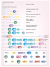
Figure 5. Domain arrangements of Sec14-like proteins
Representative Sec14-like proteins are schematized and ordered by general complexity. Domains of interest are identified. The CRAL-TRIO domain N1 lobe is depicted as a white circle.
Figure 6
Figure 6. Phosphatidylinositol transfer proteins α structures
(A) Phosphatidylinositol transfer protein (PITP)-α apo-structure depicting an open conformation (pdb 1KCM); (B) PtdIns-bound form (pdb 1UW5); (C) PtdCho-bound form (pdb 1T27). The eight β-strands (yellow) of PITPα comprise the hydrophobic cavity floor and two α-helices generate the cavity walls (blue). Additional components of PITPα include a regulatory loop (green), a C-terminal region (red) and a lipid exchange loop (gray).
Figure 7
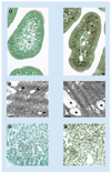
Figure 7. Intestinal and hepatic steatosis in phosphatidylinositol transfer proteins in α-deficient mice
Intestinal slices stained for neutral lipid content with osmium from (A) pitpα+/+ and (B) pitpα0/0 mice. Note the obvious accumulation of neutral lipid in mutant enterocytes. This accumulation is dependent on nursing and chases only slowly during periods of fast. The phenotype Is also obvious in electron micrographs of the villi of duodenal enterocytes from (C) pitpα+/+ and (D) pitpα0/0 mice (scale bars are 0.2 and 0.5 µM, respectively). Lipid deposits are highlighted by asterisks. Liver slices stained with osmium from (E) pitpα+/+ and (F) pitpα0/0 mice are also shown.

References

Bibliography

    1. Michell RH. Inositol derivatives: evolution and functions. Nat. Rev. Mol. Biol. 2008;9:151–161. - PubMed
    1. Irivine RF. Inositide evolution – towards turtle domination? J. Physiol. 2005;566:295–300. - PMC - PubMed
    1. Fruman DA, Meyers RE, Cantley LC. Phosphoinositide kinases. Annu. Rev. Biochem. 1998;67:481–507. - PubMed
    1. Di Paolo G, De Camilli P. Phosphoinositides in cell regulation and membrane dynamics. Nature. 2006;443:651–657. - PubMed
    1. Strahl T, Thorner J. Synthesis and function of membrane phosphoinositides in budding yeast, Saccharomyces cerevisiae. Biochim. Biophys. Acta. 2007;1771:353–404. - PMC - PubMed

Website

    1. The Scripps Research Institute – Mutagenetix home page. http://mutagenetix.scripps.edu/home.cfm.

LinkOut - more resources