

Case Reports
doi: 10.3348/kjr.2011.12.3.390.
Epub 2011 Apr 25.
MRI findings of pericardial fat necrosis: case report
Affiliations
- PMID: 21603300
- PMCID: PMC3088858
- DOI: 10.3348/kjr.2011.12.3.390
Item in Clipboard
Case Reports
MRI findings of pericardial fat necrosis: case report
Korean J Radiol.
2011 May-Jun.
Abstract
Pericardial fat necrosis is an infrequent cause of acute chest pain and this can mimic acute myocardial infarction and acute pericarditis. We describe here a patient with the magnetic resonance imaging (MRI) findings of pericardial fat necrosis and this was correlated with the computed tomography (CT) findings. The MRI findings may be helpful for distinguishing pericardial fat necrosis from other causes of acute chest pain and from the fat-containing tumors in the cardiophrenic space of the anterior mediastinum.
Keywords: Fat necrosis; Magnetic resonance (MR); Pericardial fat.
Figures
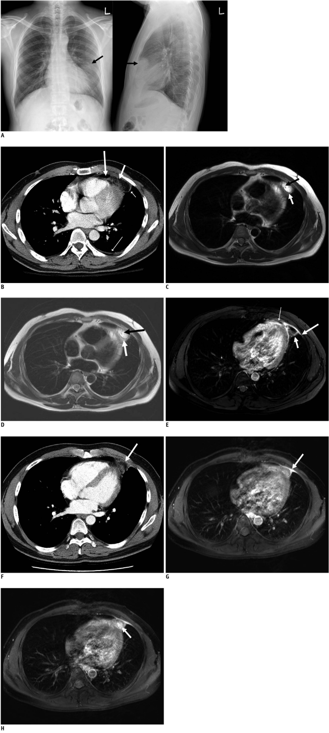
39-year-old man with sudden onset left pleuritic chest pain. A. Chest radiographs show ovoid mass (arrows) with ill-defined margins in left anterior mediastinum. B. Enhanced CT scans show that paracardiac opacity corresponded to pericardial fat surrounded by thick rim (short thin arrow). Note associated pericardial thickening (long thick arrow), linear opacity (short thick arrow) and pleural effusion (long thin arrow). C. T1-weighted breath-hold turbo spin echo images show high signal lesion with thin hypointense rim (white arrow) and hypointense linear line (black arrow). D. T2-weighted breath-hold turbo spin echo images show high signal lesion with thin hypointense rim (white arrow) and hypointense linear line (black arrow). E. T1-weighted fat suppressed breath-hold turbo spin echo image 1 minute after gadolinium administration shows enhancement of rim (short thick arrow) and adjacent pericardium (thin arrow) and pleura (long thick arrow). F. Follow-up CT scan obtained two months after A shows that pericardial lesion has decreased in size (arrow). G. Follow-up T1-weighted fat-suppressed breath-hold turbo spin echo image obtained 1 minute after gadolinium administration and two months after image C shows peripheral rim enhancement of pericardial lesion (arrow). H. Follow-up T1-weighted fat-suppressed breath-hold turbo spin echo image obtained 5 minutes after gadolinium administration and two months after image C shows enhancement of central globular pattern (arrow) of pericardial lesion.
References
-
- Pineda V, Cáceres J, Andreu J, Vilar J, Domingo ML. Epipericardial fat necrosis: radiologic diagnosis and follow-up. AJR Am J Roentgenol. 2005;185:1234–1236. - PubMed
-
- Chan LP, Gee R, Keogh C, Munk PL. Imaging features of fat necrosis. AJR Am J Roentgenol. 2003;181:955–959. - PubMed
-
- Sirvanci M, Balci NC, Karaman K, Duran C, Karakas E. Primary epiploic appendagitis: MRI findings. Magn Reson Imaging. 2002;20:137–139. - PubMed
-
- Inoue S, Fujino S, Tezuka N, Sawai S, Kontani K, Hanaoka J, et al. Encapsulated pericardial fat necrosis treated by video-assisted thoracic surgery: report of a case. Surg Today. 2000;30:739–743. - PubMed
-
- Webster MW, Jr, Bahnson HT. Pericardial fat necrosis. Case report and review. J Thorac Cardiovasc Surg. 1974;67:430–443. - PubMed
Publication types
MeSH terms
Substances
LinkOut - more resources
Full Text Sources
Medical
