

Pitfalls and important issues in the pathologic diagnosis of melanocytic tumors
- PMID: 21603360
- PMCID: PMC3096206
Pitfalls and important issues in the pathologic diagnosis of melanocytic tumors
Abstract
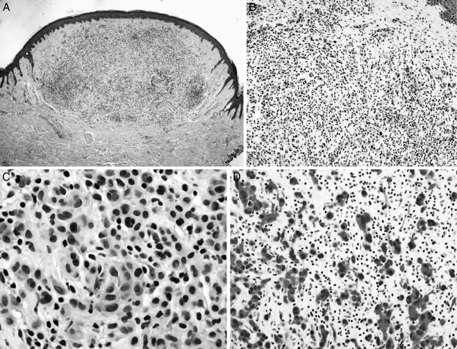
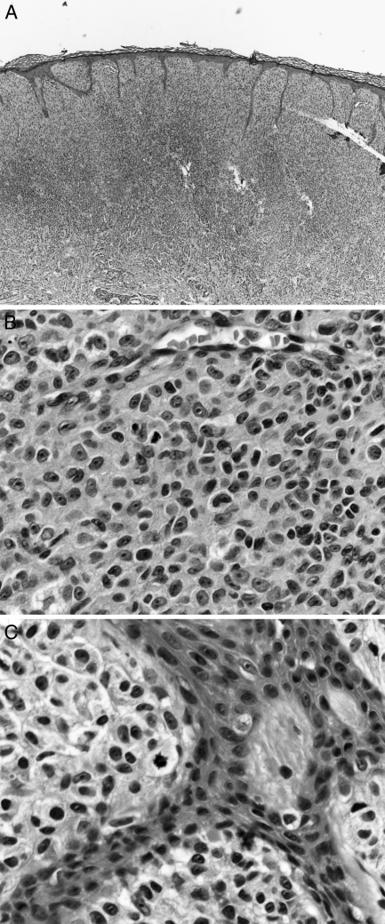
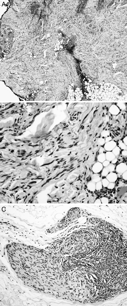
In Australia and many other countries, melanoma is a major public health problem, particularly in those individuals of Celtic ancestry. Other races are not immune, especially when acral and mucosal sites are taken into account. Accurate diagnosis requires the balancing of clinical data (including patient age and sex, family history, the anatomic site of the lesion, the history of the lesion, and other factors such as a history of trauma, sunburn, or pregnancy), histologic features (including architecture, cytology, and the host response), awareness of pitfalls, and judgment. Several types of nevi-such as regenerating nevi, combined nevi, acral nevi, deep penetrating nevi, and Spitz nevi-are prone to be misdiagnosed as melanoma. Melanomas often underdiagnosed include the nevoid, desmoplastic, Spitzoid, and regressed types. The type of biopsy and suboptimal processing may also significantly influence the diagnosis.
Keywords: Biopsy; clinical; diagnosis; melanoma; misdiagnosis; morphology; nevus; pathology; pitfalls.
Figures
Similar articles
-
Melanocytic nevi simulant of melanoma with medicolegal relevance.Virchows Arch. 2007 Sep;451(3):623-47. doi: 10.1007/s00428-007-0459-7. Epub 2007 Jul 26. Virchows Arch. 2007. PMID: 17653760 Review.
-
Analysis of mutations in B-RAF, N-RAS, and H-RAS genes in the differential diagnosis of Spitz nevus and spitzoid melanoma.Am J Surg Pathol. 2005 Sep;29(9):1145-51. doi: 10.1097/01.pas.0000157749.18591.9e. Am J Surg Pathol. 2005. PMID: 16096402
-
BAP1 and BRAFV600E expression in benign and malignant melanocytic proliferations.Hum Pathol. 2015 Feb;46(2):239-45. doi: 10.1016/j.humpath.2014.10.015. Epub 2014 Nov 4. Hum Pathol. 2015. PMID: 25479927
-
Recognizing Histopathological Simulators of Melanoma to Avoid Misdiagnosis.Cureus. 2022 Jun 20;14(6):e26127. doi: 10.7759/cureus.26127. eCollection 2022 Jun. Cureus. 2022. PMID: 35875272 Free PMC article. Review.
-
Acral Spitz Nevi: A Clinicopathologic Study of 50 Cases With Immunohistochemical Analysis of P16 and P21 Expression.Am J Surg Pathol. 2018 Jun;42(6):821-827. doi: 10.1097/PAS.0000000000001051. Am J Surg Pathol. 2018. PMID: 29537991
Cited by
-
Germ cell proteins in melanoma: prognosis, diagnosis, treatment, and theories on expression.J Skin Cancer. 2012;2012:621968. doi: 10.1155/2012/621968. Epub 2012 Nov 12. J Skin Cancer. 2012. PMID: 23209909 Free PMC article.
-
Benign Nevi Mimicking Melanoma: A Diagnostic Dilemma.Cureus. 2024 Nov 30;16(11):e74821. doi: 10.7759/cureus.74821. eCollection 2024 Nov. Cureus. 2024. PMID: 39618769 Free PMC article. Review.
-
Deep Learning Approach to Classify Cutaneous Melanoma in a Whole Slide Image.Cancers (Basel). 2023 Mar 22;15(6):1907. doi: 10.3390/cancers15061907. Cancers (Basel). 2023. PMID: 36980793 Free PMC article.
-
Representativeness of initial skin biopsies showing pure desmoplastic melanoma: implications for management.Pathology. 2023 Mar;55(2):214-222. doi: 10.1016/j.pathol.2022.12.346. Epub 2023 Jan 6. Pathology. 2023. PMID: 36646575 Free PMC article.
-
One Step Melanoma Surgery for Patient with Thick Primary Melanomas: "To Break the Rules, You Must First Master Them!".Open Access Maced J Med Sci. 2018 Feb 9;6(2):367-371. doi: 10.3889/oamjms.2018.084. eCollection 2018 Feb 15. Open Access Maced J Med Sci. 2018. PMID: 29531606 Free PMC article.
References
-
- Thompson J. F., Scolyer R. A., Kefford R. F. Cutaneous melanoma. Lancet. 2005;365((9460)):687–701. - PubMed
-
- Murali R., Thompson J. F., Scolyer R. A. Sentinel lymph node biopsy for melanoma: aspects of pathologic assessment. Future Oncol. 2008;4((4)):535–551. - PubMed
-
- Scolyer R. A., McCarthy S. W., Elder D. E. Frontiers in melanocytic pathology. Pathology. 2004;36((5)):385–386. - PubMed
-
- Scolyer R. A., Thompson J. F., McCarthy S. W., Strutton G. M., Elder D. E. Incomplete biopsy of melanocytic lesions can impair the accuracy of pathological diagnosis [erratum appears in Australas J Dermatol. 2006;47(4):308]. Australas J Dermatol. 2006;47((1)):71–75. - PubMed
-
- Scolyer R. A., Thompson J. F., Stretch J. R., Sharma R., McCarthy S. W. Pathology of melanocytic lesions: new, controversial, and clinically important issues. J Surg Oncol. 2004;86((4)):200–211. - PubMed
LinkOut - more resources
Full Text Sources