

Sphenoid sinus myxoma: case report and literature review
- PMID: 21603497
- PMCID: PMC3096368
Sphenoid sinus myxoma: case report and literature review
Abstract
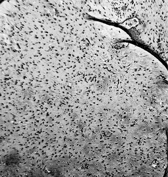
Objectives: We present the first known case in the English-language literature of a myxoma arising in the sphenoid sinus. By describing the patient's clinical course and the salient features of this rare neoplasm, we seek to increase the awareness of the presentation, histological features, and treatment considerations for myxomas of the head and neck. In the process, we intend to describe the work-up of isolated sphenoid sinus lesions and focus on the varying and evolving techniques for surgical access to the sphenoid sinus.
Study design and methods: Case report and literature review.
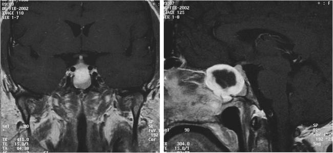
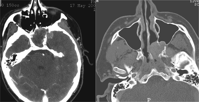
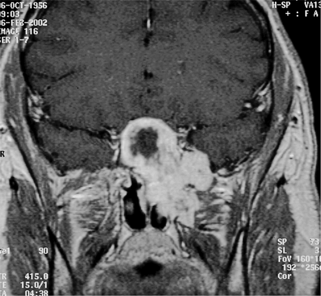
Results: We describe the clinical course of a patient with a myxoma of the sphenoid sinus. The patient underwent an external sphenoethmoidectomy through a lateral rhinotomy approach with medial maxillectomy under MRI-guidance. He remains without evidence of recurrent disease after 8 months.
Conclusions: Myxomas of the head and neck are rare neoplasms. Their infiltrative nature and tendency to recur demand an aggressive surgical approach that may be accomplished with minimal morbidity using currently available image-guided techniques.
Keywords: Myxoma; neoplasms; sphenoid sinus.
Figures
Similar articles
-
The Unusual Presentation of a Myxoma Within the Sphenoid Sinus: Case Report and Review of the Literature.World Neurosurg. 2017 Jul;103:951.e5-951.e12. doi: 10.1016/j.wneu.2017.04.019. Epub 2017 Apr 20. World Neurosurg. 2017. PMID: 28433840 Review.
-
Maxillary sinus nonodontogenic myxoma extending into the sphenoid sinus and pterygopalatine fossa: case report.Ear Nose Throat J. 2011 Sep;90(9):E28-30. doi: 10.1177/014556131109000922. Ear Nose Throat J. 2011. PMID: 21938690
-
Isolated inverting papilloma of the sphenoid sinus.Laryngoscope. 2003 Jan;113(1):41-4. doi: 10.1097/00005537-200301000-00008. Laryngoscope. 2003. PMID: 12514380
-
Isolated inverting papilloma of the sphenoid sinus: clinical presentations, imaging manifestations, and therapeutic strategies.J Craniofac Surg. 2012 Jul;23(4):1109-14. doi: 10.1097/SCS.0b013e31825434fc. J Craniofac Surg. 2012. PMID: 22777475
-
The endoscopic transnasal paraseptal approach to a sphenoid sinus osteoma: case report and literature review.Ear Nose Throat J. 2013 Dec;92(12):E7-E10. Ear Nose Throat J. 2013. PMID: 24366714 Review.
Cited by
-
Multidisciplinary Approach to Rehabilitation after Tumor Resective Jaw Surgery: A 9-Year Follow-Up.Case Rep Dent. 2020 Dec 10;2020:8867320. doi: 10.1155/2020/8867320. eCollection 2020. Case Rep Dent. 2020. PMID: 33381326 Free PMC article.
-
Sinonasal Myxoma: A Distinct Lesion of Infants.Head Neck Pathol. 2020 Mar;14(1):212-219. doi: 10.1007/s12105-018-0989-0. Epub 2018 Nov 27. Head Neck Pathol. 2020. PMID: 30484069 Free PMC article.
-
Odontogenic myxoma: ambiguous pathology of anterior maxilla.BMJ Case Rep. 2020 Aug 25;13(8):e234933. doi: 10.1136/bcr-2020-234933. BMJ Case Rep. 2020. PMID: 32843449 Free PMC article.
-
Isolated Sphenoid Sinus Lesions: Experience with a Few Rare Pathologies.J Neurosci Rural Pract. 2017 Jan-Mar;8(1):107-113. doi: 10.4103/0976-3147.193540. J Neurosci Rural Pract. 2017. PMID: 28149092 Free PMC article.
-
Odontogenic myxoma: A review with report of an uncommon case with recurrence in the mandible of a teenage male.Saudi Dent J. 2017 Jul;29(3):93-101. doi: 10.1016/j.sdentj.2017.02.003. Epub 2017 Mar 16. Saudi Dent J. 2017. PMID: 28725126 Free PMC article. Review.
References
-
- Virchow R. Die cellularpathologie in ihrer Begrundung auf physiologische und pathologische Gewebelehre. Berlin, Germany: Verlag von August Hirschwald; 1871. p. 563.
-
- Stout A. P. Myxoma: the tumor of primitive mesenchyme. Ann Surg. 1948;127:706–719. - PubMed
-
- Allphin A. L., Manigilia A. J., Gregor R. T., Sawyer R. Myxomas of the mandible and maxilla. Ear Nose Throat J. 1993;72:280–284. - PubMed
-
- Fu Y. S., Perzin K. H. Non-epithelial tumors of the nasal cavity, paranasal sinuses and nasopharynx: a clinico-pathologic study. Cancer. 1977;39:195–203. - PubMed
-
- Perzin K. H., Panyu J., Wechter S. Nonepithelial tumors of the nasal cavity, paranasal sinuses, and nasopharynx. Cancer. 1982;50:2193–2202. - PubMed
LinkOut - more resources
Full Text Sources