. 2011 Sep;60(9):1269-79.
doi: 10.1007/s00262-011-1029-z. Epub 2011 May 21.

Distinct myeloid suppressor cell subsets correlate with plasma IL-6 and IL-10 and reduced interferon-alpha signaling in CD4⁺ T cells from patients with GI malignancy

Affiliations

Distinct myeloid suppressor cell subsets correlate with plasma IL-6 and IL-10 and reduced interferon-alpha signaling in CD4⁺ T cells from patients with GI malignancy

Bethany L Mundy-Bosse et al. Cancer Immunol Immunother. 2011 Sep.

Abstract

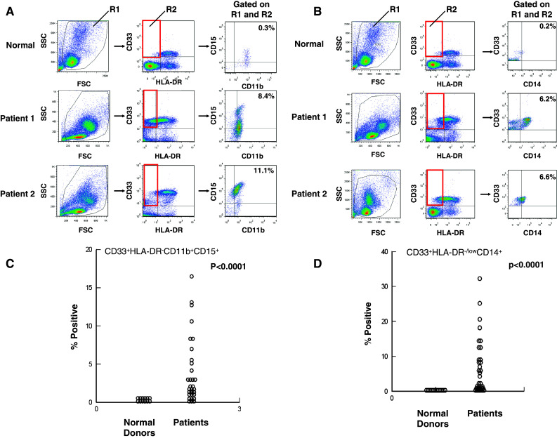
Interferon-alpha (IFN-α) promotes anti-tumor immunity through its actions on immune cells. We hypothesized that elevated percentages of myeloid-derived suppressor cells (MDSC) and increased pro-inflammatory cytokines in peripheral blood would be associated with impaired response to IFN-α in patients with gastrointestinal (GI) malignancies. This study evaluated relationships between plasma IL-6, IL-10, circulating MDSC subsets, and IFN-α-induced signal transduction in 40 patients with GI malignancies. Plasma IL-6 and IL-10 were significantly higher in patients versus normal donors. CD33(+)HLADR(-)CD11b(+)CD15(+) and CD33(+)HLADR(-/low)CD14(+) MDSC subsets were also elevated in patients versus normal donors (P < 0.0001). Plasma IL-6 was correlated with CD33(+)HLADR(-)CD15(+) MDSC (P = 0.008) and IL-10 with CD33(+)HLADR(-)CD15(-) MDSC (P = 0.002). The percentage of CD15(+) and CD15(-) but not CD14(+) MDSC subsets were inversely correlated with IFN-α-induced STAT1 phosphorylation in CD4(+) T cells, while co-culture with in vitro generated MDSC led to reduced IFN-α responsiveness in both PBMC and the CD4(+) subset of T cells from normal donors. Exploratory multivariable Cox proportional hazards models revealed that an increased percentage of the CD33(+)HLADR(-)CD15(-) MDSC subset was associated with reduced overall survival (P = 0.049), while an increased percentage of the CD33(+)HLADR(-/low)CD14(+) subset was associated with greater overall survival (P = 0.033). These data provide evidence for a unique relationship between specific cytokines, MDSC subsets, and IFN-α responsiveness in patients with GI malignancies.


Figures

Fig. 1
Fig. 1
Phenotypic analysis of MDSC subsets in patients with GI malignancies. Peripheral blood mononuclear cells (PBMCs) were obtained from n = 31 GI cancer patients for whom peripheral blood was available or normal donors (n = 12) and stained for MDSC using fluorochrome-labeled antibodies targeting CD33, HLADR, CD15, CD14, and CD11b or the appropriate isotype controls. MDSC levels were evaluated by flow cytometry based on a minimum of 20,000 live events and presented as the percentage of total cells, listed in box R2 in figure. a Representative scatter plots from two GI cancer patients as compared to PBMC obtained from a normal control. PBMCs were labeled with CD33, HLA-DR, CD15, and CD11b, box R2 indicates gated population, and arrows indicate the sequence of gating. The first dot plot depicts forward scatter (FSC) by side scatter (SSC) properties of PBMC for determining initial live cell gate. b Representative scatter plots from two GI cancer patients as compared to PBMC obtained from a normal control. PBMCs were labeled with CD33, HLA-DR, and CD14, box R2 indicates gated population, and arrows indicate the sequence of gating. c CD33+HLADRCD11b+CD15+ MDSC and d CD33+HLA-DR−/lowCD14+ MDSC and compared to levels found in normal control PBMCs
Fig. 2
Fig. 2
MDSC subsets are elevated in all types of GI malignancies. ab The levels of each MDSC subset, as determined by flow cytometry, were evaluated by type of GI malignancy and compared to normal control PBMC. The number of patients with cancer at each organ site is listed on the plot. Patients designated as ‘other’ consist of one patient each with cancer of the gallbladder, appendix, hepatocellular carcinoma, or cholangiocarcinoma. c Early myeloid cells (CD33+HLADR) were evaluated in a similar manner from all 31 patients to determine the percentage positive and negative for CD15 or positive for CD14
Fig. 3
Fig. 3
Plasma levels of IL-6 and IL-10 and the association with MDSC in patients with GI malignancies. a IL-6 and IL-10 were measured by ELISA in plasma isolated from the peripheral blood of patients with GI malignancies (n = 40) and plasma obtained from normal donors (n = 20). The levels of IL-6 and IL-10 were significantly elevated in patients with GI malignancy as compared to normal controls. All samples were assayed in duplicate and quantitated using a standardized protein curve. bc MDSC levels were obtained as described previously from n = 31 patients and compared with plasma cytokine levels from the same patient. b The percentage of CD33+HLADRCD15+ MDSC and plasma levels of IL-6 and c the percentage of CD33+HLADRCD15 MDSC and plasma levels of IL-10
Fig. 4
Fig. 4
Inverse correlation between MDSC and IFN-α-induced STAT1 phosphorylation. MDSC levels were obtained as described previously from n = 31 patients and compared with the levels of IFN-α-induced STAT1 phosphorylation (P-STAT1). a-b The percentage of both CD33+HLADRCD15+ MDSC and CD33+HLADRCD15 MDSC inversely correlated with STAT1 phosphorylation following ex vivo stimulation with IFN-α (104 U/ml for 15 min) in CD4+ T cells from GI patients. c Normal PBMC co-cultured with in vitro generated MDSC exhibited reduced IFN-α-induced STAT1 phosphorylation. In vitro generated MDSC expressed CD33 and CD11b, with variable, donor-dependent levels of HLA-DR and CD15, and showed no difference in CD15 expression in the presence or absence of IL-10 (data not shown). d CD4+ T cells from normal donors have reduced IFN-α-induced P-STAT1 when co-cultured with autologous, in vitro generated MDSC. Error bars represent standard deviation obtained from separate experiments using PBMC and autologous, in vitro generated MDSC from four unique donors

Similar articles

Cited by

References

    1. Jemal A, Siegel R, Ward E, Hao Y, Xu J, Thun MJ. Cancer statistics. CA Cancer J Clin. 2009;59:225–249. doi: 10.3322/caac.20006. - DOI - PubMed
    1. Chaudry MA, Sales K, Ruf P, Lindhofer H, Winslet MC. EpCAM an immunotherapeutic target for gastrointestinal malignancy: current experience and future challenges. Br J Cancer. 2007;96:1013–1019. doi: 10.1038/sj.bjc.6603505. - DOI - PMC - PubMed
    1. Dougan M, Dranoff G. Immune therapy for cancer. Annu Rev Immunol. 2009;27:83–117. doi: 10.1146/annurev.immunol.021908.132544. - DOI - PubMed
    1. Almand B, Clark JI, Nikitina E, van Beynen J, English NR, Knight SC, Carbone DP, Gabrilovich DI. Increased production of immature myeloid cells in cancer patients: a mechanism of immunosuppression in cancer. J Immunol. 2001;166:678–689. - PubMed
    1. Ugel S, Delpozzo F, Desantis G, Papalini F, Simonato F, Sonda N, Zilio S, Bronte V. Therapeutic targeting of myeloid-derived suppressor cells. Curr Opin Pharmacol. 2009;9:470–481. doi: 10.1016/j.coph.2009.06.014. - DOI - PubMed

Publication types