. 2011 Jul;79(7):2282-90.
doi: 10.1002/prot.23054. Epub 2011 May 20.

Binding to the open conformation of HIV-1 protease

Affiliations

Binding to the open conformation of HIV-1 protease

Katrina W Lexa et al. Proteins. 2011 Jul.

Abstract

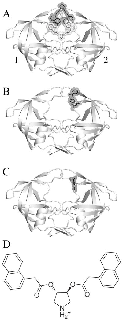
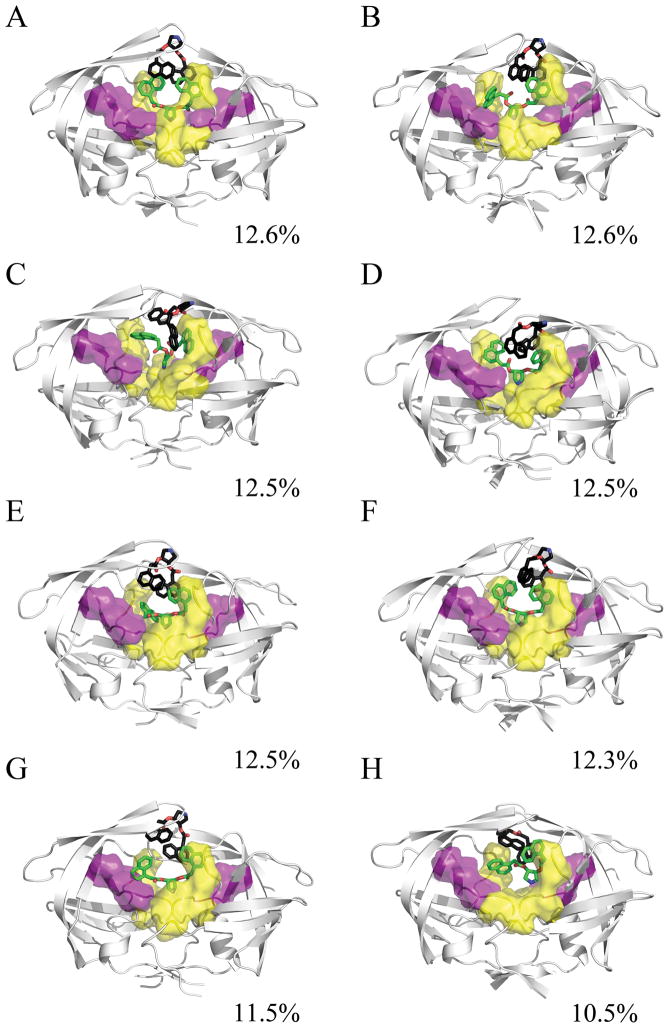
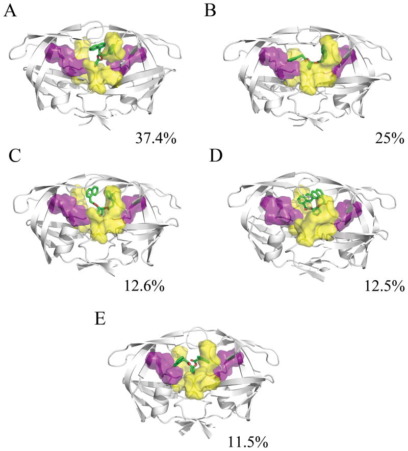
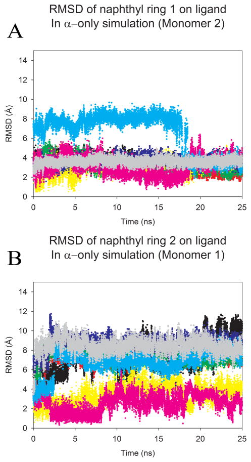
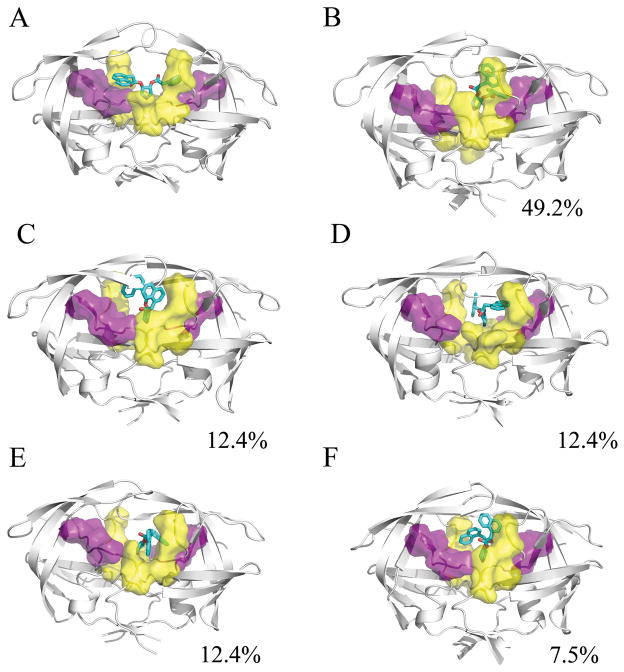
A recent crystal structure of HIV-1 protease (HIVp) was the first to experimentally observe a ligand targeting an open-flap conformation. Researchers studying a symmetric pyrrolidine inhibitor found that two ligands cocrystallized with the protease, forcing an unusual configuration and unique crystallographic contacts. One molecule is centered in the traditional binding site (α pose) and the other binds between the flaps (β pose). The ligands stack against each other in a region termed the "eye" site. Ligands bound to the eye site should prevent flap closure, but it is unclear if the pyrrolidine inhibitors or the crystal packing are causing the open state. Molecular dynamics simulations were used to examine the solution-state behavior of three possible binding modes: the ternary complex of HIVp+αβ and the binary complexes, HIVp+α and HIVp+β. We show that HIVp+α is the most stable of the three states. During conformational sampling, α takes an asymmetric binding pose, with one naphthyl ring occupying the eye site and the other reoriented down to occupy positions seen with traditional inhibitors. This finding supports previous studies that reveal a requirement for asymmetric binding at the eye site. In fact, if the α pose is modified to splay both naphthyl rings across the binding site like traditional inhibitors, one ring consistently flips to occupy the eye site. Our simulations reveal that interactions to the eye site encourage a conformationally restrained state, and understanding those contacts may aid the design of ligands to specifically target alternate conformations of the protease.


Figures

Figure 1
Figure 1
A) The crystallized HIV-1 protease uniquely bound by two identical inhibitors, with pose α colored in grey and pose β in black. B) The crystal structure 3BC4 with Damm compound 1 (black) bound at the eye site. C) The 5-Nitroindole fragment (black) crystallized in the eye site by Perryman et al. D) A 2-dimensional representation of the pyrrolidine inhibitor that was co-crystallized with 3BC4. The affinity (Ki) of the compound for HIVp was measured by Klebe and coworkers as 20μM (WT), 41μM (I50V), and 4.5μM (I84V). For the following figures, we have used a convention of orienting the complex so that a naphthyl occupies the eye position on the right (ie. Monomer 2). We are labeling the monomers as “1” and “2” instead of “A” and “B” to avoid confusion with the α and β notation for the ligands.
Figure 2
Figure 2
Representative structures from the MD simulation of the HIVp+αβ complex, taken from the last 5ns of each 25ns trajectory. The α ligand is shown in green, the β ligand is shown in black, the S1/S1′ site is shown in yellow, and the S2/S2′ site is shown in purple. The conformational families demonstrate the instability of the 2:1 bound complex. The pyrrolidine ligands find a wide variety of ways to interact with the protease flaps, S1/S1′, S2/S2′, and/or the eye site.
Figure 3
Figure 3
Representative structures from the MD simulation of the HIVp+α complex, taken from the last 5ns of each 25ns trajectory. The α ligand is shown in green, the S1/S1′ site is shown in yellow, and the S2/S2′ site is shown in purple. The conformational families for the α ligand illustrate its strong preference for forming one interaction between the naphthyl ring and the eye site, while the other naphthyl ring flips to interact at the S1/S1′ or S2/S2′ site, and the pyrrole maintains a hydrogen-bonding interaction with the catalytic aspartic acids.
Figure 4
Figure 4
The overall RMSD from the crystal pose calculated for each naphthyl ring of the ligand in HIVp+α over the length of the production run. Trajectories were first fit to the Cα core of the 3BC4 crystal structure. Each color represents a single production run; and denotes the same run for each plot. A) highlights the RMSD of the first naphtyhl ring over time, (B) highlights the RMSD of the second naphtyhl ring over time. As noted in figure 1, we have used the convention of labeling monomers 1 and 2 based on the behavoir of the ligand, where better agreement with the initial position in the eye is oriented to the right in the figures and labeled as monomer 2 in the graphs. An RMSD of 6.2–7.9 Å indicates occupation of the S2/S2′ site, while an RMSD of 7.8–10.1 Å indicates occupation of the S1/S1′ site.
Figure 5
Figure 5
A) The initial minimized conformation of the α′ ligand. B–F) Representative structures from the 200ns MD simulation of the HIVp-α′ complex. The α′ ligand is shown in cyan, the S1/S1′ site is shown in yellow, and the S2/S2′ site is shown in purple. Although the simulations were initiated with the naphthyl rings occupying traditional subsites of the active site, one naphthyl ring moves to form interactions at the eye site over the course of all eight independent simulations. The second ring remains in contact with the S1 or S2 site.

References

    1. Eder J, Hommel U, Cumin F, Martoglio B, Gerhartz B. Aspartic proteases in drug discovery. Curr Pharm Des. 2007;13(3):271–285. - PubMed
    1. McKeage K, Perry CM, Keam SJ. Darunavir: a review of its use in the management of HIV infection in adults. Drugs. 2009;69(4):477–503. - PubMed
    1. Mehellou Y, De Clercq E. Twenty-six years of anti-HIV drug discovery: where do we stand and where do we go? J Med Chem. 53(2):521–538. - PubMed
    1. Hornak V, Simmerling C. Targeting structural flexibility in HIV-1 protease inhibitor binding. Drug Discov Today. 2007;12(3–4):132–138. - PMC - PubMed
    1. Freedberg DI, Ishima R, Jacob J, Wang YX, Kustanovich I, Louis JM, Torchia DA. Rapid structural fluctuations of the free HIV protease flaps in solution: relationship to crystal structures and comparison with predictions of dynamics calculations. Protein Sci. 2002;11(2):221–232. - PMC - PubMed

Publication types

Substances