

Impact of Francisella tularensis pilin homologs on pilus formation and virulence
- PMID: 21605655
- PMCID: PMC3120926
- DOI: 10.1016/j.micpath.2011.05.001
Impact of Francisella tularensis pilin homologs on pilus formation and virulence
Abstract
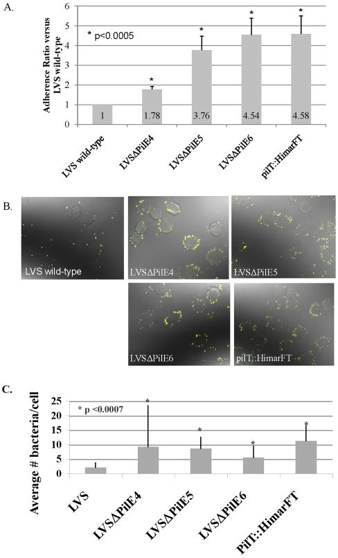
Francisella tularensis is a facultative intracellular bacterium and the causative agent of tularemia. Virulence factors for this bacterium, particularly those that facilitate host cell interaction, remain largely uncharacterized. However, genes homologous to those involved in type IV pilus structure and assembly, including six genes encoding putative major pilin subunit proteins, are present in the genome of the highly virulent Schu S4 strain. To analyze the roles of three putative pilin genes in pili structure and function we constructed individual pilE4, pilE5, and pilE6 deletion mutants in both the F. tularensis tularensis strain Schu S4 and the Live Vaccine Strain (LVS), an attenuated derivative strain of F. tularensis holarctica. Transmission electron microscopy (TEM) of Schu S4 and LVS wild-type and deletion strains confirmed that pilE4 was essential for the expression of type IV pilus-like fibers by both subspecies. By the same method, pilE5 and pilE6 were dispensable for pilus production. In vitro adherence assays with J774A.1 cells revealed that LVS pilE4, pilE5, and pilE6 deletion mutants displayed increased attachment compared to wild-type LVS. However, in the Schu S4 background, similar deletion mutants displayed adherence levels similar to wild-type. In vivo, LVS pilE5 and pilE6 deletion mutants were significantly attenuated compared to wild-type LVS by intradermal and subcutaneous murine infection, while no Schu S4 deletion mutant was significantly attenuated compared to wild-type Schu S4. While pilE4 was essential for fiber expression on both Schu S4 and LVS, neither its protein product nor the assembled fibers contributed significantly to virulence in mice. Absent a role in pilus formation, we speculate PilE5 and PilE6 are pseudopilin homologs that comprise, or are associated with, a novel type II-related secretion system in Schu S4 and LVS.
Copyright © 2011 Elsevier Ltd. All rights reserved.
Figures


Similar articles
-
Contributions of TolC Orthologs to Francisella tularensis Schu S4 Multidrug Resistance, Modulation of Host Cell Responses, and Virulence.Infect Immun. 2019 Mar 25;87(4):e00823-18. doi: 10.1128/IAI.00823-18. Print 2019 Apr. Infect Immun. 2019. PMID: 30670554 Free PMC article.
-
Differential ability of novel attenuated targeted deletion mutants of Francisella tularensis subspecies tularensis strain SCHU S4 to protect mice against aerosol challenge with virulent bacteria: effects of host background and route of immunization.Vaccine. 2010 Feb 17;28(7):1824-31. doi: 10.1016/j.vaccine.2009.12.001. Epub 2009 Dec 16. Vaccine. 2010. PMID: 20018266 Free PMC article.
-
Type IV pili in Francisella tularensis: roles of pilF and pilT in fiber assembly, host cell adherence, and virulence.Infect Immun. 2008 Jul;76(7):2852-61. doi: 10.1128/IAI.01726-07. Epub 2008 Apr 21. Infect Immun. 2008. PMID: 18426883 Free PMC article.
-
Live Attenuated Tularemia Vaccines for Protection Against Respiratory Challenge With Virulent F. tularensis subsp. tularensis.Front Cell Infect Microbiol. 2018 May 15;8:154. doi: 10.3389/fcimb.2018.00154. eCollection 2018. Front Cell Infect Microbiol. 2018. PMID: 29868510 Free PMC article. Review.
-
A comprehensive guide to pilus biogenesis in Gram-negative bacteria.Nat Rev Microbiol. 2017 May 12;15(6):365-379. doi: 10.1038/nrmicro.2017.40. Nat Rev Microbiol. 2017. PMID: 28496159 Review.
Cited by
-
Production of outer membrane vesicles and outer membrane tubes by Francisella novicida.J Bacteriol. 2013 Mar;195(6):1120-32. doi: 10.1128/JB.02007-12. Epub 2012 Dec 21. J Bacteriol. 2013. PMID: 23264574 Free PMC article.
-
Identifying key soil characteristics for Francisella tularensis classification with optimized Machine learning models.Sci Rep. 2024 Jan 19;14(1):1743. doi: 10.1038/s41598-024-51502-z. Sci Rep. 2024. PMID: 38242908 Free PMC article.
-
The type IV pili component PilO is a virulence determinant of Francisella novicida.PLoS One. 2022 Jan 25;17(1):e0261938. doi: 10.1371/journal.pone.0261938. eCollection 2022. PLoS One. 2022. PMID: 35077486 Free PMC article.
-
Type IV pilin proteins: versatile molecular modules.Microbiol Mol Biol Rev. 2012 Dec;76(4):740-72. doi: 10.1128/MMBR.00035-12. Microbiol Mol Biol Rev. 2012. PMID: 23204365 Free PMC article. Review.
-
From the Outside-In: The Francisella tularensis Envelope and Virulence.Front Cell Infect Microbiol. 2015 Dec 23;5:94. doi: 10.3389/fcimb.2015.00094. eCollection 2015. Front Cell Infect Microbiol. 2015. PMID: 26779445 Free PMC article. Review.
References
-
- Sjostedt A. Tularemia: history, epidemiology, pathogen physiology, and clinical manifestations. Ann N Y Acad Sci. 2007;1105:1–29. - PubMed
-
- Tarnvik A, Berglund L. Tularaemia. Eur Respir J. 2003;21:361–73. - PubMed
-
- Saslaw S, Eigelsbach HT, Prior JA, Wilson HE, Carhart S. Tularemia vaccine study. II. Respiratory challenge. Arch Intern Med. 1961;107:702–14. - PubMed
-
- Dennis DT, Inglesby TV, Henderson DA, Bartlett JG, Ascher MS, Eitzen E, et al. Tularemia as a biological weapon: medical and public health management. JAMA. 2001;285:2763–73. - PubMed
-
- Khan AS, Morse S, Lillibridge S. Public-health preparedness for biological terrorism in the USA. Lancet. 2000;356:1179–82. - PubMed
Publication types
MeSH terms
Substances
Grants and funding
LinkOut - more resources
Full Text Sources
