

doi: 10.1109/TMI.2011.2156806.
Epub 2011 May 19.
A supervised patch-based approach for human brain labeling
Affiliations
- PMID: 21606021
- PMCID: PMC3318921
- DOI: 10.1109/TMI.2011.2156806
Item in Clipboard
A supervised patch-based approach for human brain labeling
IEEE Trans Med Imaging.
2011 Oct.
Abstract
We propose in this work a patch-based image labeling method relying on a label propagation framework. Based on image intensity similarities between the input image and an anatomy textbook, an original strategy which does not require any nonrigid registration is presented. Following recent developments in nonlocal image denoising, the similarity between images is represented by a weighted graph computed from an intensity-based distance between patches. Experiments on simulated and in vivo magnetic resonance images show that the proposed method is very successful in providing automated human brain labeling.
© 2011 IEEE
Figures
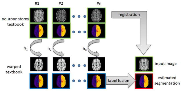
Principle of registration-based label propagation methods. The input data (shown with green borders) are an anatomy textbook (i.e. a set of N anatomical images with the corresponding label maps), and one anatomical image I. The set of anatomical images of the textbook is (non-linearly) registered to the input image I, and each label map is deformed with respect to the estimated transformation Hi. The final image segmentation (shown with red borders) is then obtained by fusing all the deformed label maps (shown with blue borders).
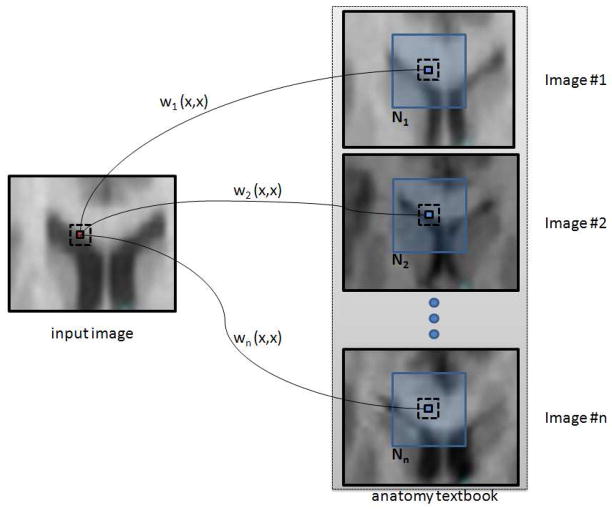
Weighted graph building. The set of graphs {wi}i=1, ···,
n is a representation of non-local interactions between the input image I and the images {
 }i=1, ···,
n of the textbook.
}i=1, ···,
n of the textbook.
 (x) is the neighborhood of the voxel x in the image
.
(x) is the neighborhood of the voxel x in the image
.
}i=1, ···,
n of the textbook.
(x) is the neighborhood of the voxel x in the image
.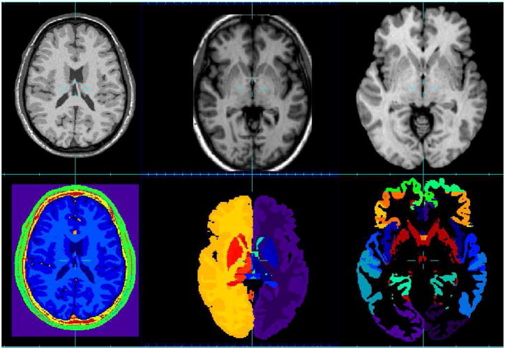
MR image datasets used for the evaluation and the corresponding segmentation. First column: Brainweb database, second column: IBSR database, third column: NA0-NIREP database.
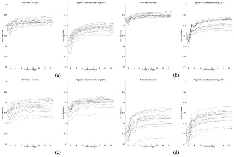
Mean dice index with respect to the number of images used for label propagation Left: patch-based approach, right: registration-based approach using ANTS (using, for each case, majority voting to fuse labels). (a): left white matter, (b): left cortex, (c): left thalamus, (d): left hyppocampus.
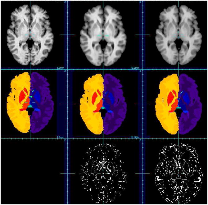
Visual evaluation of brain segmentation results (IBSR, image #7). First row: T1-weighted images, second row: corresponding segmentation, third row: misclassified voxels. Left: ground-truth, middle: patch-based technique, right: non-rigid registration-based approach (using ANTs).
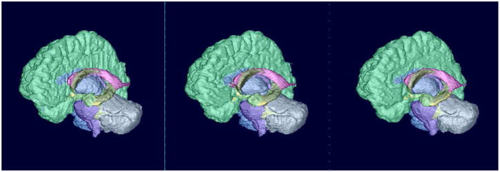
Surface rendering of segmentation results (IBSR, image #7). Left: ground-truth, middle: patch-based technique, right: non-rigid registration-based approach (using ANTs).
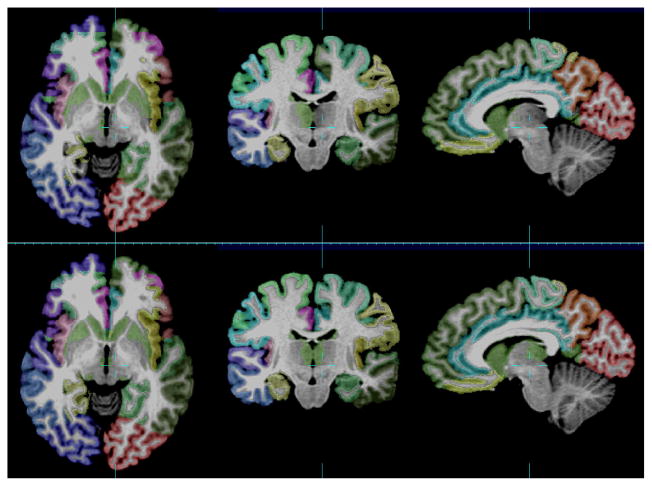
Visual evaluation of cortex parcellation results (NA0-NIREP, image #6). First row: ground truth, second row: combination technique (ANTs + patch + STAPLE).
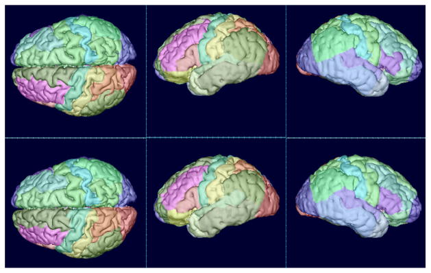
Surface rendering of cortex parcellation results (NA0-NIREP, image #6). First row: ground truth, second row: combination technique (ANTs + patch + STAPLE).
References
-
- Akselrod-Ballin A, Galun M, Gomori JM, Brandt A, Basri R. Prior knowledge driven multiscale segmentation of brain MRI. Medical Image Computing and Computer-Assisted Intervention: MICCAI. 2007;10(Pt 2):118–126. - PubMed
-
- Aljabar P, Heckemann RA, Hammers A, Hajnal JV, Rueckert D. Multi-atlas based segmentation of brain images: atlas selection and its effect on accuracy. NeuroImage. 2009 Jul;46(3):726–738. - PubMed
-
- Artaechevarria X, Munoz-Barrutia A, Ortiz-de-Solorzano C. Combination strategies in multi-atlas image segmentation: application to brain MR data. IEEE Transactions on Medical Imaging. 2009 Aug;28(8):1266–1277. - PubMed
-
- Aubert-Broche B, Evans AC, Collins L. A new improved version of the realistic digital brain phantom. Neuroimage. 2006 Aug;32(1):138145. - PubMed
Publication types
MeSH terms
Grants and funding
LinkOut - more resources
Full Text Sources
Other Literature Sources
Medical
