. 2013 Jun;34(22):1681-90.
doi: 10.1093/eurheartj/ehr131. Epub 2011 May 23.

Increased expression of pigment epithelium-derived factor in aged mesenchymal stem cells impairs their therapeutic efficacy for attenuating myocardial infarction injury

Affiliations

Increased expression of pigment epithelium-derived factor in aged mesenchymal stem cells impairs their therapeutic efficacy for attenuating myocardial infarction injury

Hongliang Liang et al. Eur Heart J. 2013 Jun.

Abstract

Aims: Mesenchymal stem cells (MSCs) can ameliorate myocardial infarction (MI) injury. However, older-donor MSCs seem less efficacious than those from younger donors, and the contributing underlying mechanisms remain unknown. Here, we determine how age-related expression of pigment epithelium-derived factor (PEDF) affects MSC therapeutic efficacy for MI.

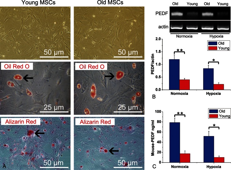
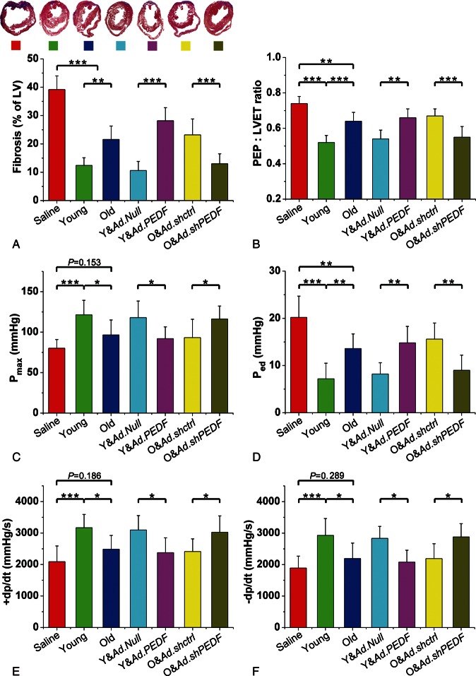
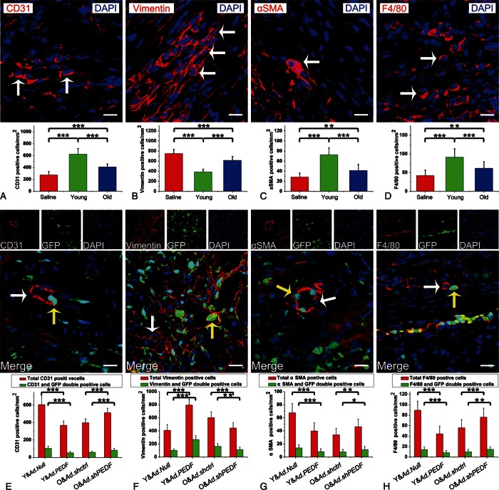
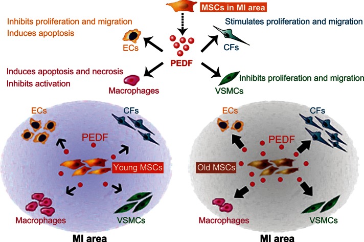
Methods and results: Reverse transcriptase-polymerized chain reaction and enzyme-linked immunosorbent assay analyses revealed dramatically increased PEDF expression in MSCs from old mice compared to young mice. Morphological and functional experiments demonstrated significantly impaired old MSC therapeutic efficacy compared with young MSCs in treatment of mice subjected to MI. Immunofluorescent staining demonstrated that administration of old MSCs compared with young MSCs resulted in an infarct region containing fewer endothelial cells, vascular smooth muscle cells, and macrophages, but more fibroblasts. Pigment epithelium-derived factor overexpression in young MSCs impaired the beneficial effects against MI injury, and induced cellular profile changes in the infarct region similar to administration of old MSCs. Knocking down PEDF expression in old MSCs improved MSC therapeutic efficacy, and induced a cellular profile similar to young MSCs administration. Studies in vitro showed that PEDF secreted by MSCs regulated the proliferation and migration of cardiac fibroblasts.

Conclusions: This is the first evidence that paracrine factor PEDF plays critical role in the regulatory effects of MSCs against MI injury. Furthermore, the impaired therapeutic ability of aged MSCs is predominantly caused by increased PEDF secretion. These findings indicate PEDF as a promising novel genetic modification target for improving aged MSC therapeutic efficacy.

Keywords: Mesenchymal stem cells; Myocardial infarction; Paracrine; Pigment epithelium-derived factor.


Figures

Figure 1
Figure 1
Characterization of mesenchymal stem cells and expression of pigment epithelium-derived factor. (A) Mesenchymal stem cells (passage 3) from both young and older donor mice display fibroblast-like morphology in culture. In vitro adipocyte differentiation was confirmed by Oil Red O staining (arrows show accumulation of lipid droplets in vacuoles). In vitro osteogenic differentiation was demonstrated by alizarin red staining, demonstrating Ca2+ deposition (arrows). (B) Reverse transcriptase–polymerized chain reaction results demonstrated pigment epithelium-derived factor mRNA levels in older mesenchymal stem cells and young mesenchymal stem cells under both normoxic/hypoxic conditions (n= 5 mice/group). (C ) Enzyme-linked immuno sorbent assay experiment demonstrated pigment epithelium-derived factor protein levels in older mesenchymal stem cells and young mesenchymal stem cells culture supernatants (n= 5 mice/group) Data expressed as means ± SEM. *P< 0.01, **P< 0.001.
Figure 2
Figure 2
Localization of engrafted mesenchymal stem cells in the infarcted heart. (A) The representative image of infarcted heart 1 day post-injection of adenoviral vector transduced mesenchymal stem cells (haematoxylin and eosin staining). (B) The fluorescent image of consecutive sections of Figure 3A. Many green cells were dispersed in the infarct region. (C) Bar graph shows no significant differences (P= 0.708) in the green fluorescent intensity of infarcted hearts among Y&Ad.Null, Y&Ad.PEDF, O&Ad.shctrl, and O&Ad.shPEDF groups. Data expressed as means ± SEM (n= 5 mice/group).
Figure 3
Figure 3
Measurement of left ventricular fibrosis and function. (A) Representative Masson trichrome-stained myocardial sections and bar graph showing fibrotic region measurement (n= 5 mice/group). (B–E) Bar graphs illustrating left ventriculum function parameters (n= 5 mice/group). Data expressed as means ± SEM. *P< 0.05, **P< 0.01, ***P≤ 0.001. Pre-ejection period: left ventricular ejection time ratio, the ratio of the pre-ejection period/left ventricular ejection time. Pmax, maximum pressure. Ped, end-diastolic pressure.
Figure 4
Figure 4
Mesenchymal stem cells alter the cellular profile in the myocardial infarction area through pigment epithelium-derived factor. (A–D) Representative confocal microscopic images of CD31- or vimentin-, or αSMA- or F4/80-positive cells (red fluorescence, white arrows point to representative positive cells) in infarct region 7 days after administration of saline, young mesenchymal stem cells, or older mesenchymal stem cells (bar: 20 µm). Nuclei were stained by DAPI (blue fluorescence). The bar graph shows the cell density (n= 5 mice/group). (E–H) Representative confocal microscopic images of CD31- or vimentin- or αSMA- or F4/80-positive cells (red fluorescence) in infarct region 7 days after administration of Y&Ad.Null, Y&Ad.PEDF, O&Ad.shctrl, or O&Ad.shPEDF groups (bar: 20 µm). Injected mesenchymal stem cells were green fluorescent protein-positive (green fluorescence). Nuclei were stained by DAPI (blue fluorescence). White arrows point to representative CD31-, vimentin-, αSMA-, or F4/80 positive cells. Yellow arrows show representative CD31- or vimentin- or αSMA- or F4/80 and green fluorescent protein double positive cells. Bar graph shows the cell density (n= 5 mice/group). All data are expressed as means ± SEM. *P< 0.05, **P< 0.01, ***P≤ 0.001.
Figure 5
Figure 5
Co-culture of cardiac fibroblasts with mesenchymal stem cells and the direct effects of pigment epithelium-derived factor on cardiac fibroblasts. (A) Bar graph illustrates proliferation assays of cardiac fibroblasts co-cultured with mesenchymal stem cells. Cardiac fibroblasts co-cultured with cardiac fibroblasts served as control. (B) Bar graph illustrates migration assays of cardiac fibroblasts co-cultured with mesenchymal stem cells. Cardiac fibroblasts co-cultured with cardiac fibroblasts served as control. (C) Bar graph shows the direct effect of pigment epithelium-derived factor on cardiac fibroblasts proliferation. (D) Representative flow cytometry analyses for cell cycle and the bar graph shows the effect of pigment epithelium-derived factor on cardiac fibroblasts' cell cycle. (E) Representative western blotting analyses and bar graphs demonstrate that pigment epithelium-derived factor regulated cardiac fibroblasts expression of cyclin D1 and P27. (F) The effect of pigment epithelium-derived factor on cardiac fibroblasts apoptosis. Representative Hoechest stained images and bar graphs illustrating degree of apoptosis for cardiac fibroblasts analysed by Hoechest staining or flow cytometry. White arrows show representative apoptotic cells with nuclear fragmentation. (G) The effect of pigment epithelium-derived factor on cardiac fibroblast migration. Representative crystal violet staining of migrated cardiac fibroblasts in the lower compartments of the transwell system and bar graph illustrating the effect of pigment epithelium-derived factor on cardiac fibroblasts migration in transwell system. All data expressed as means ± SEM. n = 6 per group. *P< 0.05, **P< 0.01, ***P≤ 0.001.
Figure 6
Figure 6
Schematic illustrating mesenchymal stem cells can regulate the cellular profile in the myocardial infarction area via paracrine factor pigment epithelium-derived factor. Older mesenchymal stem cells express and secrete more pigment epithelium-derived factor in infarct region than young mesenchymal stem cells, leading to fewer endothelial cells, vascular smooth muscle cells, and macrophages, but more fibroblasts in the infarct region after older mesenchymal stem cell administration. (Biological effects of pigment epithelium-derived factor upon endothelial cells, vascular smooth muscle cells, macrophages, and fibroblasts mentioned in schematic are based on present study results and previous literatures).

References

    1. Sutton MG, Sharpe N. Left ventricular remodeling after myocardial infarction: pathophysiology and therapy. Circulation. 2000;101:2981–2988. - PubMed
    1. Passier R, van Laake LW, Mummery CL. Stem-cell-based therapy and lessons from the heart. Nature. 2008;453:322–329. - PubMed
    1. Pittenger MF, Martin BJ. Mesenchymal stem cells and their potential as cardiac therapeutics. Circ Res. 2004;95:9–20. - PubMed
    1. Psaltis PJ, Zannettino AC, Worthley SG, Gronthos S. Concise review: mesenchymal stromal cells: potential for cardiovascular repair. Stem Cells. 2008;26:2201–2210. - PubMed
    1. Meirelles Lda S, Fontes AM, Covas DT, Caplan AI. Mechanisms involved in the therapeutic properties of mesenchymal stem cells. Cytokine Growth Factor Rev. 2009;20:419–427. - PubMed

Publication types

MeSH terms