

Review
doi: 10.1083/jcb.201010024.
Epub 2011 May 23.
Mitochondrial DNA mutations in disease and aging
Affiliations
- PMID: 21606204
- PMCID: PMC3105550
- DOI: 10.1083/jcb.201010024
Item in Clipboard
Review
Mitochondrial DNA mutations in disease and aging
J Cell Biol.
.
Abstract
The small mammalian mitochondrial DNA (mtDNA) is very gene dense and encodes factors critical for oxidative phosphorylation. Mutations of mtDNA cause a variety of human mitochondrial diseases and are also heavily implicated in age-associated disease and aging. There has been considerable progress in our understanding of the role for mtDNA mutations in human pathology during the last two decades, but important mechanisms in mitochondrial genetics remain to be explained at the molecular level. In addition, mounting evidence suggests that most mtDNA mutations may be generated by replication errors and not by accumulated damage.
Figures

Schematic representation of mammalian mtDNA. The double-stranded circular mammalian mtDNA molecule of ∼16.5 kb contains a single longer noncoding region, the displacement loop (D loop) region, harboring the promoters for transcription of both mtDNA strands (HSP and LSP) and the origin of leading strand replication (OH). The origin of lagging strand replication (OL) is embedded in a cluster of tRNA genes. The genes for the two rRNAs (12S and 16S rRNA), 13 mRNAs (ND1–6, ND4L, Cyt b, COI–III, ATP6, and ATP8), and 22 tRNAs (F, V, L1, I, M, W, D, K, G, R, H, S1, L2, T, P, E, S2, Y, C, N, A, and Q) are indicated by boxes. Illustration by Annika Röhl.
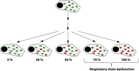
Mitotic segregation of mtDNA. A single mutational event creates heteroplasmy in a cell, but the level of mutated mtDNA (red dots) is very low in comparison with normal mtDNA (green dots). There is no synchronization between cell division and mtDNA replication, and a particular mtDNA molecule may be replicated many times or not at all during a single cell cycle. Repeated cell division will lead to mitotic segregation of normal and mutated mtDNA, and accumulation of mutated mtDNA above a certain threshold level will lead to impaired respiratory chain function.
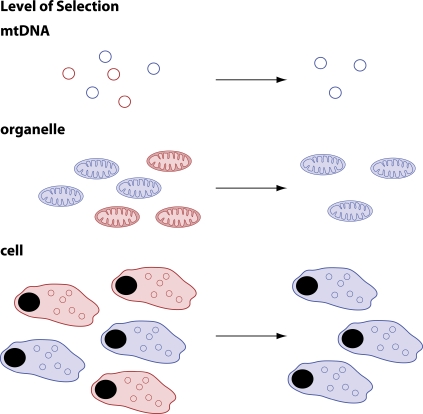
Different levels at which purifying selection can occur in the maternal germline. (top) Genomes with mutations could be blocked from replication or selectively destroyed without the need for gene expression. (middle) A fragmented mitochondrial network would allow functional testing of individual mtDNA molecules. The presence of a mutated mtDNA molecule would result in a mitochondrion with deficient respiratory chain function, which, in turn, would lead to selection against and/or destruction of this mitochondrion. (bottom) Cells with high levels of mutated mtDNA may fail to compete with respiratory chain–competent cells and may be selected against or undergo apoptosis. The colors indicate mutant (red) and wild-type (blue) mtDNA (top); respiratory chain–deficient (red) and normal (blue) mitochondria (middle); and respiratory chain–deficient (red) and normal (blue) cells (bottom).
Similar articles
-
Oxidative stress is not a major contributor to somatic mitochondrial DNA mutations.PLoS Genet. 2014 Feb 6;10(2):e1003974. doi: 10.1371/journal.pgen.1003974. eCollection 2014 Feb. PLoS Genet. 2014. PMID: 24516391 Free PMC article.
-
Somatic mitochondrial DNA mutations in mammalian aging.Annu Rev Biochem. 2010;79:683-706. doi: 10.1146/annurev-biochem-060408-093701. Annu Rev Biochem. 2010. PMID: 20350166 Review.
-
Mitochondrial DNA mutations and oxidative damage in aging and diseases: an emerging paradigm of gerontology and medicine.Proc Natl Sci Counc Repub China B. 1998 Apr;22(2):55-67. Proc Natl Sci Counc Repub China B. 1998. PMID: 9615468 Review.
-
Cause or casualty: The role of mitochondrial DNA in aging and age-associated disease.Biochim Biophys Acta Mol Basis Dis. 2019 Feb 1;1865(2):285-297. doi: 10.1016/j.bbadis.2018.09.035. Epub 2018 Nov 9. Biochim Biophys Acta Mol Basis Dis. 2019. PMID: 30419337 Free PMC article. Review.
-
Mitochondrial DNA mutations in human disease.Nat Rev Genet. 2005 May;6(5):389-402. doi: 10.1038/nrg1606. Nat Rev Genet. 2005. PMID: 15861210 Free PMC article. Review.
Cited by
-
Reprogramming: Emerging Strategies to Rejuvenate Aging Cells and Tissues.Int J Mol Sci. 2021 Apr 13;22(8):3990. doi: 10.3390/ijms22083990. Int J Mol Sci. 2021. PMID: 33924362 Free PMC article. Review.
-
Aging Hallmarks and Progression and Age-Related Diseases: A Landscape View of Research Advancement.ACS Chem Neurosci. 2024 Jan 3;15(1):1-30. doi: 10.1021/acschemneuro.3c00531. Epub 2023 Dec 14. ACS Chem Neurosci. 2024. PMID: 38095562 Free PMC article. Review.
-
Do we age because we have mitochondria?Protoplasma. 2014 Jan;251(1):3-23. doi: 10.1007/s00709-013-0515-x. Epub 2013 Jun 22. Protoplasma. 2014. PMID: 23794102 Review.
-
Naive T cell maintenance and function in human aging.J Immunol. 2015 May 1;194(9):4073-80. doi: 10.4049/jimmunol.1500046. J Immunol. 2015. PMID: 25888703 Free PMC article. Review.
-
Burns in the Elderly: Potential Role of Stem Cells.Int J Mol Sci. 2020 Jun 29;21(13):4604. doi: 10.3390/ijms21134604. Int J Mol Sci. 2020. PMID: 32610474 Free PMC article. Review.
References
-
- Akman H.O., Dorado B., López L.C., García-Cazorla A., Vilà M.R., Tanabe L.M., Dauer W.T., Bonilla E., Tanji K., Hirano M. 2008. Thymidine kinase 2 (H126N) knockin mice show the essential role of balanced deoxynucleotide pools for mitochondrial DNA maintenance. Hum. Mol. Genet. 17:2433–2440 10.1093/hmg/ddn143 - DOI - PMC - PubMed
-
- Alpers B.J. 1931. Diffuse progressive degeneration of the gray matter of the cerebrum. Arch. Neurol. Psychiatry. 25:469–505
Publication types
MeSH terms
Substances
LinkOut - more resources
Full Text Sources
Other Literature Sources
Medical
