. 2011 Jul 1;187(1):350-60.
doi: 10.4049/jimmunol.1004144. Epub 2011 May 23.

IL-6 mediates the susceptibility of glycoprotein 130 hypermorphs to Toxoplasma gondii

Affiliations

IL-6 mediates the susceptibility of glycoprotein 130 hypermorphs to Toxoplasma gondii

Jonathan S Silver et al. J Immunol. .

Abstract

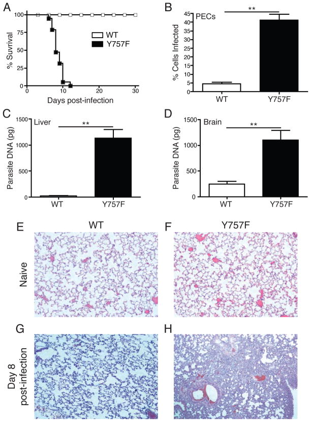
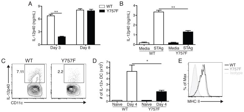
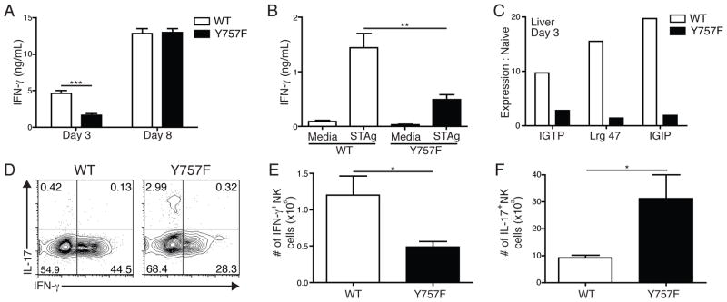
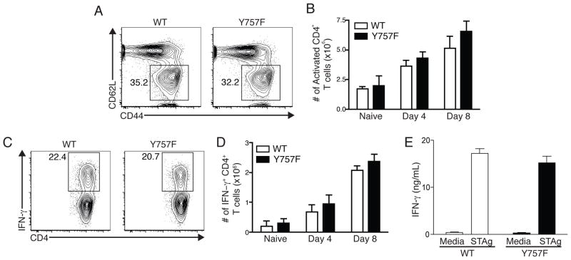
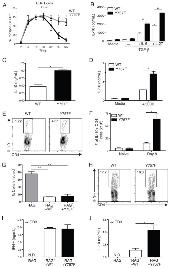
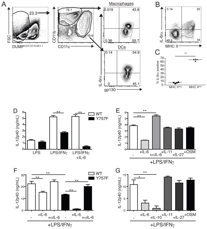
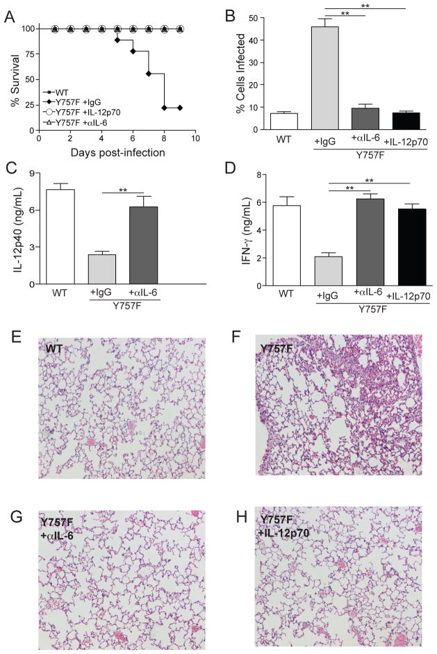
IL-6 and IL-27 are closely related cytokines that play critical but distinct roles during infection with Toxoplasma gondii. Thus, IL-6 is required for the development of protective immunity to this pathogen, whereas IL-27 is required to limit infection-induced pathology. Paradoxically, these factors both signal through gp130, but little is known about how the signals downstream of gp130 are integrated to coordinate the immune response to infection. To better understand these events, gp130 Y757F mice that have a mutation in gp130 at the binding site for suppressor of cytokine signaling 3, a critical negative regulator of gp130 signaling, were infected with T. gondii. These mutant mice were acutely susceptible to this challenge, characterized by an early defect in the production of IL-12 and IFN-γ and increased parasite burdens. Consistent with the reduced IL-12 levels, IL-6, but not other gp130 cytokines, was a potent antagonist of IL-12 production by gp130 Y757F macrophages and dendritic cells in vitro. Moreover, in gp130 Y757F mice, blocking IL-6 in vivo, or administration of rIL-12, during infection restored IFN-γ production and protective immunity. Collectively, these studies highlight that a failure to abbreviate IL-6-mediated gp130 signaling results in a profound anti-inflammatory signal that blocks the generation of protective immunity to T. gondii.


Figures

Figure 1
Figure 1
Gp130 Y757F mice are susceptible to infection with Toxoplasma gondii. A) Survival of WT (n=15) and gp130 Y757F mice (n=12) infected intraperitoneally with 20 cysts of the Me49 strain of T. gondii. B) Percentage of T. gondii-infected peritoneal exudate cells (PECs) at day 8 post infection in WT (n=14) and gp130 Y757F (n=12) mice. **p<.001. Enumeration of parasite DNA by real-time PCR in livers (C) and brains (D) of infected WT (n=10) and gp130 Y757F (n=14) mice at day 8 post-infection. **p<.001. Histopathology of lungs from naïve and infected WT (E and G) and gp130 Y757F (F and H) mice, analyzed by staining with hematoxylin and eosin at day 8 post-infection.
Figure 2
Figure 2
Gp130 Y757F mice have an early defect in the ability to produce IL-12 during infection with T. gondii. A) Kinetics of IL-12p40 production in the serum of infected WT (n=8) or gp130 Y757F mice (n=10), as measured by ELISA.**p<.001. B) ELISA of IFN-g levels in the supernatants of splenocytes isolated from WT and gp130 Y757F mice that were incubated with STAg for 48 hours. Data are representative of three independent experiments. **p<.001. C) Flow cytometry of intracellular IL-12p40 in DC isolated from PECs of infected WT or gp130 Y757F mice at day 4 post-infection; cells were incubated with BFA for 6 hours before staining. D) Total number of IL-12+ DC in the PECs at day 4 post-infection in WT or gp130 Y757F mice, calculated from percentages based on flow cytometry. *p<.01 E) Representative histrogram of MHC II expression on DC from the PEC of WT (solid line) or gp130 Y757F (dotted line) mice at day 4 post-infection. Data are representative of two independent experiments.
Figure 3
Figure 3
Gp130 Y757F mice have lower levels of IFN-γ early after T. gondii infection. A) Kinetics of IFN-γ production in the serum of infected WT (n=10) or gp130 Y757F mice (n=11), as measured by ELISA. Data are pooled from 4 independent experiments, ***p<.001. B) At day 3 post-infection, whole splenocytes from WT and Y757F mice were stimulated with STAg for 48 hours and assayed for IFN-γ. Data are representative of 4 independent experiments. **p<.01. C) Real time PCR of total cellular RNA isolated from the livers of WT and Y757F mice at day 3 post-infection to detect IGTP, Lrg 47 and IGIP mRNA. Data are representative of two independent experiments. D) Flow cytometry of IL-17A vs. IFN-γ from splenocytes isolated from WT or gp130 Y757F mice at day 4 post-infection. Plots are gated on NK1.1+ CD3- cells; cells were incubated with PMA, Ionomycin and BFA for 4 hours before being stained. Total numbers of of IFN-γ+ (E) and IL-17+ (F) splenic NK cells from WT (n=5) and Y757F mice (n=7) at day 4 post-infection. Data are pooled from two independent experiments. *p<.05.
Figure 4
Figure 4
Infection-induced T cell activation is normal in Y757F mice. A) Flow cytometry of splenocytes from WT or gp130 Y757F mice at day 8 post-infection showing expression of CD62L and CD44. Gated on CD3+ CD4+ cells. B) The number of CD44hi CD62Llow CD4+ T cells was enumerated in naïve WT and gp130 Y757F mice, and at day 4 and day 8 post-infection. n≥5 mice per time point. C) Flow cytometry of IFN-γ in splenocytes isolated from WT or gp130 Y757F mice at day 8 post-infection. Gated on CD3+ CD4+ events; cells were incubated with PMA, Ionomycin and BFA for 4 hours before being stained. D) Enumeration of IFN-γ+ CD4+ T cells in naïve or infected WT or gp130 Y757F mice. n≥5 mice per time point. E) ELISA of IFN-γ levels from cultures of splenocytes isolated at day 8 post-infection and incubated with STAg for 48 hours. Data are representative of 4 independent experiments.
Figure 5
Figure 5
Gp130 Y757F T cells produce augmented levels of IL-10 in response to IL-6 and IL-27. Purified CD4+ T cells from naïve WT and Y757F mice were stimulated in vitro with IL-6 (A) for various time points before being fixed, permeabilized and stained for intracellular phosphorylated STAT3. Data are pooled from 5 independent experiments. *p<.01. B) Purified CD4+ T cells from naïve WT or gp130 Y757F mice were activated with anti-CD3 and anti-CD28 for 48 hours in the presence of TGF-β with IL-6 or IL-27. IL-10 levels in culture supernatants were determined by ELISA. Data are representative of 4 independent experiments with similar results. **p<.001. C) IL-10 levels in the serum of infected WT (5) or gp130 Y757F (8) mice were determined by ELISA at day 8 post-infection. *p<.01. Data are pooled from two independent experiments. D) At day 8 post-infection, whole splenocytes from WT and Y757F mice were stimulated with media or anti-CD3 for 48 hours and assayed for IL-10 by ELISA. Data are representative of three independent experiments. *p<.01. D) Flow cytometry of IL-10 from splenocytes isolated from WT or gp130 Y757F mice at day 8 post-infection. Gated on CD3+ CD4+ events, cells were incubated with PMA, Ionomycin and BFA for 4 hours before being stained. F) The total number of IL-10+ CD4+ T cells was calculated in WT (n=5) and Y757F mice (n=6) at 8 days post-infection. Data are pooled from two independent experiments. *p<.01. G) 10 million splenic CD4+ and CD8+ T cells were isolated from WT and Y757F mice and transferred into RAG KO mice. The recipient RAG mice that received T cells from WT (n=5) or gp130 Y757F mice (n=5) were infected with T. gondii and at 8 days post-infection the percentage of infected PECs was enumerated. **p<.001. H) Flow cytometry of IFN-g from splenocytes isolated from RAG KO mice that received WT or gp130 Y757F mice at day 8 post-infection. Gated on CD3+ CD4+ events; cells were incubated with PMA, Ionomycin and BFA for 4 hours before being stained. I) At 8 days post-infection, whole splenocytes from recipient RAG KO mice were stimulated with anti-CD3 for 48 hours and assayed for IFN-γ by ELISA. J) At 8 days post-infection, whole splenocytes from recipient RAG KO mice were stimulated with anti-CD3 for 48 hours and assayed for IL-10 by ELISA. Data are representative of 3 independent experiments with similar results. *p<.01.
Figure 6
Figure 6
IL-6 blocks the production of IL-12 by gp130 Y757F APC. A) Flow cytometry of IL-6 receptor alpha and gp130 expression on PECs from WT mice at day 4 post-infection. Gated on CD3/CD19/NK1.1neg, CD11c or CD11b+ cells. Representative FACS plot (B) and enumeration (C) of the percentage of MHC II negative or MHC II positive DCs that express the IL-6 receptor alpha. Data are representative of two independent experiments with similar results. D) Purified splenic DC from naïve WT and Y757F mice were stimulated in vitro with LPS, IFN-γ and IL-6 for 24 hours before IL-12p40 was measured in culture supernatants by ELISA. E) Purified splenic DC from naïve gp130 Y757F mice were stimulated in vitro with LPS, IFN-γ and IL-6, IL-11, IL-27, Oncostatin M or anti-IL-6 antibodies for 24 hours before IL-12p40 was measured in culture supernatants by ELISA. N=3 samples per condition. Data are representative of 3 independent experiments. F) Bone marrow-derived macrophages from WT and Y757F mice were stimulated in vitro with LPS, IFN-γ and IL-6 or anti-IL-6 antibodies for 24 hours before IL-12p40 was measured in culture supernatants by ELISA. G) Bone marrow-derived macrophages from gp130 Y757F mice were stimulated in vitro with LPS, IFN-γ and IL-6, IL-10, IL-11, IL-27 or Oncostatin M for 24 hours before IL-12p40 was measured in culture supernatants by ELISA.
Figure 7
Figure 7
Anti-IL-6 treatment, or administration of IL-12p70, rescues gp130 Y757F mice. A) Survival of T. gondii-infected WT (n=5) or gp130 Y757F mice that were treated with Rat IgG (n=12), anti-IL-6 antibodies (n=9) or recombinant IL-12p70 (n=6). B) Percentage of PECs infected in WT (n=5) or gp130 Y757F mice that were treated with Rat IgG (n=5), anti-IL-6 antibodies (n=8) or recombinant IL-12p70 (n=5). C) At day 3 post-infection, WT (n=5) or gp130 Y757F mice treated with Rat IgG (n=7) or anti-IL-6 blocking antibodies (n=8) were bled and the levels of circulating IL-12p40 was determined by ELISA. **p<.01. D) At day 3 post-infection, WT (n=5) or gp130 Y757F mice treated with Rat IgG (n=7), anti-IL-6 blocking antibodies (n=8), or recombinant IL-12p70 (n=5) were bled and the levels of circulating IFN-γ was determined by ELISA. **p<.01. E-H) Histopathology of lungs from infected WT or gp130 Y757F mouse that was treated with Rat IgG, anti-IL-6 or recombinant IL-12p70 and stained with hemotoxylin and eosin.

References

    1. Taga T, Kishimoto T. Gp130 and the interleukin-6 family of cytokines. Annu Rev Immunol. 1997;15:797–819. - PubMed
    1. O’Shea JJ, Gadina M, Schreiber RD. Cytokine signaling in 2002: new surprises in the Jak/Stat pathway. Cell. 2002;109(Suppl):S121–31. - PubMed
    1. O’Shea JJ, Murray PJ. Cytokine signaling modules in inflammatory responses. Immunity. 2008;28:477–487. - PMC - PubMed
    1. De Souza D, Fabri LJ, Nash A, Hilton DJ, Nicola NA, Baca M. SH2 domains from suppressor of cytokine signaling-3 and protein tyrosine phosphatase SHP-2 have similar binding specificities. Biochemistry. 2002;41:9229–9236. - PubMed
    1. Nicholson SE, De Souza D, Fabri LJ, Corbin J, Willson TA, Zhang JG, Silva A, Asimakis M, Farley A, Nash AD, Metcalf D, Hilton DJ, Nicola NA, Baca M. Suppressor of cytokine signaling-3 preferentially binds to the SHP-2-binding site on the shared cytokine receptor subunit gp130. Proc Natl Acad Sci U S A. 2000;97:6493–6498. - PMC - PubMed

Publication types

MeSH terms