

Side-chain hydrophobicity scale derived from transmembrane protein folding into lipid bilayers
- PMID: 21606332
- PMCID: PMC3121867
- DOI: 10.1073/pnas.1103979108
Side-chain hydrophobicity scale derived from transmembrane protein folding into lipid bilayers
Abstract
The transfer free energies of the twenty natural amino acid side chains from water to phospholipid bilayers make a major contribution to the assembly and function of membrane proteins. Measurements of those transfer free energies will facilitate the identification of membrane protein sequences and aid in the understanding of how proteins interact with membranes during key biological events. We report the first water-to-bilayer transfer free energy scale (i.e., a "hydrophobicity scale") for the twenty natural amino acid side chains measured in the context of a native transmembrane protein and a phospholipid bilayer. Our measurements reveal parity for apolar side-chain contributions between soluble and membrane proteins and further demonstrate that an arginine side-chain placed near the middle of a lipid bilayer is accommodated with much less energetic cost than predicted by molecular dynamics simulations.
Conflict of interest statement
The authors declare no conflict of interest.
Figures
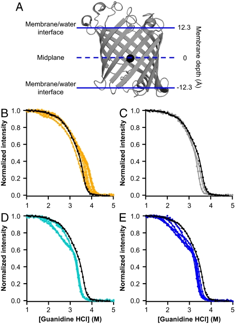
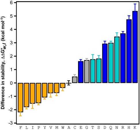
 ) of each amino acid variant at position 210 is compared to the wild-type OmpLA. Error bars represent standard errors of the mean from individual titration experiments. The color scheme is the same as in Fig. 1 B–E.
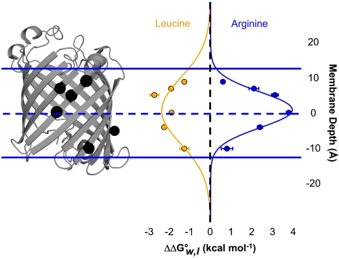
 of leucine and arginine variants (compared to alanine variants) is shown aligned with the OmpLA image. Normal distributions fit to the leucine and arginine data are also shown. Error bars represent standard errors of the mean from individual titration experiments.
Comment in
-
We choose to go to the membrane.Proc Natl Acad Sci U S A. 2011 Jun 21;108(25):10027-8. doi: 10.1073/pnas.1107322108. Epub 2011 Jun 13. Proc Natl Acad Sci U S A. 2011. PMID: 21670273 Free PMC article. No abstract available.
References
-
- Tanford C. The hydrophobic effect and the organization of living matter. Science. 1978;200:1012–1018. - PubMed
-
- White SH, Wimley WC. Membrane protein folding and stability: Physical principles. Annu Rev Biophys Biomol Struct. 1999;28:319–365. - PubMed
-
- Kyte J, Doolittle RF. A simple method for displaying the hydropathic character of a protein. J Mol Biol. 1982;157:105–132. - PubMed
Publication types
MeSH terms
Substances
Grants and funding
LinkOut - more resources
Full Text Sources
Other Literature Sources
Molecular Biology Databases
