. 2011 Jun 7;108(23):9384-9.
doi: 10.1073/pnas.1017200108. Epub 2011 May 23.

Structure of a bacterial cell surface decaheme electron conduit

Affiliations

Structure of a bacterial cell surface decaheme electron conduit

Thomas A Clarke et al. Proc Natl Acad Sci U S A. .

Abstract

Some bacterial species are able to utilize extracellular mineral forms of iron and manganese as respiratory electron acceptors. In Shewanella oneidensis this involves decaheme cytochromes that are located on the bacterial cell surface at the termini of trans-outer-membrane electron transfer conduits. The cell surface cytochromes can potentially play multiple roles in mediating electron transfer directly to insoluble electron sinks, catalyzing electron exchange with flavin electron shuttles or participating in extracellular intercytochrome electron exchange along "nanowire" appendages. We present a 3.2-Å crystal structure of one of these decaheme cytochromes, MtrF, that allows the spatial organization of the 10 hemes to be visualized for the first time. The hemes are organized across four domains in a unique crossed conformation, in which a staggered 65-Å octaheme chain transects the length of the protein and is bisected by a planar 45-Å tetraheme chain that connects two extended Greek key split β-barrel domains. The structure provides molecular insight into how reduction of insoluble substrate (e.g., minerals), soluble substrates (e.g., flavins), and cytochrome redox partners might be possible in tandem at different termini of a trifurcated electron transport chain on the cell surface.


Conflict of interest statement

The authors declare no conflict of interest.

Figures
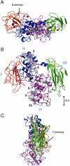
Fig. 1.
Fig. 1.
Crystal structure of MtrF with the four domains colored sequentially red, blue, green, and purple from the N terminus to the C terminus. The 10 heme cofactors are colored blue. (A) A view of MtrF showing the positioning of all 10 hemes on one side of the molecule and the two split β-barrel domains. (B) A view of MtrF rotated 90° on the longitudinal axis compared to A. It shows the arrangement of hemes within the protein that are numbered according to their position in the peptide sequence. The disulfide bond between Cys 428 and Cys 437 is shown with sulfurs colored as yellow spheres. (C) A view of MtrF rotated 90° on the vertical axis compared to B.
Fig. 2.
Fig. 2.
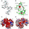
Heme packing motifs within the MtrF molecule. (A) Arrangement of hemes within the MtrF molecule. The orientation corresponds to Fig. 1B, and the distances between the porphyrin rings are indicated. (B) Calcium binding site on the surface of MtrF with the heme and peptide displayed as sticks, the heme iron and calcium displayed as spheres. The calcium is coordinated by the carbonyl backbones of Pro 257, Leu 259, and Arg262, as well as the carboxy side chain of Asp255. (C and D) Electrostatic surface of MtrF calculated and displayed using CCP4mg. The surface potentials displayed scale from -0.5 V (red, negatively charged) to +0.5 V (blue, positively charged). (C) Surface of MtrF showing the charge associated with the heme propionate groups. (D) Opposite surface of MtrF to that shown in C.
Fig. 3.
Fig. 3.
Cartoon showing the possible integration of MtrF into the respiratory electron transport system. The MtrF is oriented so that domains I and IV interact with the outer-membrane MtrDE cytochrome-porin electron delivery module. This orientation positions heme 10 in domain IV to accept electrons from MtrD and heme 5 in domain II to be the solvent-exposed terminus for electron output to solid substrates, soluble substrates, or electron shuttles as suggested in the main text. The more buried hemes 2 and 7 in the domain I/II and III/IV interfaces are possible sites for electron exchange with soluble substrate or electron shuttles that could be particularly important if the heme 5 terminus is occluded by interaction with a solid surface. Electron delivery to MtrD from the inner membrane (IM) quinol pool is via the tetraheme CymA, which may be direct or via other periplasmic cytochromes that are omitted for clarity. In this illustrative cartoon electron input is shown via the formate dehydrogenase (Fdh). A transmembrane electrochemical gradient is generated across the inner membrane, whereas the extracellular respiratory substrates serve as electron sinks to recycle the menaquinone (MQ) pool. The extent to which MtrF and MtrD extend into the MtrE barrel is not known but the terminal hemes of the two proteins must come within 14 Å to allow for efficient electron transfer because the homologous MtrCAB complex has been reconstituted in proteoliposomes and shown to conduct electrons across the membrane (4).
Fig. 4.
Fig. 4.
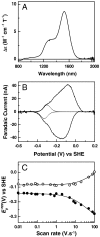
Spectroscopic and voltammetric properties of MtrF. (A) Room temperature near infrared MCD of 95 μM MtrF in 50 mM Hepes buffer, pH* 7.0, in D2O. The spectrum was recorded using a magnetic field of 6 T and intensity is given per protein. (B) PFV of MtrF: a cyclic voltammogram recorded using a freshly polished PGE electrode and immersed in 50 mM Hepes, 100 mM NaCl, pH 7.0 at 20 °C, scan rate 30 mV s-1, electrode rotation 3,000 rpm. The background current was subtracted using previously described procedures (31). Fitting each peak to 10 equal n = 1 responses (Fig. S4) showed the low-potential flanks could be described by a single process (dotted lines) with Em (average peak potential) of -312 mV. (C) Scan-rate dependence of MtrF PFV. Variation of the oxidative (open circles) and reductive (closed circles) apparent peak potentials (formula image) with scan rate. A best fit trumpet plot is shown (solid lines) with an interfacial electron exchange rate constant of 220 s-1.
Fig. 5.
Fig. 5.
(A) EPR monitored potentiometric titration of MtrF. CW X-band EPR (perpendicular mode) of 95 μM MtrF in 50 mM Hepes, 100 mM NaCl, 0.5% CHAPS, pH 7.5. Samples were poised in an anaerobic glove box at the potentials indicated. Spectra were recorded at 10 K, with microwave frequency, 9.68 GHz; microwave power, 2 mW; modulation amplitude, 0.1 mT (10 G). (B) Deconvolution of the MtrF spectrum recorded at -100 mV. Baseline subtracted (solid line) and simulated (broken line) spectra are offset for clarity. Shown below are the individual simulated line shapes for LGM, LS1, and LS2 heme populations that sum to give the simulated spectrum. (C) Potential dependence of the observed heme signals, LS1 (squares), LS2 (circles), and LGM (triangles). Signal area of each signal was normalized to that observed for the LS2 population. Integration of the simulated LS2 line shape from the 200-mV spectrum, relative to a 1 mM Cu2+-EDTA standard, gave approximately 0.9 spins per protein. * indicates the methylviologen radical the arises from the redox mediator cocktail.

References

    1. Shi L, Squier TC, Zachara JM, Fredrickson JK. Respiration of metal (hydr)oxides by Shewanella and Geobacter: A key role for multihaem c-type cytochromes. Mol Microbiol. 2007;65:12–20. - PMC - PubMed
    1. Coursolle D, Gralnick JA. Modularity of the Mtr respiratory pathway of Shewanella oneidensis strain MR-1. Mol Microbiol. 2010;77:995–1008. - PubMed
    1. Ross DE, et al. Characterization of protein-protein interactions involved in iron reduction by Shewanella oneidensis MR-1. Appl Environ Microbiol. 2007;73:5797–7808. - PMC - PubMed
    1. Hartshorne RS, et al. Characterization of an electron conduit between bacteria and the extracellular environment. Proc Natl Acad Sci USA. 2009;106:22169–22174. - PMC - PubMed
    1. McLean JS, et al. Oxygen-dependent autoaggregation in Shewanella oneidensis MR-1. Environ Microbiol. 2008;10:1861–1876. - PubMed

Publication types

MeSH terms

Associated data