. 2011 Jun 7;108(23):9578-82.
doi: 10.1073/pnas.1106383108. Epub 2011 May 23.

Noninvasive measurement of androgen receptor signaling with a positron-emitting radiopharmaceutical that targets prostate-specific membrane antigen

Affiliations

Noninvasive measurement of androgen receptor signaling with a positron-emitting radiopharmaceutical that targets prostate-specific membrane antigen

Michael J Evans et al. Proc Natl Acad Sci U S A. .

Abstract

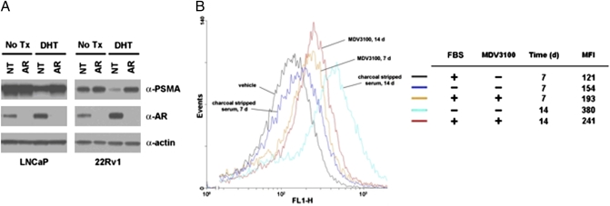
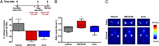
Despite encouraging clinical results with next generation drugs (MDV3100 and abiraterone) that inhibit androgen receptor (AR) signaling in patients with castration-resistant prostate cancer (CRPC), responses are variable and short-lived. There is an urgent need to understand the basis of resistance to optimize their future use. We reasoned that a radiopharmaceutical that measures intratumoral changes in AR signaling could substantially improve our understanding of AR pathway directed therapies. Expanding on previous observations, we first show that prostate-specific membrane antigen (PSMA) is repressed by androgen treatment in multiple models of AR-positive prostate cancer in an AR-dependent manner. Conversely, antiandrogens up-regulate PSMA expression. These expression changes, including increased PSMA expression in response to treatment with the antiandrogen MDV3100, can be quantitatively measured in vivo in human prostate cancer xenograft models through PET imaging with a fully humanized, radiolabeled antibody to PSMA, (64)Cu-J591. Collectively, these results establish that relative changes in PSMA expression levels can be quantitatively measured using a human-ready imaging reagent and could serve as a biomarker of AR signaling to noninvasively evaluate AR activity in patients with CRPC.


Conflict of interest statement

Conflict of interest statement: The article describes a new radiotracer to monitor androgen receptor signaling noninvasively. The various studies that demonstrate the utility of this radiotracer include an experiment using the antiandrogen drug MDV3100. C.L.S. is a coinventor of MDV3100 and owns stock in the company (Medivation) that is developing the drug for prostate cancer treatment. The article does not make any claims about the efficacy of MDV3100; it merely uses it as tool to evaluate the new radiotracer. N.H.B. is the inventor of patents related to PSMA antibodies assigned to Cornell Research Foundation.

Figures

Fig. 1.
Fig. 1.
Androgens suppress PSMA in multiple prostate cancer cell lines and xenografts. (A) Incubation of the hormone-responsive prostate cancer cell lines LNCaP and CWR22Rv1 (22Rv1) for 72 h with the endogenous androgens testosterone (Test., 10 nM) and DHT (10 nM) or the synthetic androgen R1881 (0.1 nM) decreases PSMA protein levels compared with low androgen conditions (FBS and CSS). In CWR22Rv1 cells, AR levels increase after androgen treatment compared with vehicle control, suggesting agonist-mediated receptor stabilization and minimal hormone degradation during incubation. (B) PSMA mRNA levels are suppressed after 72 h of treatment with androgens in LNCaP and CWR22Rv1, consistent with transcriptional regulation of PSMA by hormone treatment. As anticipated, mRNA levels of PSA, an AR target gene, increase in response to androgen challenge, confirming the bioactivity of hormone treatment at this time point. (C) Subcutaneous xenografts of LNCaP and CWR22Rv1 derived in castrate male mice were harvested 7 d after implantation of a DHT pellet or no surgical treatment (No Tx). Immunoblot (Upper) and quantitative PCR analysis (Lower) shows that PSMA is expressed in PCa xenografts, and expression is reduced by DHT treatment. The androgen-stimulated gene product TMPRSS2 is up-regulated by DHT, confirming the bioactivity of the pellet dose.
Fig. 2.
Fig. 2.
Genetic and pharmacological inhibition of AR blocks androgen suppression of PSMA. (A) LNCaP and CWR22Rv1 (22Rv1) were transfected with nontargeted (NT) or AR-directed (AR) siRNA pools and treated with vehicle (EtOH) or DHT (10 nM). Whereas basal expression of PSMA is unaffected by AR knockdown in either cell line at this time point, AR silencing inhibits PSMA suppression by DHT, thus suggesting a role for AR in this process. Cells were transfected with 100 nM of siRNA, DHT challenge was initiated 24 h after transfection, and cells were harvested 48 h after androgen treatment. (B) Hormone withdrawal and antiandrogen treatment increase PSMA expression in LNCaP-AR in vitro. Cells were plated in media containing 10% (vol/vol) FBS and treated with vehicle, MDV3100 (10 μM), or the media was replaced with 10% (vol/vol) CSS (indicated as “-”). After 7 or 14 d, cells were harvested and incubated with Alexa Fluor 488-labeled J591, and PSMA expression was analyzed by FACS. Mean fluorescent intensities (MFI) were calculated and show that MDV3100 up-regulates PSMA expression by 7 d, whereas the effects of hormone withdrawal were observed between 7 and 14 d.
Fig. 3.
Fig. 3.
Detection of androgen repression of PSMA in prostate cancer xenografts by PET. (A) Bilateral s.c. CWR22Rv1 xenografts were established in castrate male mice and imaged by PET with 64Cu-J591 24 h before manipulation (scan 1). Animals were again injected with 64Cu-J591 6 d after hormonal manipulation (Test., DHT) or no treatment (No Tx), and scan 2 was acquired 16 h after injection on day 7. The ratio of SUVmean values (scan 2/scan 1) shows substantial reduction in 64Cu-J591 incorporation in the xenografts exposed to testosterone (Test.) and DHT treatment. (B) Representative transverse and coronal slices (scan 2) of animals bearing CWR22Rv1 xenografts showing visibly reduced uptake in tumors exposed to androgens compared with no treatment. The positions of the tumors are indicated with arrows. (C) Ex vivo biodistribution data (%ID/g) of the tumors and selected host tissues show that the tumors alone, and not androgen-insensitive tissues, respond to hormonal manipulation. Fig. S5 shows a graphical representation of the correlation between calculated SUVmean values and activity measurements acquired ex vivo, Table S1 lists the SUVmean and biodistribution data from the full cohort of mice, and Table S2 lists the activity measurements (%ID/g) from the host tissues. *P < 0.01 compared with No Tx controls.
Fig. 4.
Fig. 4.
64Cu-J591 PET detects up-regulation of PSMA expression in prostate cancer xenografts in response to androgen deprivation therapies. (A) Percentage change in tumor volumes show that LNCaP-AR xenografts, established in intact male mice, regress after castration (Orch.) or daily oral gavage of MDV3100 (10 mg/kg). Tumor volume measurements were recorded at day 0 and after the final treatment on day 7. (B) To evaluate the effect of antiandrogen therapy on 64Cu-J591 incorporation, mice were imaged 24 h before the initiation of therapy (scan 1), and after 6 d of therapy mice were again injected with 64Cu-J591 and imaged by PET 16 h after radiotracer injection (scan 2). SUVmean values were calculated, and the ratio for each tumor (scan 2/scan 1) is reported. The change in 64Cu-J591 uptake associated with vehicle treatment and castration was minimal, but MDV3100 induced a measurable increase in 64Cu-J591 binding, consistent with the in vitro data. (C) Representative transverse and coronal slices (scan 2) of animals bearing LNCaP-AR xenografts showing increased intratumoral uptake of 64Cu-J591 in tumors exposed to MDV3100 compared with castration or vehicle. The positions of the tumors are indicated with arrows. Fig. S5 shows a graphical representation of the correlation between calculated SUVmean values and activity measurements of tumor tissue ex vivo, and Table S3 lists the SUVmean and biodistribution data from the full cohort of mice. ADTs, androgen deprivation therapies. *P < 0.05 compared with vehicle.

References

    1. Scher HI, Sawyers CL. Biology of progressive, castration-resistant prostate cancer: directed therapies targeting the androgen-receptor signaling axis. J Clin Oncol. 2005;23:8253–8261. - PubMed
    1. Chen Y, Sawyers CL, Scher HI. Targeting the androgen receptor pathway in prostate cancer. Curr Opin Pharmacol. 2008;8:440–448. - PMC - PubMed
    1. Scher HI, et al. Prostate Cancer Foundation/Department of Defense Prostate Cancer Clinical Trials Consortium Antitumour activity of MDV3100 in castration-resistant prostate cancer: A phase 1-2 study. Lancet. 2010;375:1437–1446. - PMC - PubMed
    1. Reid AH, et al. Significant and sustained antitumor activity in post-docetaxel, castration-resistant prostate cancer with the CYP17 inhibitor abiraterone acetate. J Clin Oncol. 2010;28:1489–1495. - PMC - PubMed
    1. Lilja H, Ulmert D, Vickers AJ. Prostate-specific antigen and prostate cancer: Prediction, detection and monitoring. Nat Rev Cancer. 2008;8:268–278. - PubMed

Publication types

MeSH terms