. 2011 Jun 7;108(23):9478-83.
doi: 10.1073/pnas.1019418108. Epub 2011 May 23.

Cholinergic chemosensory cells in the trachea regulate breathing

Affiliations

Cholinergic chemosensory cells in the trachea regulate breathing

Gabriela Krasteva et al. Proc Natl Acad Sci U S A. .

Abstract

In the epithelium of the lower airways, a cell type of unknown function has been termed "brush cell" because of a distinctive ultrastructural feature, an apical tuft of microvilli. Morphologically similar cells in the nose have been identified as solitary chemosensory cells responding to taste stimuli and triggering trigeminal reflexes. Here we show that brush cells of the mouse trachea express the receptors (Tas2R105, Tas2R108), the downstream signaling molecules (α-gustducin, phospholipase C(β2)) of bitter taste transduction, the synthesis and packaging machinery for acetylcholine, and are addressed by vagal sensory nerve fibers carrying nicotinic acetylcholine receptors. Tracheal application of an nAChR agonist caused a reduction in breathing frequency. Similarly, cycloheximide, a Tas2R108 agonist, evoked a drop in respiratory rate, being sensitive to nicotinic receptor blockade and epithelium removal. This identifies brush cells as cholinergic sensors of the chemical composition of the lower airway luminal microenvironment that are directly linked to the regulation of respiration.


Conflict of interest statement

The authors declare no conflict of interest.

Figures
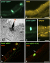
Fig. 1.
Fig. 1.
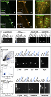
Tracheal ChAT-eGFP cells are cholinergic brush cells. (A and A′) ChAT-eGFP–expressing cells are immunoreactive for villin, a structural protein of microvilli of brush cells. (Scale bar, 20 μm.) (B) Ultrastructural preembedding anti-eGFP immunohistochemistry. ChAT-eGFP cell with micovilli tuft (mt) typical for brush cells, flanked by secretory cells (SC). (Scale bar, 1 μm.) (C and C′) Epifluorescence. ChAT-eGFP–expressing cell is immunoreactive for the vesicular ACh transporter (VAChT), further validating its cholinergic nature. (Scale bar, 20 μm.) (D and E) CLSM analysis, merged images. In addition to double-labeled cells, a villin-positive cell (arrow) with triangular shape is negative for ChAT (D), also when the eGFP signal is enhanced by immunolabeling (E). (Scale bar, 20 μm.)
Fig. 2.
Fig. 2.
Tracheal ChAT-eGFP–expressing cells are chemosensory. (A–D) Immunohistochemistry. (A and B) Trachea. ChAT-eGFP cells (arrowheads) are positive for α-gustducin (A and A′) and PLC-β2 (B and B′). Inset: Preabsorbtion (preabs.) of the antibodies with the corresponding peptides abolishes immunoreactivity for α-gustducin and PLC-β2 in ChAT-eGFP cells (arrows). (C and D) Tongue, merged images. In the vallate papilla used as a positive control for antibody specificities, no colocalization was found between α-gustducin (C) and PLC-β2 (D) and ChAT-eGFP, respectively. (Scale bar, 20 μm.) (E) RT-PCR. Alpha-gustducin, PLC-β2, and bitter taste receptors 105 (Tas2R105) and 108 (Tas2R108) are expressed in whole-trachea homogenate and abraded tracheal epithelial cells (TE); tongue served as positive control. Negative controls included samples in absence of reverse transcriptase (Ø RT) and without template (H2O). Marker = 100 base pairs. (F) RT-PCR of ChAT-eGFP cells isolated by FACS. (1) FACS analysis of isolated tracheal cells. P1 represents a subgroup of cells that includes the population of ChAT-eGFP expressing cells. (2) Representative dot plot showing gating on the ChAT-eGFP cells (GFP+) and CD45+ cells, and nonfluorescent cells (neg), used for sorting. (3) Representative dot plot of postsort analysis. Highly purified fraction of ChAT-eGFP cells after FACS sorting (97.5%) was collected and processed in RT-PCR experiments (Right). Tracheal homogenate was applied as a positive control for all investigated genes except eGFP, for which we used eGFP+ tissue biopsy (control DNA). mRNA for eGFP, α-gustducin, PLC-β2, for the cycloheximide receptor Tas2R105, and the denatonium receptor Tas2R108 is detected in the ChAT/eGFP+ fraction; respective lanes are highlighted by the blue label. Contamination of this cell fraction with other cell types is excluded by the lack of mRNA for tubulin (tub), a marker for ciliated cells, for protein gene product 9.5 (PGP), a marker for neuroendocrine cells, and for myosin heavy chain (Myh), a marker for smooth muscle cells. Corresponding cell numbers of the ChAT/eGFP+ fraction do not express taste-related genes. β-Microglobulin (β-MG) served as housekeeping gene to control for mRNA quality and PCR efficiency. All control reactions including samples in absence of reverse transcriptase (Ø RT) and without template (H2O) were negative.
Fig. 3.
Fig. 3.
Tracheal ChAT-eGFP brush cells are contacted by intraepithelial nerve fibers. Whole-mount immunohistochemistry, CLSM, 3D analysis. (A) ChAT-eGFP cells (yellow) are predominantly localized at the ligamentous parts (m) between the cartilage rings (c). (Scale bar, 1,000 μm.) (B) magnified region from A. A fine network of PGP 9.5+ fibers (red), partly colocalizing with CGRP immunoreactivity (blue), extends within and underneath the epithelium. ChAT-eGFP cells (yellow) are distinct from neuroendocrine cells (PGP 9.5+, red). (Scale bar, 50 μm.) (C and D) ChAT-GFP+ epithelial cells are seen in direct contact with PGP 9.5+/CGRP+ fibers (arrow in E; magnified Inset from B), with PGP 9.5+/CGRP fibers (arrow in C), or lack direct contact to nerve fibers (arrowheads in D and E). Neuroendocrine cells (PGP 9.5+, doubled arrows) are also seen with and without nerve fiber contacts. (Scale bar, 10 μm.)
Fig. 4.
Fig. 4.
Brush cells have contact to vagal cholinoceptive peptidergic sensory neurons. (A–C) Samples taken from Tg(Chrna3-EGFP) BAC transgenic mice. (A) An intraepithelial nerve fiber expressing eGFP in transgenic nAChR-eGFP mice (arrow) attaches to a villin+ brush cell (arrowhead). (B and C) Colocalization of CGRP and nAChR-eGFP expression can be observed in intraepithelial terminals (B) and neurons in the vagal jugular–nodose complex, which are retrogradely labeled with Fast Blue from the airways (C). (Scale bars, 20 μm.)
Fig. 5.
Fig. 5.
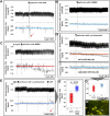
The bitter substance cycloheximide elicits an epithelium-dependent depressive respiratory reflex involving nicotinic transmission. All data from anesthetized, spontaneously breathing mice, representative traces depicted in A–E. (A and B) Model validation. Challenge of the trachea with capsaicin (CAP) evokes a rapid and transient change in respiration (arrow) resembling a single augmented breath (A), which is not seen after perfusion with Krebs buffer (baseline = blue line). (C) Perfusion with the nicotinic receptor agonist DMPP (10 μM) in the presence of atropine leads to a dramatic drop in the respiratory rate. (D and E) Cycloheximide (100 μM), a bitter substance that has affinity for Tas2R105, also causes a drop in the respiratory rate, which is completely abrogated by the nicotinic antagonist mecamylamine (MEC, 10 μM). The response to 10 μM capsaicin (CAP), however, persisted under nicotinic blockade (arrow = augmented breath). Mechanical disruption of the epithelium (D, Lower) abolishes the cycloheximide (CYC) effect in three out of four animals (box plot, ). (D″) Intravital fluorescence staining of the trachea with FM1-43 at the end of the experiment in which the animal still responded to cycloheximide revealed incomplete abrasion of the epithelial layer, and single epithelial cells (asterisks) are still contacted by capsaicin-sensitive nerve fibers (arrowhead). (D′ and E′) Statistical analyses of experiments shown representatively in E and D. Box plots: percentiles 0, 25, median, 75, and 100 (Mann-Whitney test, P ≤ 0.05 is considered significant).

References

    1. Reid L, et al. The mysterious pulmonary brush cell: A cell in search of a function. Am J Respir Crit Care Med. 2005;172:136–139. - PMC - PubMed
    1. Sbarbati A, Osculati F. The taste cell-related diffuse chemosensory system. Prog Neurobiol. 2005;75:295–307. - PubMed
    1. Rhodin J, Dalhamn T. Electron microscopy of the tracheal ciliated mucosa in rat. Z Zellforsch Mikrosk Anat. 1956;44:345–412. - PubMed
    1. Sbarbati A, Bramanti P, Benati D, Merigo F. The diffuse chemosensory system: Exploring the iceberg toward the definition of functional roles. Prog Neurobiol. 2010;91:77–89. - PubMed
    1. Merigo F, Benati D, Tizzano M, Osculati F, Sbarbati A. α-Gustducin immunoreactivity in the airways. Cell Tissue Res. 2005;319:211–219. - PubMed

Publication types

MeSH terms