. 2011 Jun 7;108(23):9613-8.
doi: 10.1073/pnas.1103187108. Epub 2011 May 23.

Endothelium-protective sphingosine-1-phosphate provided by HDL-associated apolipoprotein M

Affiliations

Endothelium-protective sphingosine-1-phosphate provided by HDL-associated apolipoprotein M

Christina Christoffersen et al. Proc Natl Acad Sci U S A. .

Abstract

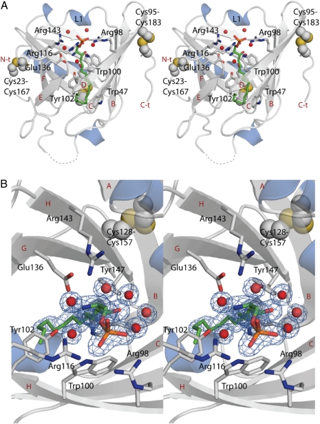
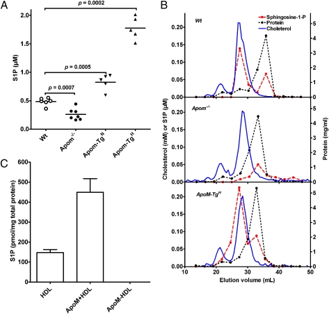
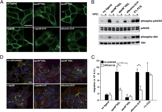
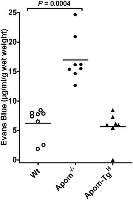
Protection of the endothelium is provided by circulating sphingosine-1-phosphate (S1P), which maintains vascular integrity. We show that HDL-associated S1P is bound specifically to both human and murine apolipoprotein M (apoM). Thus, isolated human ApoM(+) HDL contained S1P, whereas ApoM(-) HDL did not. Moreover, HDL in Apom(-/-) mice contains no S1P, whereas HDL in transgenic mice overexpressing human apoM has an increased S1P content. The 1.7-Å structure of the S1P-human apoM complex reveals that S1P interacts specifically with an amphiphilic pocket in the lipocalin fold of apoM. Human ApoM(+) HDL induced S1P(1) receptor internalization, downstream MAPK and Akt activation, endothelial cell migration, and formation of endothelial adherens junctions, whereas apoM(-) HDL did not. Importantly, lack of S1P in the HDL fraction of Apom(-/-) mice decreased basal endothelial barrier function in lung tissue. Our results demonstrate that apoM, by delivering S1P to the S1P(1) receptor on endothelial cells, is a vasculoprotective constituent of HDL.


Conflict of interest statement

The authors declare no conflict of interest.

Figures

Fig. 1.
Fig. 1.
The structure of the ApoM–S1P complex reveals the determinants of S1P-binding specificity. (A) Stereo view of the crystal structure of apoM with S1P at 1.7-Å resolution. S1P is shown as green sticks together with interacting residues. Strands B–F, the N terminus (N-t), and the C terminus (C-t) are labeled in red, and the interacting residues are labeled in black. (B) Top view of the S1P-binding site in close up. Electron density for S1P and surrounding water molecules is contoured at 1 σ and colored blue. Water molecules are shown as red spheres in both panels, and unmodeled loops toward the N terminus are shown as broken lines.
Fig. 2.
Fig. 2.
ApoM gene dosage determines plasma S1P in genetically modified mice. (A) Plasma S1P in WT, Apom−/−, and apoM-transgenic female mice with ∼twofold (Apom-TgN) and ∼10-fold (Apom-TgH) increased plasma apoM. Each point represents data from an individual mouse; horizontal lines indicate means. (B) Lipoproteins in pools of plasma from WT (Top), Apom−/− (Middle), or Apom-TgH (Bottom) mice were separated by gel filtration on serially connected Superose 6 and 12 columns. The flow rate was 0.4 mL/min. Fractions of 275 μL were collected. Aliquots of 10 consecutive fractions were pooled before measuring S1P (red filled symbols) and protein (dotted black line). Cholesterol concentration (solid blue line) was determined in each fraction. The scale bar for cholesterol (mmol/L) and S1P (μmol/L) is shown on the left y axis. Protein (mg/mL) is shown on the right y axis. (C) S1P was measured with HPLC in purified preparations of human total HDL, apoM+ HDL, and apoM HDL. Values are mean ± SEM (n = 3). S1P was not detectable in apoM HDL. Results were confirmed by LC-MS/MS.
Fig. 3.
Fig. 3.
ApoM-bound S1P activates S1P1-mediated intracellular signaling pathways. (A) Confocal microscopy of HEK293 cells stably expressing S1P1-GFP. Cells were serum starved, stimulated for 1 h with indicated ligands, fixed, and imaged. ApoM+ HDL (equivalent to ∼100 nM S1P as determined by LC/MS/MS) or ApoM HDL was used at 100 μg/mL Fatty acid-free BSA and r-apoM were complexed with S1P and used at afinal concentration of 100 nM (equimolar for both protein and lipid). (Scale bar, 20 μm.) (B) HUVEC were serum starved and pretreated with 1 μM of the S1P1 antagonist VPC44116 for 30 min before stimulated with apoM+ HDL (20 μg/mL protein, 20 nM S1P), apoMHDL (20 μg/mL protein), or albumin-S1P (100 nM S1P and equimolar protein) for 10 min. (Note that VPC44116 has no inhibitory effects on cells stimulated by FCS, because FCS can activate receptor systems other than S1P1.) Activation of p44/42 and Akt was examined by Western blot analysis using phospho-specific antibodies. (C) HUVEC were serum starved and pretreated with 1 μM VPC44116 for 30 min where indicated and subjected to migration assay with 10 μg/mL apoM+ HDL (10 nM S1P), 10 μg/mL apoM HDL, or 10 nM albumin-S1P. Data are mean ± SD (n = 3). *P < 0.01. (D) Microscopy of HUVEC that were serum starved and stimulated with 100 μg/mL apoM+ HDL, apoM HDL, 100 nM albumin-S1P, r-apoM–S1P, or 100 nM S1P-free r-apoM for 1 h. After fixation, VE-cadherin (green), nuclei (blue), and F-actin (red) were visualized with confocal microscopy. (Scale bar, 20 μm.)
Fig. 4.
Fig. 4.
The apoM–S1P complex maintains lung endothelial barrier function. WT, Apom−/−, and Apom-TgH mice were injected i.v. with Evans Blue (30 μg/g body weight). After 30 min the mice were perfused with saline; then the lungs were removed and used for extraction of Evans Blue. Each point represents the content of Evans Blue in the lungs of one mouse, and horizontal lines represent mean values.

References

    1. Garcia JG, et al. Sphingosine 1-phosphate promotes endothelial cell barrier integrity by Edg-dependent cytoskeletal rearrangement. J Clin Invest. 2001;108:689–701. - PMC - PubMed
    1. Kimura T, et al. Role of scavenger receptor class B type I and sphingosine 1-phosphate receptors in high density lipoprotein-induced inhibition of adhesion molecule expression in endothelial cells. J Biol Chem. 2006;281:37457–37467. - PubMed
    1. Aoki S, et al. Sphingosine 1-phosphate-related metabolism in the blood vessel. J Biochem. 2005;138:47–55. - PubMed
    1. Nofer JR, et al. HDL induces NO-dependent vasorelaxation via the lysophospholipid receptor S1P3. J Clin Invest. 2004;113:569–581. - PMC - PubMed
    1. Argraves KM, Argraves WS. HDL serves as a S1P signaling platform mediating a multitude of cardiovascular effects. J Lipid Res. 2007;48:2325–2333. - PubMed

Publication types

MeSH terms

Associated data

LinkOut - more resources