. 2011 Jun 7;108(23):9554-9.
doi: 10.1073/pnas.1017296108. Epub 2011 May 23.

Glucocorticoids target suppressor of cytokine signaling 1 (SOCS1) and type 1 interferons to regulate Toll-like receptor-induced STAT1 activation

Affiliations

Glucocorticoids target suppressor of cytokine signaling 1 (SOCS1) and type 1 interferons to regulate Toll-like receptor-induced STAT1 activation

Sandip Bhattacharyya et al. Proc Natl Acad Sci U S A. .

Abstract

Endogenous and pharmacologic glucocorticoids (GCs) limit inflammatory cascades initiated by Toll-like receptor (TLR) activation. A long-standing clinical observation has been the delay between GC administration and the manifestation of GC's anti-inflammatory actions. We hypothesized that the GCs would have inhibitory effects that target late temporal pathways that propagate proinflammatory signals. Here we interrogated signal transducer and activator of transcription 1 (STAT1) regulation by GC and its consequences for cytokine production during activation of macrophages with TLR-specific ligands. We found that robust STAT1 activation does not occur until 2-3 h after TLR engagement, and that GC suppression of STAT1 phosphorylation first manifests at this time. GC attenuates TLR4-mediated STAT1 activation only through induction of suppressor of cytokine signaling 1 (SOCS1), which increases throughout the 6-h period after treatment. Inhibition of TLR3-mediated STAT1 activation occurs via two mechanisms, impairment of type I IFN secretion and induction of SOCS1. Our data show that SOCS1 and type I interferons are critical GC targets for regulating STAT1 activity and may account for overall GC effectiveness in inflammation suppression in the clinically relevant time frame.


Conflict of interest statement

The authors declare no conflict of interest.

Figures

Fig. 1.
Fig. 1.
TLR-induced cytokine secretion in control and STAT1-null macrophages. Effect of LPS (100 ng/mL) (A), Poly (I:C) (50 μg/mL) (B), and CpG (2 μM) (C) on proinflammatory cytokine secretion in C57BL/6 (□), 129S6 (■), and STAT1−/− (formula image
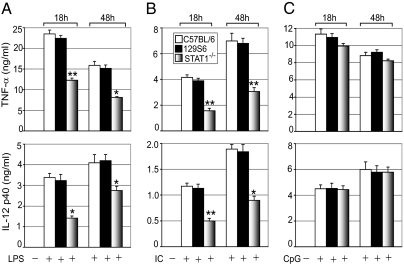
) macrophages. Cells were treated with TLR ligands for the indicated periods. Concentrations of TNF-α and IL-12p40 in the culture media were analyzed by ELISA. Data are mean ± SEM; n = 3. *P < 0.05; **P < 0.01 for STAT1−/− macrophages compared with control 129S6 macrophages.
Fig. 2.
Fig. 2.
Effect of GCs on TLR-induced STAT1 phosphorylation. Control or MGRKO macrophages were treated with or without Dex (100 nM) for 3 h, followed by LPS (A), Poly (I:C) (B), and CpG (C) treatment for the indicated periods. Cell lysates were analyzed by Western blot analysis using anti-phospho STAT1 Ser727 (P-STAT1 Ser), phospho STAT1 Tyr701 (P-STAT1 Tyr), and total STAT1 (Total-STAT1) antibodies. Representative of between three and five independent experiments.
Fig. 3.
Fig. 3.
(A) IFNAR1 activation and TLR-induced STAT1 phosphorylation. Macrophages were pretreated with anti-IFNAR1 antibody (MARI) for 1 h, followed by treatment with LPS, Poly (I:C), CpG, or control oligonucleotide (ODN) for the indicated periods. Cell lysates were analyzed by Western blot analysis as described in Fig. 2. Representative of three independent experiments. (B–F) GC modulation of TLR-induced type I IFN secretion and nuclear IRF3. Macrophages were treated with Dex, followed by treatment with Poly (I:C) (B and D) or LPS (C) for 3, 6, or 9 h for IFN-β and for 12 or 18 h for IFN-α. Concentrations of IFN-α and IFN-β in the culture media were analyzed by ELISA. Data are mean ± SEM; n = 3. *P < 0.05; **P < 0.01 for Dex-treated cells compared with the group treated with Poly (I:C) only. (E and F) Cells were treated as described previously, and nuclear extracts were analyzed by Western blot analysis using anti-IRF3 or anti–β-actin antibodies. (G and H) Effect of GCs on IFN-β–induced STAT1 phosphorylation. Cells were pretreated with Dex, followed by treatment with varying doses of IFN-β (31.25–500 U/mL) for 1 h (G) or 125 U/mL of IFN-β for the indicated periods (H). Cell lysates were analyzed as described in Fig. 2. Representative of three independent experiments.
Fig. 4.
Fig. 4.
(A–C) Effect of GCs on TLR-induced SOCS1 expression. Cells were treated with LPS (A), Poly (I:C) (B), and CpG (C) as described in Fig. 2. Cell lysates were analyzed by Western blot analysis using anti-SOCS1 or anti–β-actin antibodies. (D–G) SOCS1 mediates GC inhibition of STAT1. SOCS1+/+ or SOCS1−/− macrophages were treated with or without Dex for 3 h, followed by treatment with LPS (D) or Poly (I:C) (E) for the indicated periods. Cell lysates were analyzed by Western blot analysis using anti-phospho STAT1 Ser727 (P-STAT1 Ser), phospho STAT1 Tyr701 (P-STAT1 Tyr), and total STAT1 (Total-STAT1) antibodies. Representative of two or three independent experiments. Similar experiments were performed with LPS for 18 h (F) and Poly (I:C) for 48 h (G). Concentrations of TNF-α and IL-12p40 in the culture media were analyzed by ELISA. Data are mean ± SEM; n = 3. *P < 0.05; **P < 0.01 for Dex-treated cells compared with the group treated with TLR ligand only. (H and I) SOCS1 inhibits phospho-JAK2 abundance to inhibit STAT1 activation. SOCS1+/+ (H) or SOCS1−/− (I) macrophages were treated as described previously. Cell lysates were analyzed by Western blot analysis using anti-phospho JAK2 (P-JAK2), and total JAK2 (Total-JAK2) antibodies. Representative of two independent experiments.

References

    1. O'Neill LA. Targeting signal transduction as a strategy to treat inflammatory diseases. Nat Rev Drug Discov. 2006;5:549–563. - PubMed
    1. Rhen T, Cidlowski JA. Anti-inflammatory action of glucocorticoids—new mechanisms for old drugs. N Engl J Med. 2005;353:1711–1723. - PubMed
    1. Bhattacharyya S, Brown DE, Brewer JA, Vogt SK, Muglia LJ. Macrophage glucocorticoid receptors regulate Toll-like receptor 4–mediated inflammatory responses by selective inhibition of p38 MAP kinase. Blood. 2007;109:4313–4319. - PMC - PubMed
    1. Brewer JA, et al. T-cell glucocorticoid receptor is required to suppress COX-2–mediated lethal immune activation. Nat Med. 2003;9:1318–1322. - PubMed
    1. Kleiman A, Tuckermann JP. Glucocorticoid receptor action in beneficial and side effects of steroid therapy: Lessons from conditional knockout mice. Mol Cell Endocrinol. 2007;275:98–108. - PubMed

Publication types

MeSH terms