. 2011 Jul 21;118(3):618-26.
doi: 10.1182/blood-2010-12-324533. Epub 2011 May 23.

Perforin is a critical physiologic regulator of T-cell activation

Affiliations

Perforin is a critical physiologic regulator of T-cell activation

Jennifer E Lykens et al. Blood. .

Abstract

Individuals with impaired perforin-dependent cytotoxic function (Ctx(-)) develop a fatal inflammatory disorder called hemophagocytic lymphohistiocytosis (HLH). It has been hypothesized that immune hyperactivation during HLH is caused by heightened infection, defective apoptosis/responsiveness of Ctx(-) lymphocytes, or enhanced antigen presentation. Whereas clinical and experimental data suggest that increased T-cell activation drives HLH, potential abnormalities of T-cell activation have not been well characterized in Ctx(-) hosts. To define such abnormalities and to test these hypotheses, we assessed in vivo T-cell activation kinetics and viral loads after lymphocytic choriomeningitis virus (LCMV) infection of Ctx(-) mice. We found that increased T-cell activation occurred early during infection of Ctx(-) mice, while they had viral burdens that were identical to those of WT animals, demonstrating that T-cell hyperactivation was independent of viral load. Furthermore, cell transfer and signaling studies indicated that increased antigenic stimulation, not a cell-intrinsic defect of responsiveness, underlay heightened T-cell activation in vivo. Finally, direct measurement of viral antigen presentation demonstrated an increase in Ctx(-) mice that was proportional to abnormal T-cell activation. We conclude that perforin-dependent cytotoxicity has an immunoregulatory role that is distinguishable from its pathogen clearance function and limits T-cell activation in the physiologic context by suppressing antigen presentation.


Figures

Figure 1
Figure 1
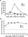
T-cell cytokine production is heightened in Ctx mice after LCMV-WE infection despite similar concurrent viral burdens. (A) Serum IFN-γ levels were assessed in WT and various Ctx mice at the indicated times after infection with LCMV-WE, a strain of virus that is acutely cleared in WT mice. CD8 depletion eliminated > 90% of serum IFN-γ in prf−/− mice (n = 8-57/curve). All data are displayed as means ± SEM. Serum IFN-γ levels at days 6 and 8 were significantly different (P < .01) between WT and all Ctx mice. (B) Virus was quantitated by plaque assay in the spleens of LCMV-WE infected animals at the indicated times. Before day 10, no significant difference (P > .05) was observed between WT and prf−/− animals (n = 6-8/point). Dashed line indicates limit of detection of the assay.
Figure 2
Figure 2
Prf−/− mice display heightened immune activation independently of virus control. (A) Serum IFN-γ levels were measured in WT and prf−/− mice infected with the persistent strain of LCMV, clone 13 (LCMV-Cl13), at the indicated time points. Serum IFN-γ levels at days 6, 8, and 10 were significantly elevated (P < .01) in prf−/− mice compared with WT. (B) Plasma viral levels were measured by plaque assay at the indicated time points in LCMV-Cl13–infected mice. Dashed line indicates limit of detection of the assay. *P < .01.
Figure 3
Figure 3
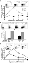
In vivo CD8+ T-cell activation is heightened in Ctx mice after LCMV infection. (A) Splenic CD8+ T cells producing IFN-γ in vivo were quantitated at the indicated times after LCMV-WE infection by direct ex vivo staining (see “Methods”). Sample dot plots of uninfected and day 8 LCMV-infected WT or prf−/− mice are shown, with the percentage of CD8+ cells that are IFN-γ+ displayed (gated on MHC II cells, 50 000 cells displayed). The numbers of IFN-γ+ CD8+ cells at day 6 and beyond were significantly different (P < .01) between WT and prf−/− mice (n = 6-12 per point). (B) Direct ex vivo IFN-γ staining of endogenous, Db-Gp33-specific CD8+ T cells was quantitated 8 days after LCMV infection by combining IFN-γ staining with MHC-tetramer staining. As a positive control, LCMV-infected mice were injected with 100 μg of Gp33 peptide 4 hours before animals were killed (+Gp33′ or peptide Rx). *P < .001. CD8+/CD4 cells are shown in dot plots (n = 4). (C) Splenic CD8+ T cells degranulating in vivo were quantitated at the indicated times after infection by direct ex vivo staining (see “Methods”). Numbers of CD107a+ cells at days 6, 8, and 10 were significantly different (P < .01) between WT and prf−/− mice (n = 6-8/point). 50 000 MHC II cells are shown in the dot plots.
Figure 4
Figure 4
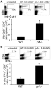
CD4+ T-cell expansion and in vivo activation are heightened in Ctx mice after LCMV infection. (A) LCMV-specific CD4+ T cells were quantitated in WT and prf−/− mice after LCMV infection by MHC-peptide tetramer (IAb-Gp61) staining of spleen cells. CD4+/CD16 cells from uninfected and day 8 LCMV-infected WT or prf−/− mice are shown in the example dot plots, with the percentage of IAb-Gp61+ of CD4+ cells listed. *P < .001. (B) Splenic CD4+ T cells spontaneously producing IFN-γ in vivo were quantitated 8 days after LCMV infection in WT and prf−/− mice, as described in Figure 3 (n = 8/group). *P < .01.
Figure 5
Figure 5
CD8+ T-cell populations from prf−/− mice display cell-signaling patterns indicating heightened antigenic stimulation after LCMV infection. (A) Phospho-c-Jun and IFN-γ staining of CD8+ T cells from prf−/− mice 8 days after LCMV infection. (B) Spleen cells from uninfected or LCMV-infected mice were fixed immediately ex vivo and analyzed for intracellular NFAT localization. CD8+/MHC II cells are displayed, plotting CD8 area (a measure of cell size) against an index of nuclear localization (NLI) of NFATc1. Examples of cells with an NLI of 1 or 3 are shown. (C) NLI of CD8+/MHC II cells from the indicated animals is plotted. (D) Percentage of CD8+ cells with nuclear-localized NFATc1 (NLI > 2) from uninfected and infected mice are displayed. P < .001 when comparing the NLI of CD8+ cells from LCMV-infected WT and prf−/− mice. Data are representative of 3 experiments.
Figure 6
Figure 6
Increased T-cell activation in Ctx mice after LCMV infection is due to heightened physiologic presentation of viral antigen. (A) Polyclonal, WT, or prf−/− (GFP+) T cells were transferred into either WT or prf−/− recipients 1 day before LCMV infection. The percentage of transferred CD8+ T cells that were producing IFN-γ+ in vivo was quantitated 6 days after infection. (B) Seven days after LCMV infection, spleens were disaggregated in collagenase and stained as indicated. Sample dot plots are shown, with the percentages of gated populations indicated (±SEM; n = 6-9 per group/stain). (C) Seven days after LCMV infection, T-cell–depleted spleen cells were plated with either LCMV-specific effector T cells (P14) or T cells of irrelevant specificity (OT1, ovalbumin) and specific IFN-γ production over background (spleen cells alone, which was < 5% of that measured in the presence of P14 T cells) was measured after 18 hours. *P < .01.

References

    1. Filipovich AH. Hemophagocytic lymphohistiocytosis and related disorders. Curr Opin Allergy Clin Immunol. 2006;6(6):410–415. - PubMed
    1. Favara BE, Feller AC, Pauli M, et al. Contemporary classification of histiocytic disorders. The WHO Committee On Histiocytic/Reticulum Cell Proliferations. Reclassification Working Group of the Histiocyte Society. Med Pediatr Oncol. 1997;29(3):157–166. - PubMed
    1. Stepp SE, Dufourcq-Lagelouse R, Le Deist F, et al. Perforin gene defects in familial hemophagocytic lymphohistiocytosis. Science. 1999;286(5446):1957–1959. - PubMed
    1. Henter JI. Biology and treatment of familial hemophagocytic lymphohistiocytosis: importance of perforin in lymphocyte-mediated cytotoxicity and triggering of apoptosis. Med Pediatr Oncol. 2002;38(5):305–309. - PubMed
    1. Fadeel B, Orrenius S, Henter JI. Familial hemophagocytic lymphohistiocytosis: too little cell death can seriously damage your health. Leuk Lymphoma. 2001;42(1–2):13–20. - PubMed

Publication types

MeSH terms