. 2011 Jun;121(6):2278-89.
doi: 10.1172/JCI46322. Epub 2011 May 23.

Angiopoietin-1 is essential in mouse vasculature during development and in response to injury

Affiliations

Angiopoietin-1 is essential in mouse vasculature during development and in response to injury

Marie Jeansson et al. J Clin Invest. 2011 Jun.

Abstract

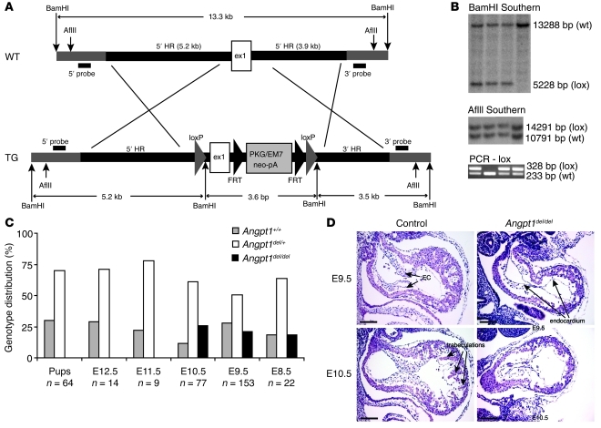
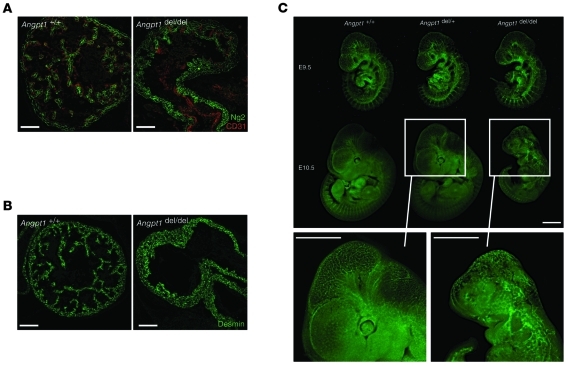
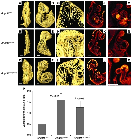
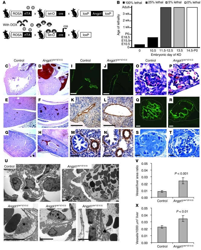
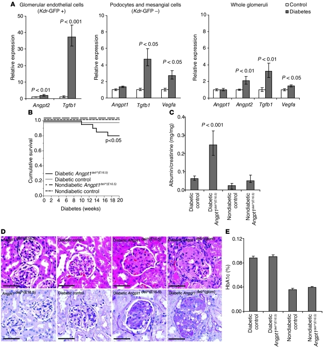
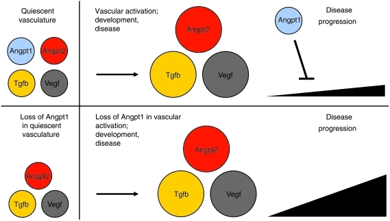
Angiopoietin-1/Tek signaling is a critical regulator of blood vessel development, with conventional knockout of angiopoietin-1 or Tek in mice being embryonically lethal due to vascular defects. In addition, angiopoietin-1 is thought to be required for the stability of mature vessels. Using a Cre-Lox conditional gene targeting approach, we have studied the role of angiopoietin-1 in embryonic and adult vasculature. We report here that angiopoietin-1 is critical for regulating both the number and diameter of developing vessels but is not required for pericyte recruitment. Cardiac-specific knockout of angiopoietin-1 reproduced the phenotype of the conventional knockout, demonstrating that the early vascular abnormalities arise from flow-dependent defects. Strikingly, deletion in the entire embryo after day E13.5 produced no immediate vascular phenotype. However, when combined with injury or microvascular stress, angiopoietin-1 deficiency resulted in profound organ damage, accelerated angiogenesis, and fibrosis. These findings redefine our understanding of the biological roles of angiopoietin-1: it is dispensable in quiescent vessels but has a powerful ability to modulate the vascular response after injury.


Figures

Figure 1
Figure 1. Generation of mice with a floxed Angpt1 allele.
(A) Targeting construct for Angpt1 locus: loxP sites were inserted around exon 1 (ex1). HR, homologous region. (B) Correctly targeted ES cell clones were identified using a 3′ probe and a 5′ probe outside of the region of homology, and genotyping of mice was done by PCR. (C) Floxed Angpt1 mice were bred to pCaggs-Cre to generate germline deletion of Angpt1, resulting in embryonic lethality in homozygous embryos (Angpt1del/del embryos) around E10.5. (D) Loss of trabeculations in the heart was observed in E9.5 and E10.5 embryos (scale bar: 100 μm). EC, endocardial cushion.
Figure 2
Figure 2. Angpt1 is critical in early vascular development.
Simplification of the cardiac trabeculation pattern of E10.5 Angpt1del/del embryos as shown by immunohistochemistry for (A) Ng2 (scale bar: 100 μm) and (B) Desmin (scale bar: 100 μm). (C) Dissection microscope photos show that Angpt1del/del embryos are markedly growth restricted at E10.5 and have disorganized vasculature as evident in embryos carrying a Kdr-GFP transgene reporter (scale bar: 1 mm).
Figure 3
Figure 3. Angpt1 is critical in cardiac development.
3D OPT data showing surface rendering of the vasculature stained with CD31 in E10.5 control, Angpt1del/del, and Angpt1del/^(heart) embryos. Shown are views from (AC) behind, (DF) the side, and (GI) enlargement of the right half of the head. (JL) Embryo sections of autofluorescence, (MO) with an enlargement of the heart in Angpt1del/del and Angpt1del/^(heart) embryos. Scale bar: 500 μm. (See also Supplemental Videos 1 and 2.) (P) Quantification of hindbrain vascular area shows a significant increase in vasculature in Angpt1del/del and Angpt1del/^(heart) embryos compared with that of controls.
Figure 4
Figure 4. Angpt1 regulates both the number and diameter of developing vessels.
(A) Breeding strategy to generate inducible whole-body deletion of Angpt1 [Angpt1del/^(DOX) mice] using the DOX-inducible ROSA-rtTA/tetO-Cre bitransgenic system. (B) Induction of Angpt1 knockout at E10.5 [Angpt1del/^(E10.5) mice] or earlier results in embryonic lethality. In mice induced with DOX at E10.5, vessels are dilated at P0 (C and D) in the heart (original magnification, ×50), (E and F) liver (original magnification, ×50), and (G and H) kidney (original magnification, ×50). Pericytes/mural cells surround the vessels as shown by α-SMA staining of (IL) liver and (M and N) lung in embryos dissected at E17.5 (See also Supplemental Figure 3). Scale bar: 10 μm (I and J); 100 μm (K and L); 50 μm (M and N). (V and X) In the same embryos, measurements of vessel number and vessel area in the liver showed a significant increase of both in Angpt1del/^(E10.5) embryos compared with those in controls. Deletion of Angpt1 at E10.5 also resulted in dilated glomerular capillary loops by E17.5, as shown by (O and P) H&E and (Q and R) podocin staining (scale bar: 10 μm), and a few glomeruli with only 1 big open capillary loop by P0, as shown by (S and T) Toluidine Blue staining (scale bar: 10 μm). (U) Electron micrographs (scale bar: 5 μm) show a folded GBM (arrow) and detachment of endothelial cells (*).
Figure 5
Figure 5. Angpt1 regulates tissue response in wound healing.
(A) Wound closure in ear punch wounds is increased in Angpt1del/^(E13.5) mice. (B) Cross sections of one of the sides of the ear punch show the cut of cartilage and the larger closed zone in Angpt1del/^(E13.5) mice (Masson Trichrome) (original magnification, ×100). (C) Quantification (mean ± SEM) of the punch area after 60 days shows a significant decrease in open area in the Angpt1del/^(E13.5) mice. (D) Z-stacks of flat mounted ears stained for CD31 (red, endothelium) and Ng2 (green, pericytes) show increased angiogenesis in the closed zone of Angpt1del/^(E13.5) mice compared with that of controls (scale bar: 100 μm). Z-stacks are composed of 8 images every 3 μm. The thick line (only in control) is the start of the closed zone (cut), and the thin line is the edge of the closed zone. (E) The close up of vessels in the healed area shows normal pericyte coverage (Desmin, green) of vessels (scale bar: 10 μm).
Figure 6
Figure 6. Angpt1 protects the glomerular vasculature in diabetic nephropathy.
(A) Expression of Angpt1, Angpt2, Tgfb1, and Vegfa in whole glomeruli or cell fractions sorted by FACS from diabetic and nondiabetic mice carrying a Kdr-GFP transgene reporter (mean ± SEM). FACS cells from glomeruli are endothelial cells in the GFP-positive fraction (Kdr-GFP +) and mainly podocytes and mesangial cells in the GFP-negative fraction (Kdr-GFP –). (B) Angpt1del/^(E16.5) mice (induced between E16.5 and P0) made diabetic show a significant decrease in survival. (C) After 20 weeks of diabetes, Angpt1del/^(E16.5) mice have a significantly higher urinary albumin/creatinine ratio compared with that of controls and nondiabetic groups. (D) Histology shows an increase in mesangial matrix expansion and sclerosis in diabetic Angpt1del/^(E16.5) mice and diabetic Angpt1del/^(glom) mice compared with that of diabetic controls (H&E, top panel; PAS bottom panel) (scale bar: 50 μm). (E) HbA1C in controls and Angpt1del/^(E16.5) mice is comparable in nondiabetic mice and after 20 weeks of diabetes.
Figure 7
Figure 7. Model for Angpt1 function during vascular activation.
Schematic model showing the importance of Angpt1 as an inhibitor of disease progression in the case of vascular activation that occurs in development or disease. In the absence of Angpt1, Angpt2, Vegfa, and Tgfb, actions are unopposed, resulting in more aggressive injury in disease or a larger number and diameter of vessels in development.

Comment in

  • The yin, the yang, and the angiopoietin-1.
    Saharinen P, Alitalo K. Saharinen P, et al. J Clin Invest. 2011 Jun;121(6):2157-9. doi: 10.1172/JCI58196. Epub 2011 May 23. J Clin Invest. 2011. PMID: 21606600 Free PMC article.

References

    1. Satchell SC, Harper SJ, Tooke JE, Kerjaschki D, Saleem MA, Mathieson PW. Human podocytes express angiopoietin 1, a potential regulator of glomerular vascular endothelial growth factor. . J Am Soc Nephrol. 2002;13(2):544–550. - PubMed
    1. Uemura A, et al. Recombinant angiopoietin-1 restores higher-order architecture of growing blood vessels in mice in the absence of mural cells. J Clin Invest. 2002;110(11):1619–1628. - PMC - PubMed
    1. Suri C, et al. Requisite role of angiopoietin-1, a ligand for the TIE2 receptor, during embryonic angiogenesis. Cell. 1996;87(7):1171–1180. doi: 10.1016/S0092-8674(00)81813-9. - DOI - PubMed
    1. Partanen J, Dumont DJ. Functions of Tie1 and Tie2 receptor tyrosine kinases in vascular development. Curr Top Microbiol Immunol. 1999;237:159–172. - PubMed
    1. Tachibana K, Jones N, Dumont DJ, Puri MC, Bernstein A. Selective role of a distinct tyrosine residue on Tie2 in heart development and early hematopoiesis. Mol Cell Biol. 2005;25(11):4693–4702. doi: 10.1128/MCB.25.11.4693-4702.2005. - DOI - PMC - PubMed

Publication types

MeSH terms