. 2011 Jun;121(6):2169-80.
doi: 10.1172/JCI45627. Epub 2011 May 23.

Disruption of intraflagellar protein transport in photoreceptor cilia causes Leber congenital amaurosis in humans and mice

Affiliations

Disruption of intraflagellar protein transport in photoreceptor cilia causes Leber congenital amaurosis in humans and mice

Karsten Boldt et al. J Clin Invest. 2011 Jun.

Abstract

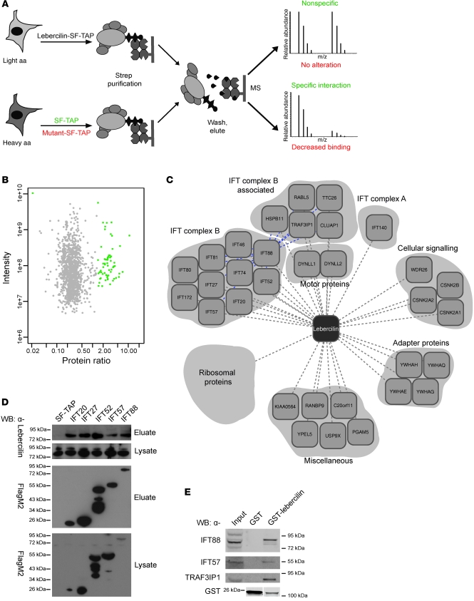
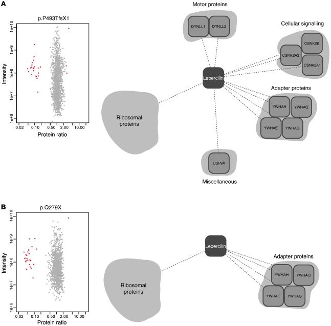
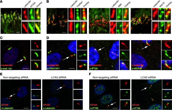
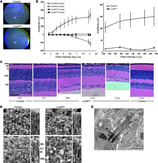
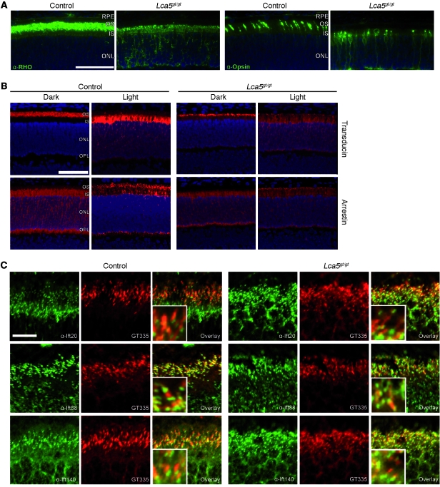
The mutations that cause Leber congenital amaurosis (LCA) lead to photoreceptor cell death at an early age, causing childhood blindness. To unravel the molecular basis of LCA, we analyzed how mutations in LCA5 affect the connectivity of the encoded protein lebercilin at the interactome level. In photoreceptors, lebercilin is uniquely localized at the cilium that bridges the inner and outer segments. Using a generally applicable affinity proteomics approach, we showed that lebercilin specifically interacted with the intraflagellar transport (IFT) machinery in HEK293T cells. This interaction disappeared when 2 human LCA-associated lebercilin mutations were introduced, implicating a specific disruption of IFT-dependent protein transport, an evolutionarily conserved basic mechanism found in all cilia. Lca5 inactivation in mice led to partial displacement of opsins and light-induced translocation of arrestin from photoreceptor outer segments. This was consistent with a defect in IFT at the connecting cilium, leading to failure of proper outer segment formation and subsequent photoreceptor degeneration. These data suggest that lebercilin functions as an integral element of selective protein transport through photoreceptor cilia and provide a molecular demonstration that disrupted IFT can lead to LCA.


Figures

Figure 1
Figure 1. Quantitative protein complex analysis of lebercilin.
(A) SILAC/AP approach to detect specific protein complex components. Identification of significantly enriched proteins compared with control (SF-TAP; green), and analysis of the effect of 2 LCA-causative mutations in lebercilin (p.P493TfsX1 and p.Q279X) on protein complex formation, by comparing the lebercilin complex with complexes formed by the mutants (red) in SILAC-labeled HEK293T cells. MS, mass spectrometry. (B) Detection of specific protein complex components. Plotted are log10 ratios and log10 intensities for each protein identified and quantified in at least 2 of 3 biological replicates. Significantly enriched proteins (P < 0.001) are plotted in green. (C) Protein complex of wild-type lebercilin. Gene names for each protein are shown (see Supplemental Table 1). Blue lines denote interconnectivity, as determined by IFT88 SF-TAP analysis (Supplemental Table 3). (D) Confirmation of IFT-lebercilin association by detecting endogenous lebercilin in eluates of IFT20, IFT27, IFT52, IFT57, and IFT88 SF-TAP experiments. Shown are the lysates as control for lebercilin input. All SF-TAP tagged constructs were expressed and purified, as demonstrated by Western blot using anti-FlagM2. Lebercilin was detected by anti-lebercilin (SN2135) in all eluates, but not in the SF-TAP control, confirming the SILAC/AP results. (E) Confirmation of the association of IFT and IFT-associated proteins with lebercilin in retina by detecting endogenous IFT88, IFT57, and TRAF3IP1 in a GST pulldown of lebercilin, but not in the GST control. Both GST-lebercilin and GST alone were expressed. Lanes for the anti-GST blot were run on the same gel but were noncontiguous (white line).
Figure 2
Figure 2. Quantitative protein complex analysis of LCA-causative lebercilin mutations.
Detection of protein complex alterations by comparison of the complexes of SF-TAP tagged lebercilin to either lebercilin-p.P493TfsX1 (A) or lebercilin-p.Q279X (B) in HEK293T cells. Plotted are log10 ratios and log10 intensities as quantified by MaxQuant from 3 independent experiments. For each experiment, label switching was done to exclude label-specific effects. Shown are only proteins quantified and identified in at least 2 of 3 experiments. Highly significant (P < 0.001) altered protein interactions are plotted in red, the majority of them being IFT proteins (Supplemental Table 1). Also shown are representations of the respective protein complexes; all proteins of the IFT protein complex, as well as WDR26, KIAA0564, PGAM5, YPEL5, C20orf11, and RANBP9, were lost for both mutants. CSNK2A1/2, CSNK2B, DYNLL1, DYNLL2, and USP9X only lost association to lebercilin-p.Q279X, not to lebercilin-p.P493TfsX1.
Figure 3
Figure 3. Endogenous localization of lebercilin and IFTs in mammalian retina and retinal cells.
(A) Immunohistochemistry of mouse BALB/c retina (P20) showed endogenous lebercilin (SN2134) expression in the connecting cilia of mouse photoreceptors, where it colocalized with the ciliary marker polyglutamylated tubulin (GT335). (B) Endogenous lebercilin (SN2134) colocalized with endogenous IFT complex B proteins Traf3ip1, Ift88, and IFT complex A protein Wdr19 in connecting cilia of mouse photoreceptors. (C) Immunocytochemistry of hTERT-RPE1 cells confirmed lebercilin (SN2134) localization in primary cilia (arrow). (D) Colocalization of endogenous lebercilin (SN2134) in the cilium with endogenous TRAF3IP1, IFT88, and WDR19 (arrows). (E) siRNA-mediated knockdown of LCA5 did not affect ciliogenesis compared with nontargeting siRNA–transfected hTERT-RPE1 cells (arrows). GT335 stained the primary cilium. SN2134 antibody was used to stain lebercilin. (F) Knockdown of LCA5 in hTERT-RPE1 cells did not affect ciliary localization of IFT88 (arrows). Scale bars: 5 μm (AF). Enlarged views are shown in the insets (A and B, ×5; CF, ×2).
Figure 4
Figure 4. Clinical and morphological assessment of Lca5gt/gt mouse retina.
(A) Fundus photography of Lca5gt/gt animals and littermate controls. At 2 months of age, depigmented patches were observed. (B) Scotopic and photopic ERG amplitudes of rod (left) and cone (right) waveforms at P25 (n = 3). Data are mean ± SEM. (C) Progressive retinal degeneration demonstrated in Lca5gt/gt mice. Retinal sections of Lca5gt/gt mice and wild-type controls, between P12 and 4 months of age, stained with hematoxylin and eosin. At P12, the OS/IS layers were remarkably thinner in Lca5gt/gt animals, whereas the ONL and INL were comparable to those of controls. At P28, the photoreceptor nuclei in the ONL were significantly reduced compared with controls. By 4 months of age, the photoreceptor layer was completely absent in Lca5gt/gt mice. (D) Scanning EM of control and Lca5gt/gt retinas at P10 and P15 from a scleral orientation (left) and fractured view (right). Control OSs at P10 were ovoid and regular in size. Vertically stacked OS discs (double asterisks) were rarely observed. OSs in Lca5gt/gt retinas were shorter and irregular in size and shape. By P15, OS discs were disorganized and did not reach the length observed in controls. Most OSs were rudimentary or abnormal in shape (asterisk). (E) Transmission EM of Lca5gt/gt retinas showed that the structure of the CC, including basal body, was preserved at P8. RPE, retinal pigment epithelium; NC, nascent cilia. Scale bars: 50 μm (C); 10 μm (D); 500 nm (E).
Figure 5
Figure 5. Localization study of phototransduction and IFT proteins in Lca5gt/gt retinas.
(A) OS proteins rhodopsin (RHO) and cone opsin were mislocalized to ISs and ONL in Lca5gt/gt animals, while normally restricted to OSs in controls. All sections were obtained at P14. (B) Localization of transducin and arrestin in dark-adapted and light-stimulated animals (P14). In dark-adapted wild-type animals, transducin localized to OSs and arrestin to ISs, ONL, and outer plexiform layer (OPL). After light stimulation, transducin was mainly found in the ISs, with weaker staining in the ONL and outer plexiform layer, whereas arrestin translocated to the OSs. In Lca5gt/gt dark-adapted animals, the majority of transducin was found in the OSs, with weaker staining in other layers. Arrestin localization in Lca5gt/gt dark-adapted animals was similar to that in dark-adapted wild-type mice. In light-stimulated Lca5gt/gt mice, transducin not only was found in the ISs, but also showed diffuse staining in the entire ONL. Arrestin staining appeared diffusely throughout the photoreceptor layer, with less pronounced OS staining than in wild-type mice. (C) Localization of Ift20, Ift88, or Ift140 in connecting cilia of photoreceptors was not affected by inactivation of Lca5 compared with wild-type at P9. GT335 specifically stained connecting cilia. Scale bars: 50 μm (A and B); 10 μm (C). Enlarged views are shown in the insets (C, ×3).

Comment in

  • Spotlight on childhood blindness.
    Sahel JA. Sahel JA. J Clin Invest. 2011 Jun;121(6):2145-9. doi: 10.1172/JCI58300. Epub 2011 May 23. J Clin Invest. 2011. PMID: 21606601 Free PMC article.

References

    1. den Hollander AI, Black A, Bennett J, Cremers FP. Lighting a candle in the dark: advances in genetics and gene therapy of recessive retinal dystrophies. . J Clin Invest. 2010;120(9):3042–3053. doi: 10.1172/JCI42258. - DOI - PMC - PubMed
    1. Estrada-Cuzcano A, et al. IQCB1 mutations in patients with Leber congenital amaurosis. Invest Ophthalmol Vis Sci. 2011;52(2):834–839. doi: 10.1167/iovs.10-5221. - DOI - PubMed
    1. den Hollander AI, Roepman R, Koenekoop RK, Cremers FP. Leber congenital amaurosis: genes, proteins and disease mechanisms. Prog Retin Eye Res. 2008;27(4):391–419. doi: 10.1016/j.preteyeres.2008.05.003. - DOI - PubMed
    1. den Hollander AI, et al. Mutations in LCA5, encoding the ciliary protein lebercilin, cause Leber congenital amaurosis. Nat Genet. 2007;39(7):889–895. doi: 10.1038/ng2066. - DOI - PubMed
    1. Blacque OE, Cevik S, Kaplan OI. Intraflagellar transport: from molecular characterisation to mechanism. Front Biosci. 2008;13:2633–2652. doi: 10.2741/2871. - DOI - PubMed

Publication types

MeSH terms