. 2011 Sep;7(9):993-1010.
doi: 10.4161/auto.7.9.16389. Epub 2011 Sep 1.

Plant NBR1 is a selective autophagy substrate and a functional hybrid of the mammalian autophagic adapters NBR1 and p62/SQSTM1

Affiliations

Plant NBR1 is a selective autophagy substrate and a functional hybrid of the mammalian autophagic adapters NBR1 and p62/SQSTM1

Steingrim Svenning et al. Autophagy. 2011 Sep.

Abstract

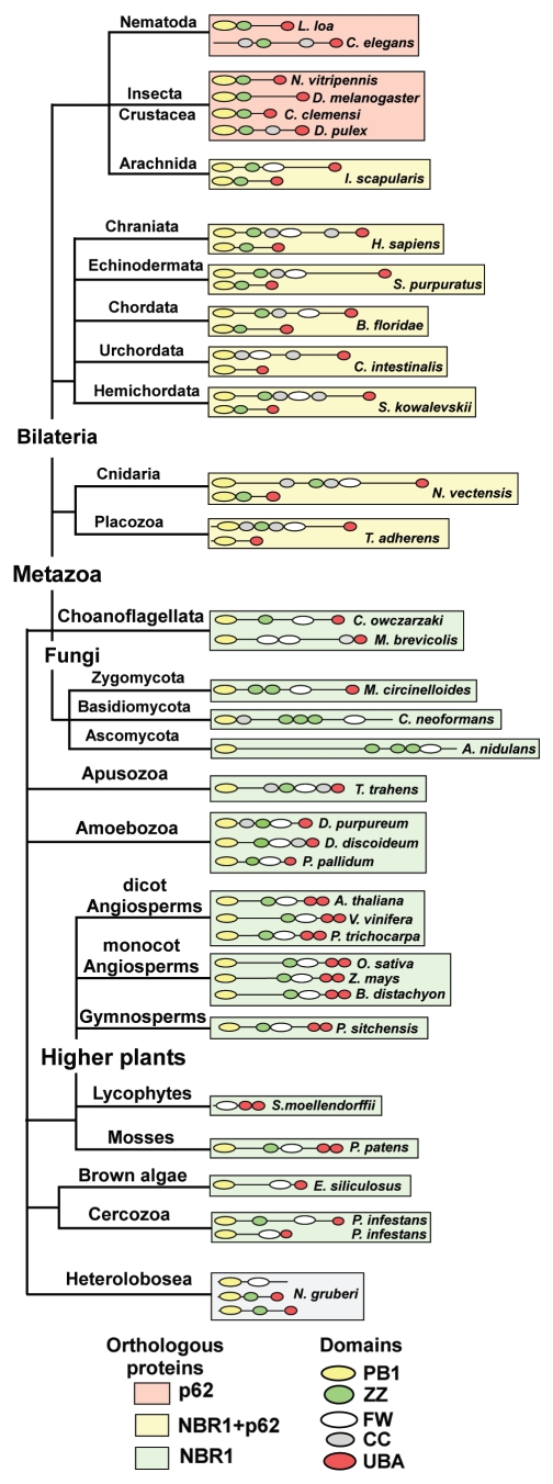
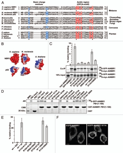
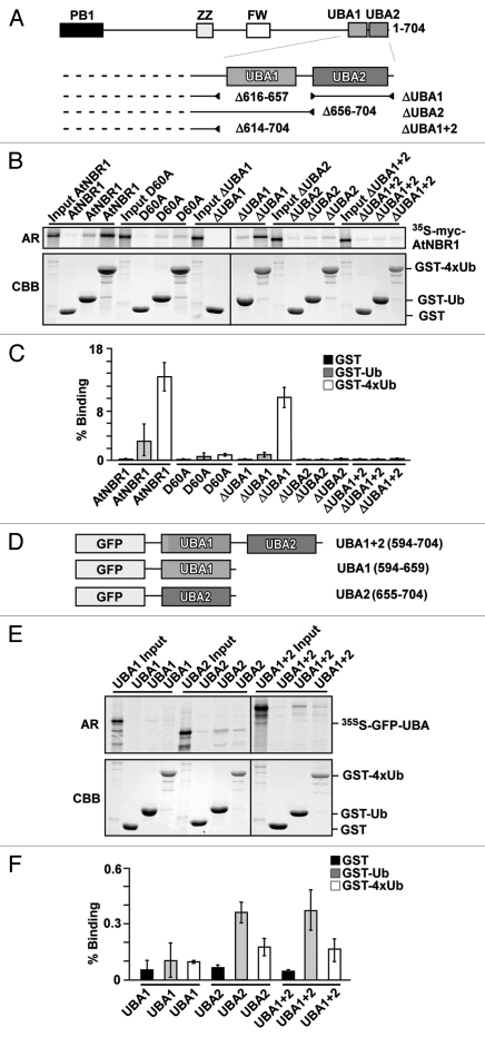
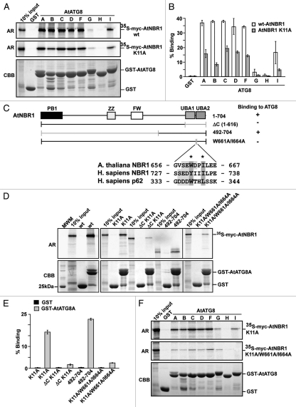
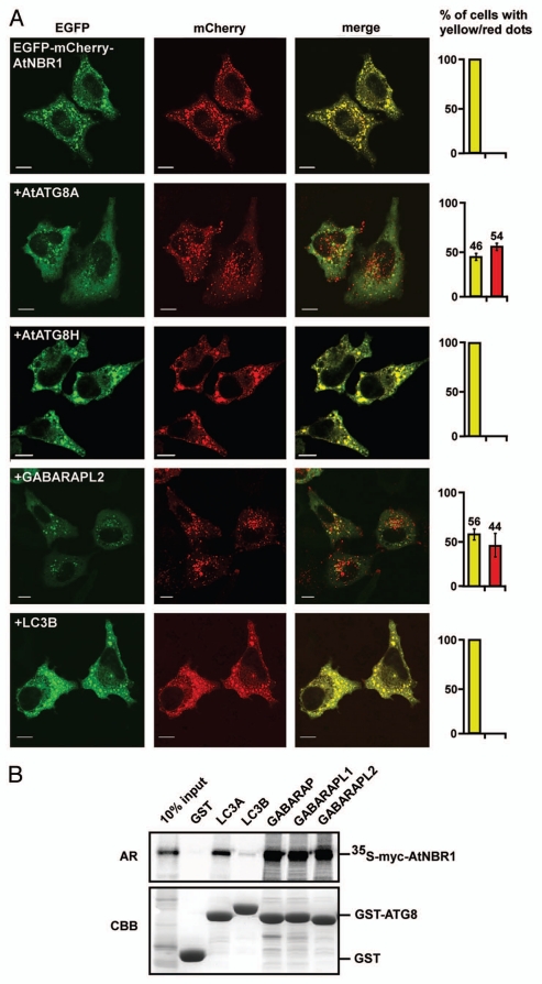
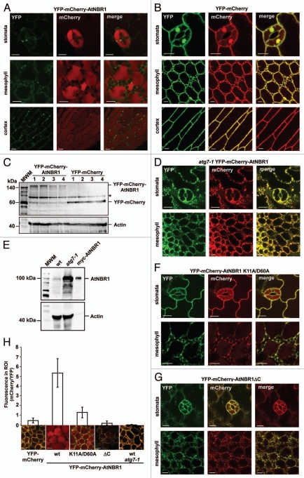
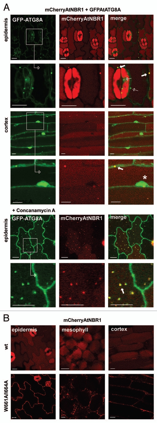
(Macro)autophagy encompasses both an unselective, bulk degradation of cytoplasmic contents as well as selective autophagy of damaged organelles, intracellular microbes, protein aggregates, cellular structures and specific soluble proteins. Selective autophagy is mediated by autophagic adapters, like p62/SQSTM1 and NBR1. p62 and NBR1 are themselves selective autophagy substrates, but they also act as cargo receptors for degradation of other substrates. Surprisingly, we found that homologs of NBR1 are distributed throughout the eukaryotic kingdom, while p62 is confined to the metazoans. As a representative of all organisms having only an NBR1 homolog we studied Arabidopsis thaliana NBR1 (AtNBR1) in more detail. AtNBR1 is more similar to mammalian NBR1 than to p62 in domain architecture and amino acid sequence. However, similar to p62, AtNBR1 homo-polymerizes via the PB1 domain. Hence, AtNBR1 has hybrid properties of mammalian NBR1 and p62. AtNBR1 has 2 UBA domains, but only the C-terminal UBA domain bound ubiquitin. AtNBR1 bound AtATG8 through a conserved LIR (LC3-interacting region) motif and required co-expression of AtATG8 or human GABARAPL2 to be recognized as an autophagic substrate in HeLa cells. To monitor the autophagic sequestration of AtNBR1 in Arabidopsis we made transgenic plants expressing AtNBR1 fused to a pH-sensitive fluorescent tag, a tandem fusion of the red, acid-insensitive mCherry and the acid-sensitive yellow fluorescent proteins. This strategy allowed us to show that AtNBR1 is an autophagy substrate degraded in the vacuole dependent on the polymerization property of the PB1 domain and of expression of AtATG7. A functional LIR was required for vacuolar import.


Figures

Figure 1
Figure 1
Homologs of NBR1 are found throughout the eukaryotic kingdom while p62 is confined to the metazoans. The evolutionary distribution of NBR1- and p62-homologs in selected species from the eukaryotic kingdoms of opisthokonts (fungi, metazoa and choanozoa), apusozoa (flagellate protozoa), amoebozoa, archeaplastida (plantae) and chromalveolata (brown algae) is shown. The domain architectures of the proteins are shown. Background color coding is used to indicate species which possess both p62 and NBR1 (yellow), species containing only p62 (orange) and species harboring only NBR1 (green). Protein sequence accession numbers are listed in Table S1.
Figure 2
Figure 2
AtNBR1 polymerizes via the N-terminal PB1-domain. (A) Alignment of PB1 domain sequences from p62- and NBR1 homologs of selected metazoan- and nonmetazoan species. Blue background color denotes basic residues and red background color denotes acidic residues of the charged clusters important for PB1 domain interactions. The OPCA-motif is indicated. Gaps are indicated with dashes and the numbers of amino acid residues not shown are specified in brackets. Residues crucial for the self-interaction of p62, and which are mutated in AtNBR1 in the analyses shown in (C–E) are indicated with asterisks. (B) Electrostatic surface potentials of the PB1 domains of NBR1 (PDB:2BKF) and p62 (PDB:2KKC) from H. sapiens and N. vectensis as well as the AtNBR1 PB1 domain. The N. vectensis and AtNBR1 PB1 domains were modeled using Swissmodel. (C) Co-immunoprecipitation experiments using full-length AtNBR1. Myc- and GFP-tagged AtNBR1 (wt or the indicated mutants) were co-translated in vitro in the presence of 35S-methionine and precipitated using an anti-GFP antibody. Immunoprecipitated and co-precipitated proteins as well as in vitro translated proteins corresponding to 15% of the input were resolved by SDS-PAGE and detected by autoradiography. The upper band corresponds to 35SGFP-AtNBR1, the lower to 35Smyc-AtNBR1. Quantifications of the interaction data are shown above the gel parts. Band intensity was measured using ImageJ (Fuji) and Y-axis values were calculated employing the following formula; (IP(myc/GFP)/input(myc/GFP)) × 100. (D) GST pulldown assays using in vitro translated 35S-labeled GFP-AtNBR1 PB1 (amino acids 1–100) (with the indicated point mutations) and GST or GST-PB1 (with indicated point mutations) constructs. Precipitated proteins were detected by autoradiography. (E) Quantitative representation of the interaction data shown in (D). Y-axis values are set to percent total binding protein; (pulldown/input) × 100. (F) AtNBR1 forms cytosolic aggregates when overexpressed with an N-terminal GFP-tag in HeLa cells, while the K11A point-mutant of AtNBR1 loses the ability to form aggregates. Results in (C and E) are mean values of three independent experiments with standard deviations indicated as bars. Bars represent 10 µm.
Figure 3
Figure 3
AtNBR1 binds ubiquitin through the most C-terminal UBA domain (UBA2). (A) Schematic illustration of AtNBR1 UBA deletion constructs. (B) GST pull-down assays using in vitro translated 35S-labeled myc-AtNBR1 (indicated deletions) and immobilized GST or indicated GST-Ub and GST-4xUb constructs. Precipitated proteins were detected by autoradiography. (C) Quantitative representation of the interaction data shown in (B). Y-axis values are set to percent total binding protein; (pulldown/input) × 100. (D) AtNBR1 GFP-UBA domain fusion constructs used for pull-down experiments. (E) GST pull-down assays using in vitro translated 35S-labeled GFP-UBA constructs and GST or indicated GST-Ub and GST-4xUb constructs. Precipitated proteins were detected by autoradiography. (F) Quantitative representation of the interaction data shown in (E). Results in (C and F) are mean values of three independent experiments with standard deviations indicated as bars.
Figure 4
Figure 4
AtNBR1 binds to Arabidopsis ATG8 family proteins via a LIR-motif located between the twin UBA domains. (A) GST pull-down assays using in vitro translated 35S-labeled myc-AtNBR1 (polymeric and monomeric K11A mutants) and immobilized GST or GST-ATG8 (indicated isoforms) constructs. Precipitated proteins were detected by autoradiography. (B) Quantitative representation of the interaction data shown in (B) (polymeric and monomeric AtNBR1). Y-axis values are set to percent total binding protein; (input/pulldown) × 100. (C) Constructs used and a summary of GST pull-down assays between full-length ATG8A fused to GST and deletion mutants of AtNBR1 (upper part). The lower part shows an alignment of the LIR in AtNBR1 to the corresponding sequences in human p62 and NBR1. The W661 and I664 residues mutated to A are indicated with asterisks. (D) GST pull-down assays using in vitro translated 35S-labeled myc-AtNBR1 (indicated deletions and mutations) and GST or indicated GST-ATG8A constructs. Precipitated proteins were detected by autoradiography. (E) Quantitative representation of the interaction data shown in (B). (F) GST pull-down assays using in vitro translated 35S-labeled, monomeric (K11A mutant) myc-AtNBR1 (indicated mutations) and immobilized GST or indicated GST-ATG8 (indicated isoforms) constructs. Precipitated proteins were detected by autoradiography. Results in (B and E) are mean values of three independent experiments with standard deviations indicated as bars.
Figure 5
Figure 5
AtNBR1 is not recognized as an autophagic substrate in HeLa cells unless AtATG8 or GABARAPL2 are co-expressed. (A) Overexpression of GFP-mCherry-AtNBR1 in HeLa cells causes accumulation of cytosolic aggregates. When co-expressed with myc-tagged AtATG8A, red punctated structures appear and the amount of cytosolic aggregates is reduced. This pattern is also found upon co-expression with myc-GABARAPL2. No red structures are found upon co-expression with myc-AtATG8H or myc-LC3B. The graphs to the right illustrate the percentage of transfected cells containing only yellow structures (yellow bar) or a mix of both yellow and red (red bar). Each graph represents the mean of three separate transfections (>100 cells counted per transfection) with standard deviation indicated. Picture bars represent 10 µm. (B) GST pull-down assays using in vitro translated 35S-labeled myc-AtNBR1 and GST or indicated GST-Ub and GST-ATG8 constructs. Precipitated proteins were detected by autoradiography.
Figure 6
Figure 6
AtNBR1 forms cytosolic, punctated bodies and is imported to the central vacuole by autophagy in vivo. (A) Transgenic Arabidopsis expressing YFP-mCherry-AtNBR1. Scattered punctate structures containing AtNBR1 can be found along the rim of the cells and the central vacuole of the cells is exhibiting diffuse red fluorescence. This pattern can be found in all tissues of the plant. The emitted fluorescence of YFP has been converted to green for visual purposes. (B) Transgenic Arabidopsis expressing YFP-mCherry. (C) Protein gel blot using an mCherry antibody to visualize the expression of YFP-mCherrry-AtNBR1 and YFP-mCherry in four different transgenic plant lines, respectively. The actin levels are shown in the lower part. The asterisk denotes an unspecific band. (D) The atg7-1 knockout mutant line of Arabidopsis expressing YFP-mCherry-AtNBR1. (E) Protein gel blot of crude protein extract from 2-week-old seedlings of Arabidopsis Col-0 and Arabidopsis atg7-1, using anti-AtNBR1 antibody. In vitro translated myc-AtNBR1 is included as positive control. Actin is included as loading control. (F and G) Transgenic Arabidopsis expressing monomeric YFP-mCherry-AtNBR1 K11A D60A construct (F) and YFP-mCherry-AtNBR1ΔC with UBA1, -2 and LIR deleted (G). (H) Quantification of vacuolar import of AtNBR1. Quantification was performed by dividing the average gray value in the cytosol (YFP) with the average gray value in vacuole (mCherry), using split channel images in ImageJ. The graphical representation is based on three separate lines per construct, >30 counted cells from three separate plants of each line (total of 100 cells), with standard deviation indicated as bars.
Figure 7
Figure 7
AtNBR1 colocalizes with AtATG8A in vivo and a functional LIR is required for vacuolar import. (A) Stable co-expression of mCherry-AtNBR1 and GFP-AtATG8A in Arabidopsis. Images collected from two different tissues with enlarged insets shown below. The central vacuole exhibits diffuse red fluorescence, while the cytosol and nucleus (marked with asterisk) exhibits diffuse green fluorescence. AtNBR1 co-localizes with AtATG8A in punctated cytosolic bodies with overlapping fluorescence (filled arrows). Punctate structures containing only GFP-AtATG8A are also found (open arrows). Upon treatment with Concanamycin A, an accumulation of punctated bodies of overlapping fluorescence can be seen within the central vacuole. (B) Transgenic Arabidopsis expressing mCherry-AtNBR1 (upper inset) and mCherry-AtNBR1 W661A I664A mutant (lower inset). Wild-type (wt) mCherryAtNBR1 is mostly localized to the central vacuole of the plants cells while the AtNBR1 LIR-mutant accumulated in the cytosol.

Similar articles

Cited by

References

    1. Mizushima N. Autophagy: process and function. Genes Dev. 2007;21:2861–2873. doi: 10.1101/gad.1599207. - DOI - PubMed
    1. Yang Z, Klionsky DJ. Eaten alive: a history of macroautophagy. Nat Cell Biol. 2010;12:814–822. doi: 10.1038/ncb0910-814. - DOI - PMC - PubMed
    1. Deretic V. Autophagy in infection. Curr Opin Cell Biol. 2010;22:252–262. doi: 10.1016/j.ceb.2009.12.009. - DOI - PMC - PubMed
    1. Levine B, Kroemer G. Autophagy in the pathogenesis of disease. Cell. 2008;132:27–42. doi: 10.1016/j.cell.2007.12.018. - DOI - PMC - PubMed
    1. Mizushima N, Levine B, Cuervo AM, Klionsky DJ. Autophagy fights disease through cellular self-digestion. Nature. 2008;451:1069–1075. doi: 10.1038/nature06639. - DOI - PMC - PubMed

Publication types

MeSH terms