

Factors influencing bone loss in paraplegia
Abstract
Background and aim: Significant bone loss develops in the first months and continues years after spinal cord injury. A cross - sectional comparative study was performed to evaluate factors influencing bone loss in spinal cord injured men with paraplegia.
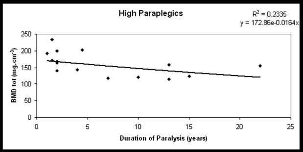
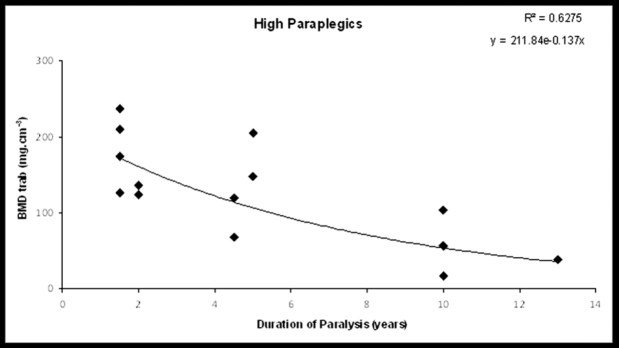
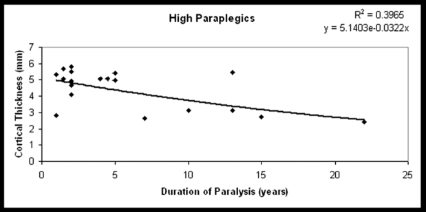
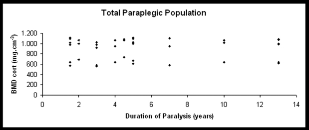
Patients and methods: We studied 31 paraplegic men in chronic stage (>1.5 years) in comparison with 30 able-bodied men of similar age, height, and weight. The paraplegic men were allocated into 2 subgroups based on the neurological level of injury; high paraplegics (n=16, T4-T7 neurological level of injury) and low paraplegics (n=15, T8-T12 neurological level of injury). The influence of positive and negative factors (spasticity, standing-therapeutic walking, and duration of paralysis) on bone structures was evaluated by pQCT measurement of the total, trabecular and cortical bone mineral density (BMDtot, BMDtrab, BMDcort, respectively) and cortical thickness (THIcort) at the distal tibial epiphysis and the tibial diaphysis at 4% and 38% proximal to the distal end of the tibia. The stress strain index (SSI) was measured at 14% (SSI(2)) and at 38% (SSI(3)) of the tibial diaphysis, and the difference SSI(3) - SSI(2) (δSSI(3-2)) was calculated.
Results: In all paraplegics, bone mineral density parameters were significantly reduced compared to the control group (BMDtot: p<0.0005, BMDtrab: p<0.0005, BMDcort: p=0.029, THIcort: p=0.019, SSI(2): p=0.009, SSI(3): p=0.003, respectively). Paraplegics who used standing frames or long brace orthoses had statistically significant higher bone mass and geometric parameters (BMDtrab: p=0.03, BMDtot: p=0.01, THIcort: p=0.013, respectively), while spasticity did not protect bone. The duration of paralysis was significantly related to trabecular bone loss (r=-0.5, p=0.05) and cortical thickness (r=-0.6, p=0.006) in high paraplegics and to δSSI(3-2) in low paraplegics (r=0.534, p=0.03).
Conclusions: The neurological level of injury adversely affects bone strength in paralyzed lower extremities such as the distal tibia. Standing or therapeutic walking could possibly have a positive effect in cortical and trabecular bone in paraplegia.
Keywords: bone loss; men; pQCT; paraplegia; spinal cord injury.
Conflict of interest statement
The authors have no conflict of interest.
Figures
Similar articles
-
Bone loss and mechanical properties of tibia in spinal cord injured men.J Musculoskelet Neuronal Interact. 2007 Jan-Mar;7(1):62-8. J Musculoskelet Neuronal Interact. 2007. PMID: 17396008
-
Relationship between the duration of paralysis and bone structure: a pQCT study of spinal cord injured individuals.Bone. 2004 May;34(5):869-80. doi: 10.1016/j.bone.2004.01.001. Bone. 2004. PMID: 15121019
-
Long-term changes in bone metabolism, bone mineral density, quantitative ultrasound parameters, and fracture incidence after spinal cord injury: a cross-sectional observational study in 100 paraplegic men.Osteoporos Int. 2004 Mar;15(3):180-9. doi: 10.1007/s00198-003-1529-6. Epub 2004 Jan 13. Osteoporos Int. 2004. PMID: 14722626
-
Assessment of anthropometric, systemic, and lifestyle factors influencing bone status in the legs of spinal cord injured individuals.Osteoporos Int. 2005 Jan;16(1):26-34. doi: 10.1007/s00198-004-1638-x. Epub 2004 May 11. Osteoporos Int. 2005. PMID: 15138665
-
Prevention of bone loss in paraplegics over 2 years with alendronate.J Bone Miner Res. 2004 Jul;19(7):1067-74. doi: 10.1359/JBMR.040313. Epub 2004 Mar 22. J Bone Miner Res. 2004. PMID: 15176988 Clinical Trial.
Cited by
-
Effects of drugs on bone metabolism in a cohort of individuals with traumatic spinal cord injury.Spinal Cord Ser Cases. 2019 Jan 16;5:3. doi: 10.1038/s41394-018-0146-8. eCollection 2019. Spinal Cord Ser Cases. 2019. PMID: 30675387 Free PMC article.
-
Anatomic changes in the macroscopic morphology and microarchitecture of denervated long bone tissue after spinal cord injury in rats.Biomed Res Int. 2014;2014:853159. doi: 10.1155/2014/853159. Epub 2014 Jul 20. Biomed Res Int. 2014. PMID: 25136632 Free PMC article.
-
Body composition in multiple sclerosis.Hippokratia. 2013 Jan;17(1):7-11. Hippokratia. 2013. PMID: 23935336 Free PMC article.
-
Bone loss and fractures in multiple sclerosis: focus on epidemiologic and physiopathological features.Int J Gen Med. 2011;4:505-9. doi: 10.2147/IJGM.S22255. Epub 2011 Jul 4. Int J Gen Med. 2011. PMID: 21845056 Free PMC article.
-
Biological basis of bone strength: anatomy, physiology and measurement.J Musculoskelet Neuronal Interact. 2020 Sep 1;20(3):347-371. J Musculoskelet Neuronal Interact. 2020. PMID: 32877972 Free PMC article. Review.
References
-
- Mamoun L, Fattal C, Micallef JP, Peruchon E, Rabischong P. Bone loss in spinal cord-injured patients: from physiopathology to therapy. Spinal Cord. 2006;44:203–210. - PubMed
-
- Jiang SD, Dai LY, Jiang LS. Osteoporosis after spinal cord injury. Osteoporos Int. 2006;17:180–192. - PubMed
-
- Wilmet E, Ismail AA, Heilporn A, Welraeds D, Bergmann P. Longitudinal study of the bone mineral content and of soft tissue composition after spinal cord section. Paraplegia. 1995;33:674–677. - PubMed
-
- Demirel G, Yilmaz H, Paker N, Onel S. Osteoporosis after spinal cord injury. Spinal Cord. 1998;36:822–825. - PubMed
-
- Lazo MG, Shirazi P, Sam M, Giobbie-Hurder A, Blacconiere MJ, Muppidi M. Osteoporosis and risk of fracture in men with spinal cord injury. Spinal Cord. 2001;39:208–214. - PubMed
LinkOut - more resources
Full Text Sources