

Spontaneous immortalization of human dermal microvascular endothelial cells
- PMID: 21607128
- PMCID: PMC3097930
- DOI: 10.4252/wjsc.v2.i5.114
Spontaneous immortalization of human dermal microvascular endothelial cells
Abstract
Aim: To establish and characterize a spontaneously immortalized human dermal microvascular endothelial cell line, iHDME1.
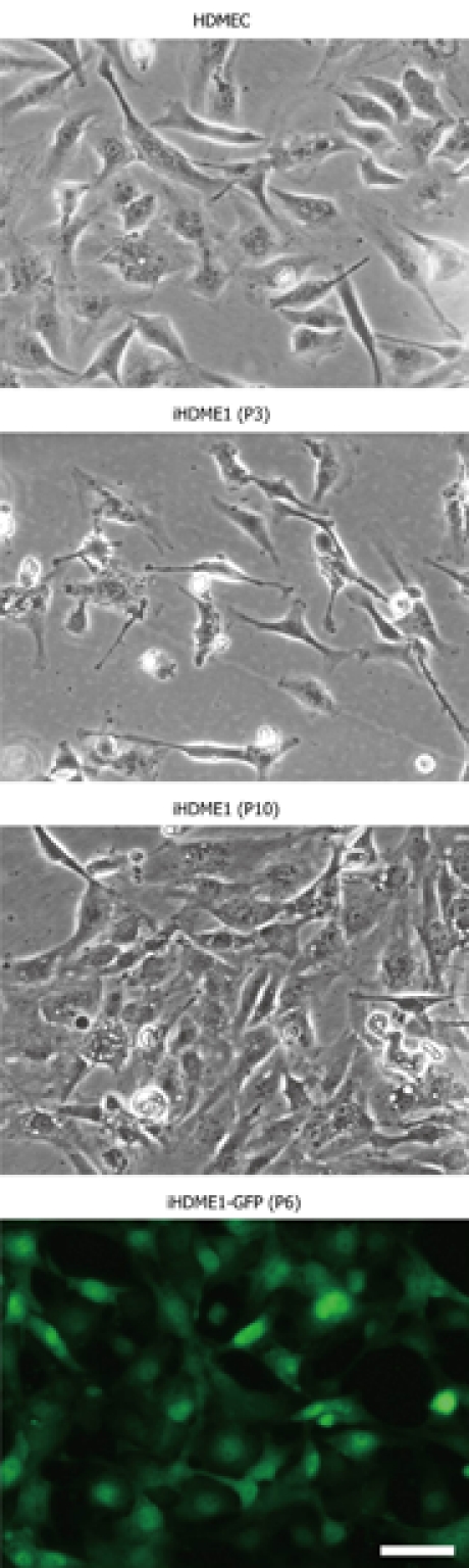
Methods: We developed a spontaneous immortalization method. This approach is based on the application of optimized culture media and culture conditions without addition of any exogenous oncogenes or carcinogens. Using this approach, we have successfully established a microvascular endothelial cell line, iHDME1, from primary human dermal microvascular endothelial cells. iHDME1 cells have been maintained in culture dishes for more than 50 passages over a period of 6 mo. Using a GFP expressing retrovirus, we generated a GFP-stable cell line (iHDME1-GFP).
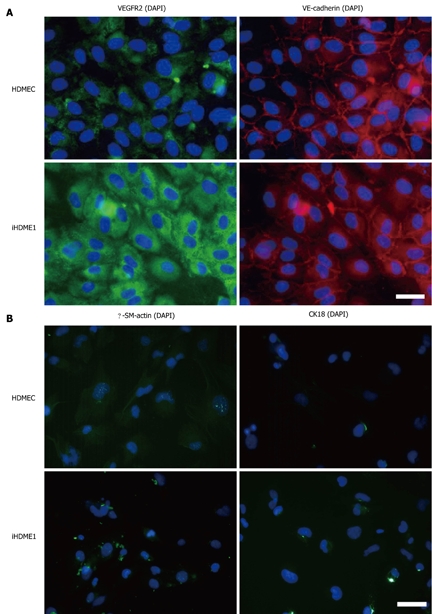
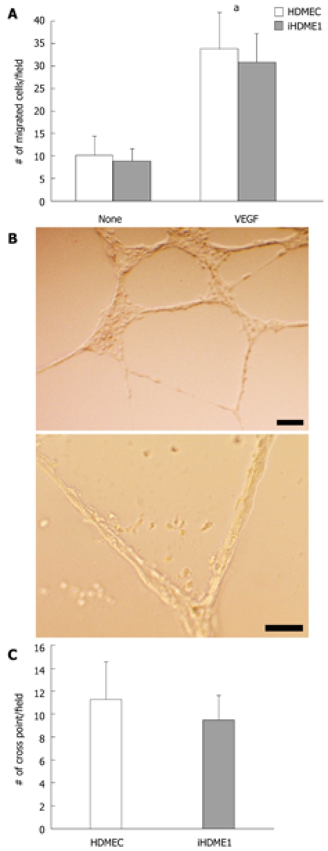
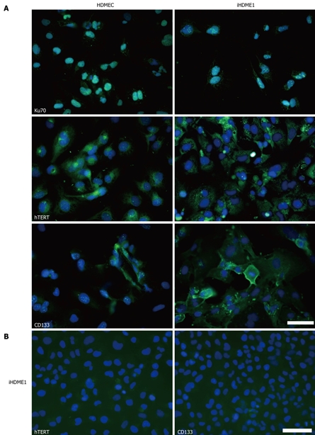
Results: iHDME1 retain endothelial morphology and uniformly express endothelial markers such as VEGF receptor 2 and VE-cadherin but not α-smooth muscle actin (α-SM-actin) and cytokeratin 18, markers for smooth muscle cells and epithelial cells respectively. These cells retain endothelial properties, migrate in response to VEGF stimulation and form 3-D vascular structures in Matrigel, similar to the parental cells. There is no significant difference in cell cycle profile between the parental cells and iHDME1 cells. Further analysis indicates enhanced stemness in iHDME1 cells compared to parental cells. iHDME1 cells display elevated expression of CD133 and hTERT.
Conclusion: iHDME1 cells will be a valuable source for studying angiogenesis.
Keywords: Angiogenesis; Endothelial cell; Spontaneous immortalization.
Figures
References
-
- Hayflick L, Moorhead PS. The serial cultivation of human diploid cell strains. Exp Cell Res. 1961;25:585–621. - PubMed
-
- Hahn WC. Immortalization and transformation of human cells. Mol Cells. 2002;13:351–361. - PubMed
-
- Dickson MA, Hahn WC, Ino Y, Ronfard V, Wu JY, Weinberg RA, Louis DN, Li FP, Rheinwald JG. Human keratinocytes that express hTERT and also bypass a p16(INK4a)-enforced mechanism that limits life span become immortal yet retain normal growth and differentiation characteristics. Mol Cell Biol. 2000;20:1436–1447. - PMC - PubMed
-
- Munro J, Stott FJ, Vousden KH, Peters G, Parkinson EK. Role of the alternative INK4A proteins in human keratinocyte senescence: evidence for the specific inactivation of p16INK4A upon immortalization. Cancer Res. 1999;59:2516–2521. - PubMed
-
- Bodnar AG, Ouellette M, Frolkis M, Holt SE, Chiu CP, Morin GB, Harley CB, Shay JW, Lichtsteiner S, Wright WE. Extension of life-span by introduction of telomerase into normal human cells. Science. 1998;279:349–352. - PubMed
Grants and funding
LinkOut - more resources
Full Text Sources
Other Literature Sources
Research Materials
