. 2011 Sep;60(9):1289-97.
doi: 10.1007/s00262-011-1033-3. Epub 2011 May 24.

Gemcitabine enhances Wilms' tumor gene WT1 expression and sensitizes human pancreatic cancer cells with WT1-specific T-cell-mediated antitumor immune response

Affiliations

Gemcitabine enhances Wilms' tumor gene WT1 expression and sensitizes human pancreatic cancer cells with WT1-specific T-cell-mediated antitumor immune response

Akitaka Takahara et al. Cancer Immunol Immunother. 2011 Sep.

Abstract

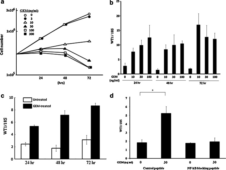
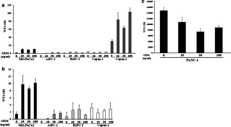
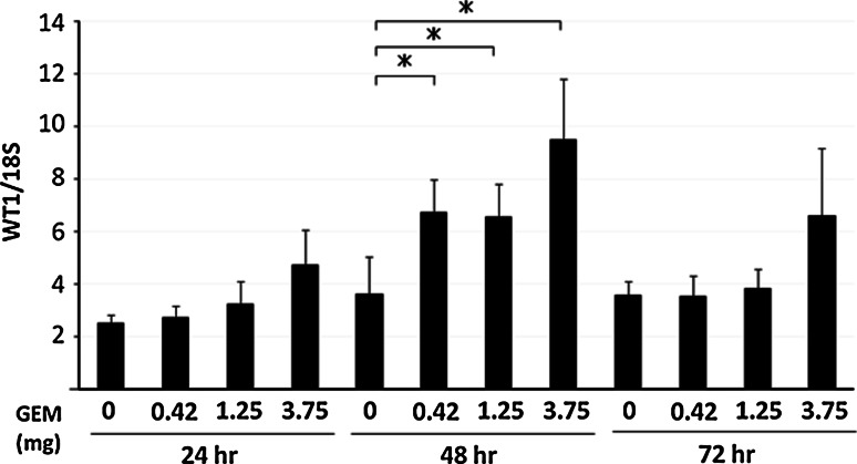
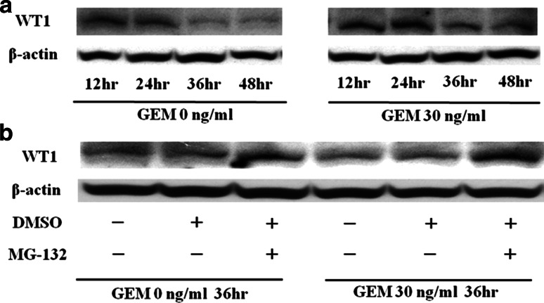
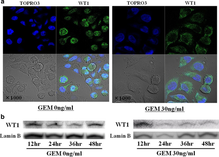
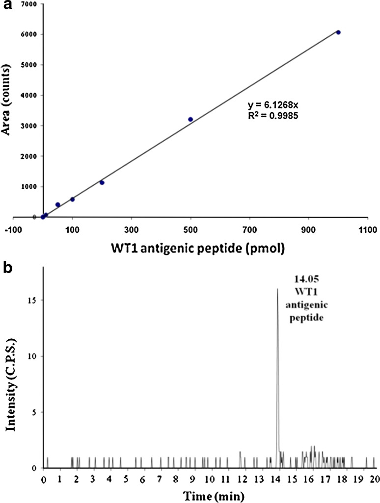
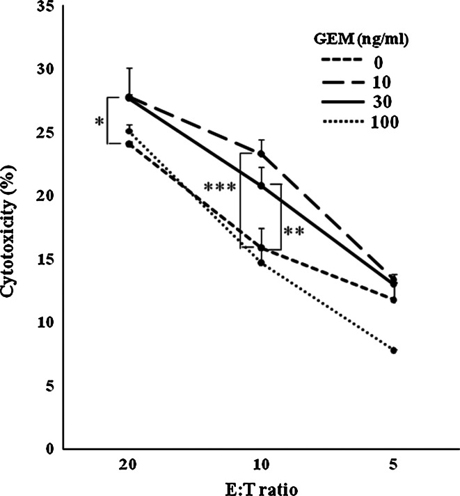
Wilms' tumor gene (WT1), which is expressed in human pancreatic cancer (PC), is a unique tumor antigen recognized by T-cell-mediated antitumor immune response. Gemcitabine (GEM), a standard therapeutic drug for PC, was examined for the regulation of WT1 expression and the sensitizing effect on PC cells with WT1-specific antitumor immune response. Expression of WT1 was examined by quantitative PCR, immunoblot analysis, and confocal microscopy. Antigenic peptide of WT1 presented on HLA class I molecules was detected by mass spectrometry. WT1-specific T-cell receptor gene-transduced human T cells were used as effecter T cells for the analysis of cytotoxic activity. GEM treatment of human MIAPaCa2 PC cells enhanced WT1 mRNA levels, and this increase is associated with nuclear factor kappa B activation. Tumor tissue from GEM-treated MIAPaCa2-bearing SCID mice also showed an increase in WT1 mRNA. Some human PC cell lines other than MIAPaCa2 showed up-regulation of WT1 mRNA levels following GEM treatment. GEM treatment shifted WT1 protein from the nucleus to the cytoplasm, which may promote proteasomal processing of WT1 protein and generation of antigenic peptide. In fact, presentation of HLA-A*2402-restricted antigenic peptide of WT1 (CMTWNQMNL) increased in GEM-treated MIAPaCa2 cells relative to untreated cells. WT1-specific cytotoxic T cells killed MIAPaCa2 cells treated with an optimal dose of GEM more efficiently than untreated MIAPaCa2 cells. GEM enhanced WT1 expression in human PC cells and sensitized PC cells with WT1-specific T-cell-mediated antitumor immune response.


Conflict of interest statement

There are no financial disclosures of any of the authors to declare.

Figures

Fig. 1
Fig. 1
a Proliferation of MIAPaCa2 cells in medium containing various concentrations of GEM. MIAPaCa2 cells (3 × 105/well) were seeded in 6-well culture plates in regular culture medium, which was then exchanged for GEM-containing medium after 24 h. At 24-h intervals, cells were detached using trypsin, and cell numbers were counted using a hemocytometer (n = 3). b Up-regulation of WT1 mRNA in MIAPaCa2 cells by GEM treatment. Twenty-four hours after plating, culture medium was exchanged to media containing GEM at indicated concentrations (0, 10, 30 and 100 ng/ml). MIAPaCa2 cells were harvested at 24-h intervals, and WT1 mRNA in cell homogenates was analyzed using qRT-PCR. WT1mRNA levels were normalized relative to those of 18S ribosomal RNA (18S). c Up-regulation of WT1 mRNA in MIAPaCa2 cells after short treatment with GEM. Twenty-four hours after plating, MIAPaCa2 cells were untreated or treated with 100 ng/ml of GEM for 2 h. MIAPaCa2 cells did not proliferate but kept alive for following 72 h by this treatment with GEM. After GEM treatment, cells were washed well, cultured in regular culture medium, and harvested at 24-h intervals. WT1 mRNA in cell homogenates was analyzed using qRT-PCR, and WT1mRNA levels were normalized relative to those of 18S ribosomal RNA (18S). d NF-kB suppresses GEM-induced up-regulation of WT1 mRNA. MIAPaCa2 cells (6 × 104/well) were seeded in 24-well culture plates. After 24 h, medium was exchanged for media containing GEM (0 or 30 ng/ml) and/or NF-kB blocking peptide (50 μM) or control peptide (50 μM). WT1 mRNA levels were quantified after 24-h incubation using qRT-PCR. *P < 0.01
Fig. 2
Fig. 2
a Up-regulation of WT1 mRNA levels in various human PC cell lines following GEM treatment. Human PC cells (1 × 106 MIAPaCa2, AsPC-1, BxPC-3, Capan-1 or Capan-2) were seeded in 10-cm culture plates. After 24-h incubation, medium was changed to media containing GEM (10, 30 or 100 ng/ml). After 48 h, we used qRT-PCR to quantify the relative ratio of WT1 to 18S mRNA levels in each cell line (n = 3). b GEM-induced up-regulation of WT1 mRNA in human PC cells with low basal levels of WT1 mRNA (MIAPaCa2, AsPC-1, BxPC-3 and Capan-1). To illustrate these results, we replotted data from (a) to represent a considerably narrower range of mRNA level ratios (0–14) on the y-axis. (c) Expression of WT1 mRNA in human PC cells with high basal levels of WT1 mRNA (PANC-1). To illustrate the results, we plotted data to represent a considerably wider range of mRNA level ratios (0–18,000) on the y-axis
Fig. 3
Fig. 3
Tumors in PC-bearing SCID mice treated with GEM show increased WT1 mRNA levels. Ten days after subcutaneous inoculation of SCID mice with 5 × 106 MIAPaCa2 cells (formation of approximately 1-cm diameter tumors), mice were injected intraperitoneally with GEM (0, 0.42, 1.25 and 3.75 mg/mouse). Tumors were resected every 24 h thereafter, and relative levels of WT1 mRNA were quantified using qRT-PCR (n = 3). Duplicate trials of the same protocol showed similar results. *P < 0.01
Fig. 4
Fig. 4
a WT1 protein is degraded by proteasomal enzymes. Twenty-four hours after 3 × 105 MIAPaCa2 cells/well were seeded in 6-well culture plates, medium was exchanged from untreated to media containing GEM (0 or 30 ng/ml). Expression of WT1 protein in the cells was analyzed every 12 h thereafter from immunoblots described in Sect. “Materials and methods”. b Protease inhibitors block WT1 degradation. Twenty-four hours after incubating MIAPaCa2 cells with GEM (0 or 30 ng/ml), MG-132 in DMSO or DMSO alone was added to each well at a concentration of 5 μM and 0.05%, respectively. Treated and control cells (in 0.05% DMSO alone) were incubated for 12 h before harvesting cells for immunoblot analysis of WT1 and beta-actin proteins
Fig. 5
Fig. 5
a GEM treatment shifts WT1 protein localization from nucleus to cytoplasm. Twenty-four hours after seeding 3 × 105 MIAPaCa2 cells/well in 6-well culture plates, untreated medium was exchanged for fresh medium with or without GEM (0 or 30 ng/ml). After 24-h incubation, cells were fixed with paraformaldehyde, followed by nuclear staining with TO-PRO-3 iodide (blue color) and detection of WT1 with rabbit anti-WT1 polyclonal antibody and anti-rabbit IgG conjugated with fluorescein isothiocyanate (green color). Stained cells were observed using confocal microscopy (original magnification ×1,000). b GEM treatment diminishes nuclear localization of WT1 protein. Twenty-four hours after seeding 3 × 105 MIAPaCa2 cells/well in 6-well culture plates, medium was exchanged for fresh medium with or without GEM (0 or 30 ng/ml). At 12-hour intervals thereafter, nuclei were isolated and WT1 protein levels of nuclear extracts were analyzed on immunoblots as described in Sect. “Materials and methods”
Fig. 6
Fig. 6
a Standard curve for HLA-A*2402 restricted WT1 antigenic peptide. b Trace of MRM signal during LC–MS/MS analysis of spiked HLA-A*2402-restricted WT-1 antigenic standard peptide (10 pmol) in MIAPaCa2 cells
Fig. 7
Fig. 7
WT1-specific CTLs kill GEM-treated MIAPaCa2 cells efficiently. MIAPaCa2 cells pretreated with 0, 10, 30, or 100 ng/ml GEM for 48 h were labeled with 51Cr. 51Cr release assays were used to measure the cytotoxic activity of WT1-specific effector cells against untreated or GEM-pretreated MIAPaCa2 cells. *P < 0.05; **P < 0.01; ***P < 0.001

Similar articles

Cited by

References

    1. Jemal A, Siegel R, Ward E, Hao Y, Xu J, Murray T. Cancer statistics, 2008. CA Cancer J Clin. 2008;58:71–96. doi: 10.3322/CA.2007.0010. - DOI - PubMed
    1. Heinemann V. Gemcitabine in the treatment of advanced pancreatic cancer: a comparative analysis of randomized trials. Semin Oncol. 2002;29(6 Suppl20):9–16. - PubMed
    1. Jemal A, Murray T, Ward E, Samuels A, Tiwari RC, Ghafoor A, Feuer EJ, Thun MJ. Cancer statistics, 2005. CA Cancer J Clin. 2005;55:10–30. doi: 10.3322/canjclin.55.1.10. - DOI - PubMed
    1. Cleary SP, Gryfe R, Guindi M, Greig P, Smith L, Mackenzie R, Strasberg S, Hanna S, Taylor B, Langer B, Gallinger S. Prognostic factors in resected pancreatic adenocarcinoma: analysis of actual 5-year survivors. J Am Coll Surg. 2004;198:722–731. doi: 10.1016/j.jamcollsurg.2004.01.008. - DOI - PubMed
    1. Sohn TA, Yeo CJ, Cameron JL, Koniaris L, Kaushal S, Abrams RA, Sauter PK, Coleman J, Hruban RH, Lillemoe KD. Resected adenocarcinoma of the pancreas-616 patients: results, outcomes, and prognostic indicators. J Gastrointest Surg. 2000;4:567–579. doi: 10.1016/S1091-255X(00)80105-5. - DOI - PubMed

Publication types

MeSH terms