Case Reports
. 2011 Nov;45(3):390-401.
doi: 10.1007/s12031-011-9545-z. Epub 2011 May 24.

TDP-43 variants of frontotemporal lobar degeneration

Affiliations
Case Reports

TDP-43 variants of frontotemporal lobar degeneration

Eileen H Bigio. J Mol Neurosci. 2011 Nov.

Abstract

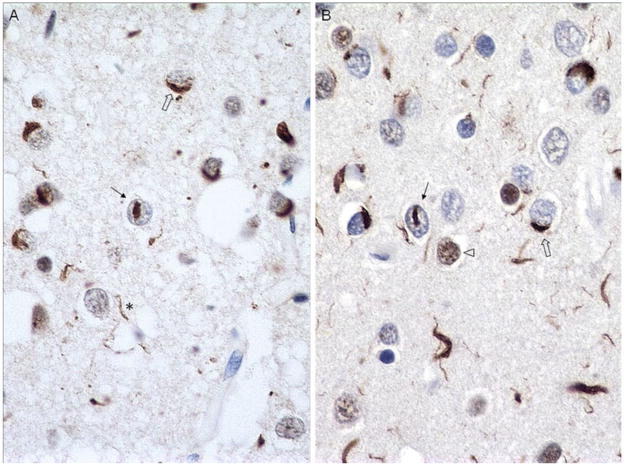
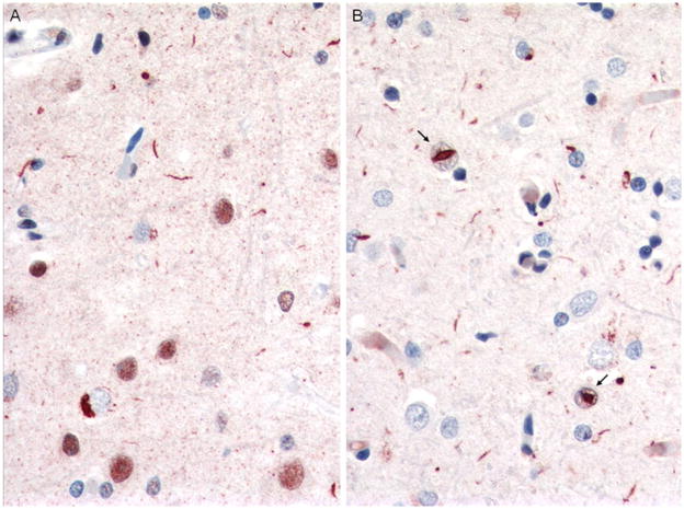
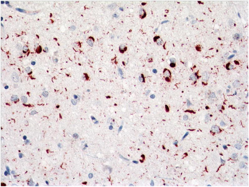
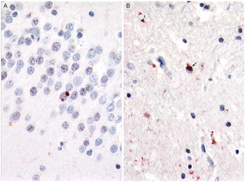
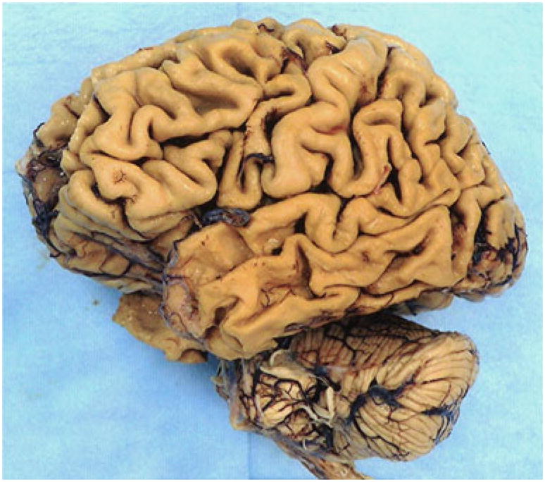
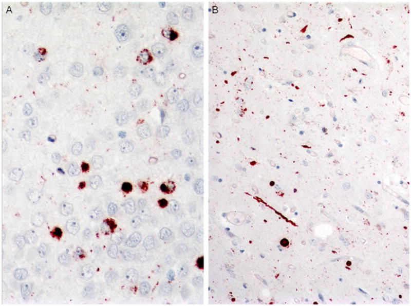
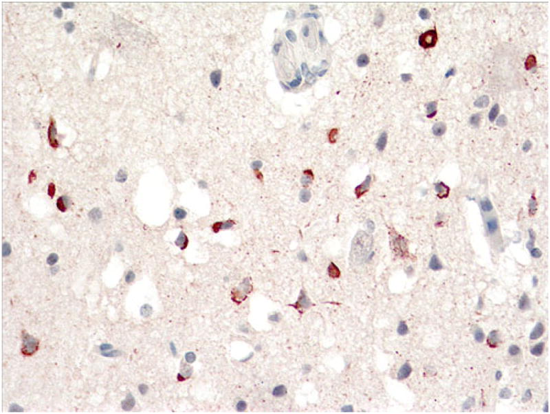
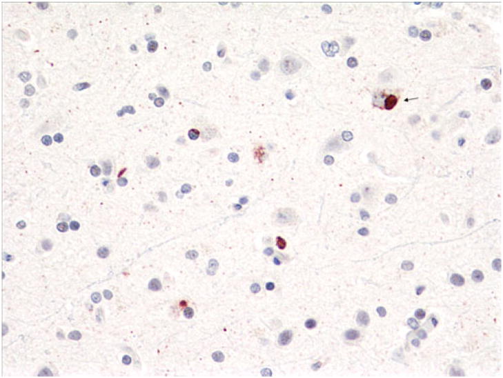
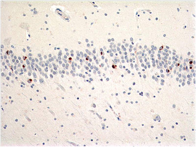
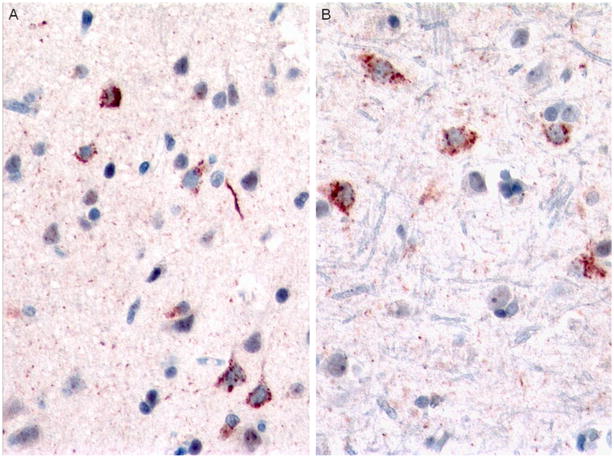
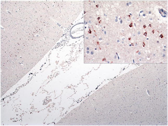
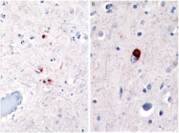
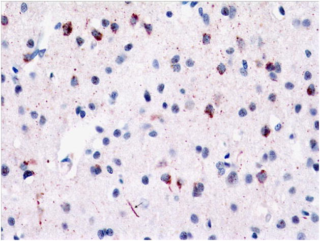
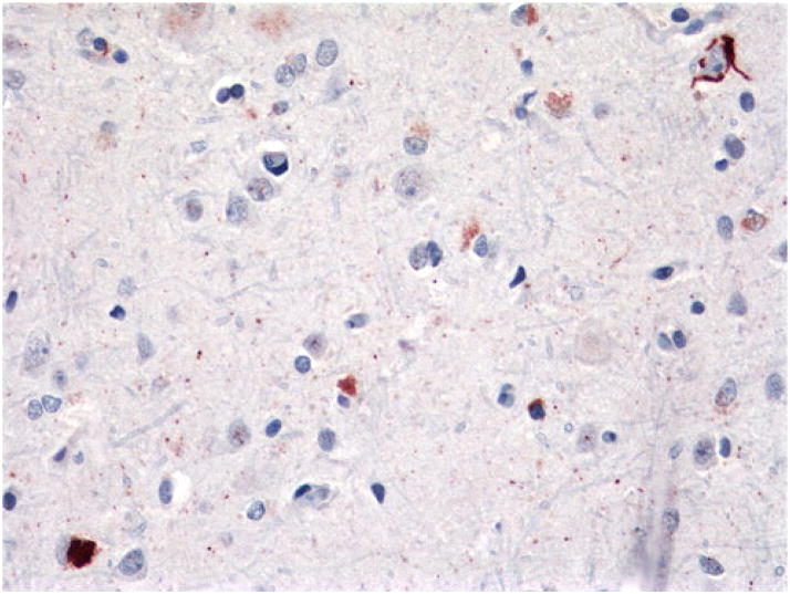
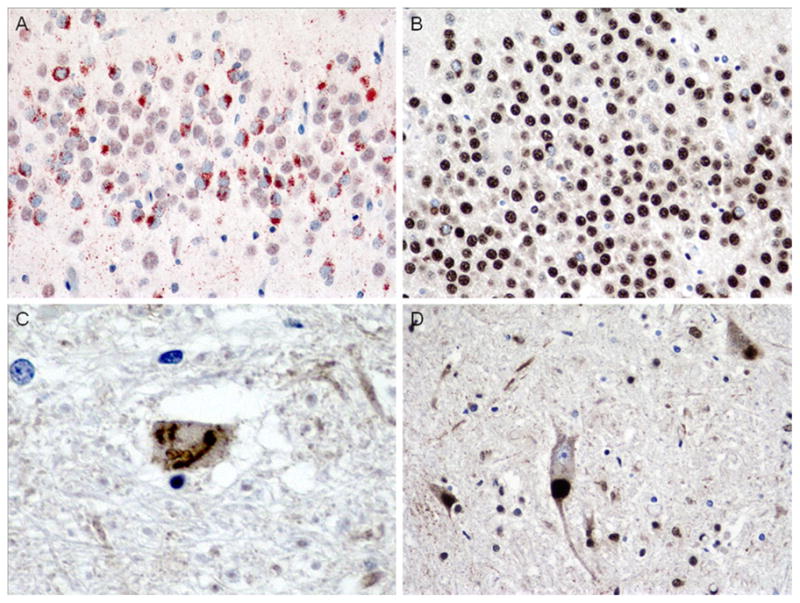
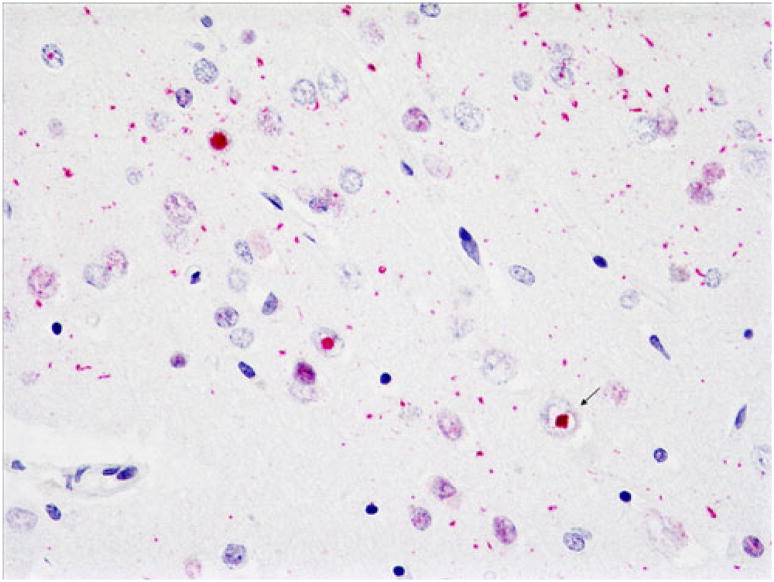
It has been only 5 years since the identification of TDP-43 as the major protein component of the ubiquitinated inclusions in FTLD-U. At that time, there were approximately a dozen papers about TDP-43; today, a "TDP-43" search reveals almost 600 papers. It is now clear that the majority of FTLD cases containing tau- and alpha-synuclein-negative, ubiquitin-positive inclusions (FTLD-U) are FTLD-TDP. The spectrum of TDP-43 proteinopathies includes FTLD-TDP with or without ALS, with or without mutations in GRN, VCP, or TARDBP, with or without chromosome 9p linkage, and sporadic and non-SOD1 familial ALS with or without FTLD-TDP. There are four sub-types of FTLD-TDP, and these correlate with specific clinical and genetic profiles. Sub-types are determined by the presence, predominance, and distribution of the various TDP-43 immunopositive insoluble aggregates-neuronal cytoplasmic inclusions, neuronal intranuclear inclusions, and dystrophic neurites. In this paper, FTLD-TDP pathologic sub-types will be described, and examples of each sub-type will be shown, and implications for future research will be discussed.


Figures

Fig. 1
Fig. 1
Ubiquitin (a) and TDP-43 (b) immunohistochemistry of superficial frontal cortex of an FTLD-U case. Note neuronal cytoplasmic inclusions (NCIs) (open arrows), neuronal intra-nuclear inclusions (NIIs) (arrows), and dystrophic neurites (DNs) (asterisks). This TDP-43 antibody labels normal as well as abnormal TDP-43, so nuclei of neurons without abnormal TDP-43 aggregates are also labeled in b (arrowhead). Note that TDP-43 reveals slightly greater pathology than does ubiquitin. Dako polyclonal ubiquitin and ProteinTech polyclonal TDP-43 antibodies. Magnification, ×600
Fig. 2
Fig. 2
Case 1, FTLD-TDP type 1. Immunostains comparing labeling with polyclonal TDP-43 antibody (a) and monclonal antibody to phosphorylated TDP-43 (p-TDP-43) (b). Note frequent NCIs and DNs, with more DNs demonstrated with the phosphorylated TDP-43 antibody. NIIs indicated by arrows. Magnification, ×400
Fig. 3
Fig. 3
Case 1. Sparse to moderate NCIs, DNs, and NIIs in deep cortex with polyclonal TDP-43 (a) and monoclonal p-TDP-43 antibodies (b). NIIs indicated by arrows. Magnification, ×400
Fig. 4
Fig. 4
Case 1. Frequent NCIs and DNs in superficial motor cortex. p-TDP-43 antibody. Magnification, ×400
Fig. 5
Fig. 5
Case 1. Sparse dentate gyrus NCIs (a) and sparse to moderate NCIs, DNs, and NIIs in the striatum (b). p-TDP-43 antibody. Magnification, ×400
Fig. 6
Fig. 6
Case 2. Left aspect of gross brain, showing severe frontal, temporal, and parietal atrophy, greatest in temporal regions, where it is “knife edge-like,” with relative sparing of the posterior superior temporal gyrus
Fig. 7
Fig. 7
Case 2. Long neurites predominate in cortex and are present in superficial as well as deep layers. Superficial cortex (a), ×200 magnification. Low power (b) showing full-thickness cortex (b), ×40 magnification. p-TDP-43 antibody
Fig. 8
Fig. 8
Case 3A. Frequent dense and granular dentate gyrus inclusions (a) and frequent long and short neurites in the caudate nucleus (b). p-TDP-43 antibody, a ×400, b ×200 magnification
Fig. 9
Fig. 9
Case 3A. Frequent filamentous, dense, and granular NCIs and sparse DNs in superficial cortex. p-TDP-43 antibody. Magnification, ×400
Fig. 10
Fig. 10
Case 3A. Sparse dense NCIs and DNs, deep cortex. Arrow points to Pick-like body. p-TDP-43 antibody. Magnification, ×400
Fig. 11
Fig. 11
Case 3A. Moderate dense NCIs in dentate gyrus. Some are large, round, and Pick-like. p-TDP-43 antibody. Magnification, ×200
Fig. 12
Fig. 12
Case 3B. Moderate granular and sparse dense NCIs and sparse DNs in superficial cortex (a) and moderate granular inclusions in deep cortex (b). p-TDP-43 antibody. Magnification for both, ×400
Fig. 13
Fig. 13
Case 3C. Frequent NCIs in superficial motor cortex (upper left) but none in sensory cortex (lower right). Inset shows superficial NCIs at higher power. p-TDP-43 antibody, ×40 magnification; inset, ×400 magnification
Fig. 14
Fig. 14
Case 3C. Sparse to moderate filamentous and dense NCIs and sparse DNs in deep layers of motor cortex (a). Pick-like dense NCI shown in (b). p-TDP-43 antibody. Magnifications, a ×200 and b ×400
Fig. 15
Fig. 15
Case 3D. Frequent granular NCIs and sparse long DNs (arrow), superficial frontal cortex. p-TDP-43 antibody. Magnification, ×400
Fig. 16
Fig. 16
Case 3D. Sparse dense and filamentous NCIs, deep frontal cortex. p-TDP-43 antibody. Magnification, ×400
Fig. 17
Fig. 17
Case 3D. Comparison of NCIs in dentate gyrus (a, b) and SLIs and LBLIs in hypoglossal nucleus (c, d) in case 3C (a, c) compared to brother (b, d). Note frequent granular NCIs in case 3 with bvFTD (a) but only sparse NCIs in brother with clinical ALS (b). Additionally, hypoglossal SLIs were rare in case 3 (c), but LBLIs were frequent (and SLIs sparse, not shown) in brother (d). p-TDP-43 antibody (a), ×400 magnification, TDP-43 antibody (b, d), ×400 magnification and c ×600 magnification
Fig. 18
Fig. 18
Case 4. Frequent NIIs (arrows) and DNs in cortex. Rare NCIs not shown. p-TDP-43 antibody. Magnification, ×400

References

    1. Amador-Ortiz C, Lin W-L, Ahmed Z, Personett D, Davies P, Duara R, Graff-Radford NR, Hutton ML, Dickson DW. TDP-43 immunoreactivity in hippocampal sclerosis and Alzheimer’s disease. Ann Neurol. 2007;61:435–445. - PMC - PubMed
    1. Arai T, Hasegawa M, Akiyama H, Ikeda K, Nonaka T, Mori H, Mann D, Tsuchiya K, Yoshida M, Hashizume Y, Oda T. TDP-43 is a component of ubiquitin-positive tau-negative inclusions in frontotemporal lobar degeneration and amyotrophic lateral sclerosis. Biochem Biophys Res Commun. 2006;351:602–611. - PubMed
    1. Armstrong RA, Ellis W, Hamilton RL, Mackenzie IRA, Hedreen J, Gearing M, Montine T, Vonsattel J-P, Head E, Lieberman AP, Cairns NJ. Neuropathological heterogeneity in frontotemporal lobar degeneration with TDP-43 proteinopathy: a quantitative study of 94 cases using principal components analysis. J Neural Transm. 2010;117:227–239. - PMC - PubMed
    1. Baker M, Mackenzie IR, Pickering-Brown SM, Gass J, Rademakers R, Lindholm C, Snowden J, Adamson J, Sadovnick D, Rollinson S, Cannon A, Dwosh E, Neary D, Melquist S, Richardson A, Dickson D, Berger Z, Eriksen J, Robinson T, Zehr C, Dickey CA, Crook R, McGowan E, Mann D, Boeve B, Feldman H, Hutton M. Mutations in progranulin cause tau-negative frontotemporal dementia linked to chromosome 17. Nature. 2006;442:916–919. - PubMed
    1. Borroni B, Bonvicini C, Alberici A, Buratti E, Agosti C, Archetti S, Papetti A, Stuani C, Di Luca M, Gennarelli M, Padovani A. Mutation within TARDBP leads to frontotemporal dementia without motor neuron disease. Human Mutat. 2009;30:E974–E983. - PubMed

Publication types

Substances

LinkOut - more resources