. 2011 Jul;33(7):538-46.
doi: 10.1002/bies.201000131. Epub 2011 May 24.

Retinal determination genes function along with cell-cell signals to regulate Drosophila eye development: examples of multi-layered regulation by master regulators

Affiliations

Retinal determination genes function along with cell-cell signals to regulate Drosophila eye development: examples of multi-layered regulation by master regulators

Nicholas E Baker et al. Bioessays. 2011 Jul.

Abstract

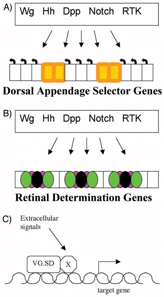
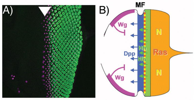
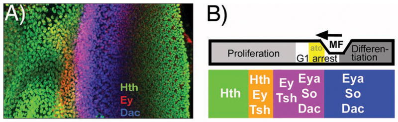
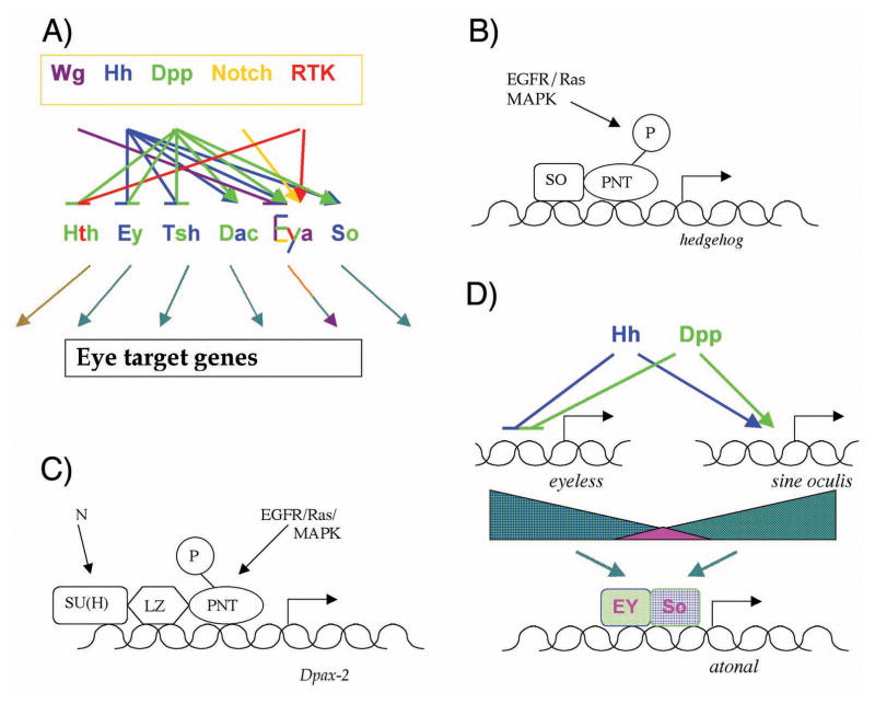
It is thought that retinal determination (RD) gene products define the response made to cell-cell signals in the field of eye development by binding to enhancers of genes that are also regulated by cell-cell signaling pathways. In Drosophila, RD genes, including eyeless, teashirt, eyes absent, dachsous, and sine oculis, are required for normal eye development and can induce ectopic eyes when mis-expressed. Characterization of the enhancers responsible for eye expression of the hedgehog, shaven, and atonal genes, as well as the dynamics of RD gene expression themselves, now suggest a multilayered network whereby transcriptional regulation by either RD genes or cell-cell signaling pathways can sometimes be indirect and mediated by other transcription factor intermediates. In this updated view of the interaction between extracellular information and cell intrinsic programs during development, regulation of individual genes might sometimes be several steps removed from either the RD genes or the cell-cell signaling pathways that nevertheless govern their expression.


Figures

Figure 1
Figure 1. Integration of developmental identity and extracellular signals in developmental patterning
Cell-cell signaling pathways that are used widely to confer spatio-temporal information are interpreted differently in each developmental field. The Drosophila wing and eye are each epithelia that are patterned by many of the same signals. A) Typical wing cell fates include wing vein (orange), and wing-hair producing intervein cells (uncolored). All these fates depend on Dorsal Appendage selector genes Scalloped and vestigial, as well as other selector genes such as the Hox gene Antennapedia, but the fates of individual cells within the wing differ due to positional information from cell-cell signaling pathways. B) Typical eye cell fates include clusters of photoreceptor neurons and various retinal support cells (black, green and magenta). These fates all depend on the Retinal Determination genes including Eyeless and its paralog twin of eyeless. Individual eye cells take different fates in response to positional information from the same cell-cell signaling pathways that are active in the wing. C) The prevailing model is that positional information is integrated with developmental identity at combinatorial enhancers[62]. In the example of a wing target gene, enhancer requiring binding by the Vg/Sd complex proteins and by transducers of cell-cell signaling pathways could confer spatio-temporal regulation[12].
Figure 2
Figure 2. Outline of retinal differentiation
A) Photoreceptor differentiation begins at the posterior of the Drosophila eye imaginal disc, the larval primordium from which the retinal epithlium differentiates. Anterior is shown to the left, and photoreceptor differentiation labeled in green. The first founder R8 photoreceptor cell, specified slightly before the others, is labelled in magenta with an antibody against Senseless, a target of the proneural gene Atonal[63]. Atonal expression begins just before the morphogenetic furrow (arrowhead) where R8 cells are specified[51]. B) Cartoon diagraming the main cell-cell signals acting in the developing eye. The morphogenetic furrow is driven anteriorly by Hh and Dpp diffusing from differentiating photoreceptor cells and from the morphogenetic furrow respectively. These positive signals are antagonized by Wg, emanating from the anterior dorsal and ventral boundaries. Posterior to the morphogenetic furrow, episodes of signaling through the Ras and Notch pathways regulates the proliferation, recruitment and differentiation of many retinal cell types[4,17,18].
Figure 3
Figure 3. Complex changes in RD gene expression accompanies eye differentiation
A) Examples of RD gene expression in the Drosophila eye imaginal disc as the wave of differentiation crosses the retinal epithelium. This eye disc has been immune-labelled to reveal overlapping, dynamic expression of three proteins, Homothorax (Hth: green), Eyeless (Ey: red), and Dachsous (Dac: blue). B) Summary diagram correlating dynamic RD gene expression patterns with developmental processes in the eye imaginal disc. Cells mature from an undifferentiated, proliferative state through a cell cycle arrest that accompanies expression of the first fate specification genes such as the proneural gene atonal (ato) and is followed by progressive differentiation of the various retinal cell types. During this period, cells evolve from expression of the antennal selector gene homothorax and RD genes ey and tsh to expression of the retinal determination genes eya, so, and dac. Figure adapted from[40].
Figure 4
Figure 4
A) Recent studies describe a complex network of cell-cell signaling that controls the spatiotemporal pattern of RD gene expression during eye development. The point of the color-coded summary shown here is not to follow the individual interactions in detail, but simply to note that all of these five cell-cell signaling pathways contribute to the spatiotemporal regulation of the RD genes that was summarized in Figure 3. This summary is probably incomplete, for example Dpp and Wg signaling pathways often act antagonistically, but most of the targets of Dpp have not been tested for regulation by Wg[40]. The figure also vertically compresses regulation by extracellular signals into a single layer, although in reality regulation is rendered multi-layered by regulatory relationships between the RD genes themselves. For example, Dac is necessary and sufficient for tsh repression, so it is possible that Hh and Dpp may regulate tsh indirectly as a secondary target of dac, which requires Hh and Dpp signaling for its expression[40]. As a result of the extensive regulation, the dynamic RD gene expression patterns contain information about the extracellular signaling pathway activities, and which could be conferred on further target genes in turn. B) Regulation of hedgehog. The enhancer responsible for eye expression is bound directly by Pnt, an Ets-domain transcription factor regulated by the EGFR/Ras/MAPK pathway of extracellular signals, and the RD protein SO[44]. This combinatorial scheme resembles the paradigm established for dorsal appendages (see Figure 1). C) Regulation of shaven (dPax-2). The sparkling (spa) enhancer responsible for eye expression is bound directly by Pnt and by Su(H), the transcription factor regulated by Notch signaling, as well as the Runx-family transcription factor Lozenge{[47]. Lz is not an RD gene, but its expression in the eye is itself regulated by So, bringing spa under RD control indirectly[49]. D) Regulation of atonal. Enhancer sequences that would be direct targets of Hh and Dpp signaling have not been found[54,55]; the observed dependence of ato expression on Hh and Dpp may instead be explained by the Hh- and Dpp-dependence of ey and so expression. Hh and Dpp regulate both ey and so expression, repressing ey and turning so on [40]. This regulation is itself likely to be multi-layered, since Ey appears to be a direct regulator of so expression (not diagrammed)[31,33], and since it is not known whether Mad (or Brinker) and Ci proteins bind directly to enhancers of the ey and so genes, or act indirectly through intermediate transcription factors. The Ey and So proteins are coexpressed ahead of the morphogenetic furrow, Ey declining as So rises. Transcription of ato initiates within the overlap. It is interesting that ato is activated by transcription factors that are co-expressed only transiently; in principle this provides a mechanism for a transient stripe of ato activation. The ato enhancer is active in only a subset of the cells coexpressing Ey and So, however, and other data also suggest that additional factors are also likely to limit ato transcription[40].

References

    1. Halder G, Callaerts P, Gehring WJ. Induction of ectopic eyes by targeted expression of the eyeless gene in Drosophila. Science. 1995;267:1788–92. - PubMed
    1. Curtiss J, Halder G, Mlodzik M. Selector and signalling molecules cooperate in organ patterning. Nat Cell Biol. 2002;4:E48–51. - PubMed
    1. Mann RS, Carroll SB. Molecular mechanisms of selector gene function and evolution. Curr Opin Genet Dev. 2002;12:592–600. - PubMed
    1. Roignant J-Y, Treisman JE. Pattern formation in the Drosophila eye disc. Int J Dev Biol. 2009;53:795–804. - PMC - PubMed
    1. Gerhart J. Warkany lecture: signaling pathways in development. Teratology. 1999;60:226–39. - PubMed

Publication types

MeSH terms

Substances

LinkOut - more resources