. 2011 Aug;20(8):1367-79.
doi: 10.1002/pro.663. Epub 2011 Jun 8.

Biochemical and structural studies on the high affinity of Hsp70 for ADP

Affiliations

Biochemical and structural studies on the high affinity of Hsp70 for ADP

Akihiko Arakawa et al. Protein Sci. 2011 Aug.

Abstract

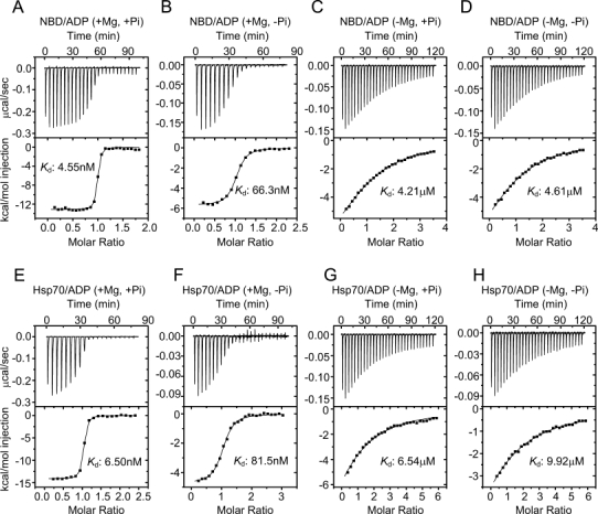
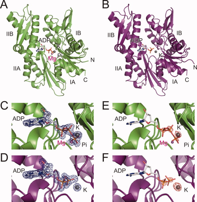
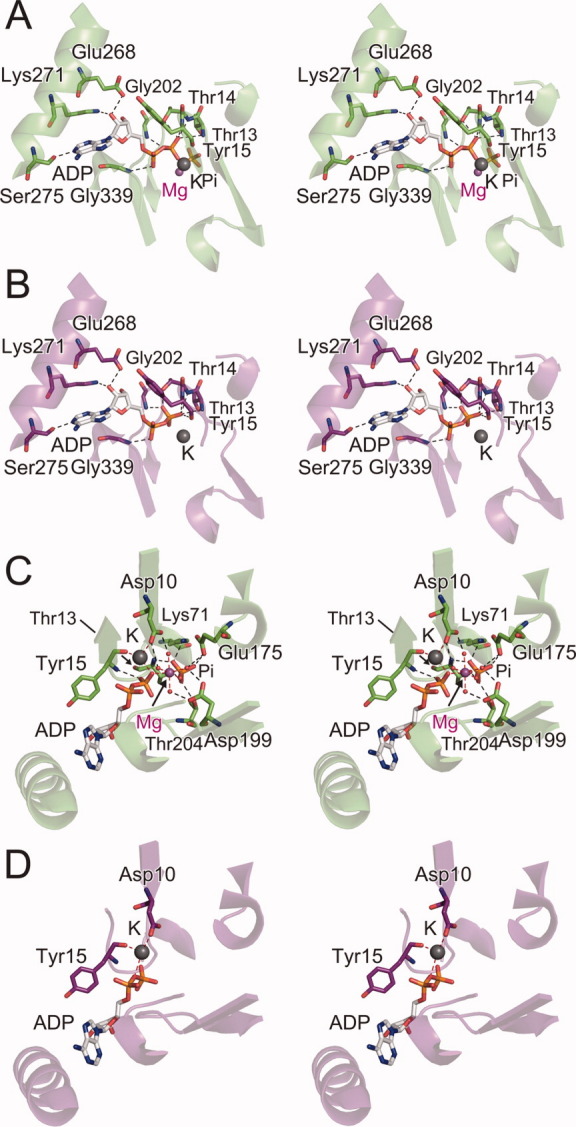
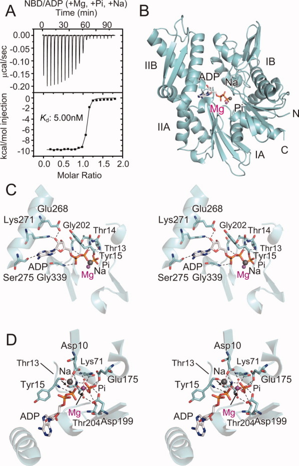
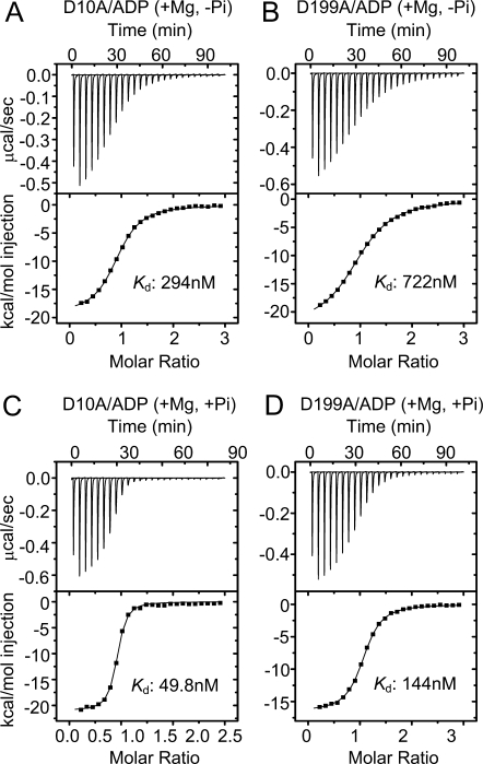
The molecular chaperone 70-kDa heat shock protein (Hsp70) is driven by ATP hydrolysis and ADP-ATP exchange. ADP dissociation from Hsp70 is reportedly slow in the presence of inorganic phosphate (P(i) ). In this study, we investigated the interaction of Hsp70 and its nucleotide-binding domain (NBD) with ADP in detail, by isothermal titration calorimetry measurements and found that Mg(2+) ion dramatically elevates the affinity of Hsp70 for ADP. On the other hand, P(i) increased the affinity in the presence of Mg(2+) ion, but not in its absence. Thus, P(i) enhances the effect of the Mg(2+) ion on the ADP binding. Next, we determined the crystal structures of the ADP-bound NBD with and without Mg(2+) ion. As compared with the Mg(2+) ion-free structure, the ADP- and Mg(2+) ion-bound NBD contains one Mg(2+) ion, which is coordinated with the β-phosphate group of ADP and associates with Asp10, Glu175, and Asp199, through four water molecules. The Mg(2+) ion is also coordinated with one P(i) molecule, which interacts with Lys71, Glu175, and Thr204. In fact, the mutations of Asp10 and Asp199 reduced the affinity of the NBD for ADP, in both the presence and the absence of P(i) . Therefore, the Mg(2+) ion-mediated network, including the P(i) and water molecules, increases the affinity of Hsp70 for ADP, and thus the dissociation of ADP is slow. In ADP-ATP exchange, the slow ADP dissociation might be rate-limiting. However, the nucleotide-exchange factors actually enhance ADP release by disrupting the Mg(2+) ion-mediated network.


Figures

Figure 1
Figure 1
ITC analysis of the interactions of the Hsp70 NBD (A–D) and the full-length Hsp70 (E–H) with ADP, in the presence and absence of Mg2+ ion and Pi. (A) The NBD in the Pi- and Mg2+ ion-containing buffer. (B) The NBD in the Pi-free and Mg2+ ion-containing buffer. (C) The NBD in the Pi-containing and Mg2+ ion-free buffer. (D) The NBD in the Pi- and Mg2+ ion-free buffer. (E) Hsp70 in the Pi- and Mg2+ ion-containing buffer. (F) Hsp70 in the Pi-free and Mg2+ ion-containing buffer. (G) Hsp70 in the Pi-containing and Mg2+ ion-free buffer. (H) Hsp70 in the Pi- and Mg2+ ion-free buffer.
Figure 2
Figure 2
Crystal structures of the ADP-bound NBD, with and without Mg2+ ion. (A) Crystal structure of the ADP- and Mg2+ ion-bound NBD (green). The stick models represent ADP and Pi, and the balls represent Mg2+ (magenta) and K+ (gray) ions. (B) Crystal structure of the ADP-bound and Mg2+ ion-free NBD (purple). The stick model represents ADP, and the gray ball represents the K+ ion. (C) The nucleotide-bound pocket of the ADP- and Mg2+ ion-bound NBD. The electron density indicates the FoFc omit map (3σ level). (D) The nucleotide-bound pocket in the ADP-bound and Mg2+ ion-free NBD. The electron density indicates the FoFc omit map (3σ level). (E) Anomalous difference Fourier map (4σ level) in the nucleotide-binding pocket of the ADP- and Mg2+ ion-bound NBD. (F) Anomalous difference Fourier map (4σ level) in the nucleotide-binding pocket of the ADP-bound and Mg2+ ion-free NBD. Molecular graphics were generated and rendered with the PyMOL program (DeLano Scientific, Palo Alto, CA).
Figure 3
Figure 3
Interactions between the NBD and ADP, in the presence and absence of Mg2+ ion. (A) The direct interactions in the presence of Mg2+ ion (stereo view). (B) The direct interactions in the absence of Mg+ ion (stereo view). (C) The indirect interactions in the presence of Mg2+ ion (stereo view). (D) The indirect interactions in the absence of Mg2+ ion (stereo view). The interacting residues are shown as stick models, with the translucent ribbon model of the NBD in green (A and C) and purple (B and D). ADP and Pi are also shown as stick models. The Mg2+ ion is colored magenta, and the K+ ion is represented by a gray ball. Water molecules are shown as red dots. Black and red dashed lines represent hydrogen bonds and coordinate bonds, respectively.
Figure 4
Figure 4
Interactions between the NBD and ADP, in the presence of Na+ ion. (A) ITC analysis of the interactions between the NBD and ADP, in buffer containing Pi-, Mg2+, and Na+ ions. (B) Crystal structure of the ADP- and Mg2+ ion-bound NBD containing Na+ ion. (C) The direct interactions between the NBD and ADP (stereo view). (D) The indirect interactions between the NBD and ADP (stereo view). The interacting residues are shown as stick models, with the ribbon model of the NBD in cyan. ADP and Pi are also shown as stick models. The Mg2+ ion is colored magenta, and the Na+ ion is represented by a gray ball. Water molecules are shown as red dots. Black and red dashed lines represent hydrogen bonds and coordinate bonds, respectively.
Figure 5
Figure 5
ITC analysis of the interactions of the NBD mutants with ADP. (A) The D10A mutant in the Pi-free and Mg2+ ion-containing buffer. (B) The D199A mutant in the Pi-free and Mg2+ ion-containing buffer. (C) The D10A mutant in the Pi- and Mg2+ ion-containing buffer. (D) The D199A mutant in the Pi- and Mg2+ ion-containing buffer.
Figure 6
Figure 6
Amino acid sequence alignment of the NBDs of five Hsp70-family proteins. Sequences are the NBDs of human (Homo sapiens) Hsp70 (NCBI Gene ID: 3303), Hsc70 (NCBI Gene ID: 3312), Bip/Grp78 (NCBI Gene ID: 3309), Grp75/mtHsp70 (NCBI Gene ID: 3313), and E. coli DnaK (NCBI Gene ID: 944750). Above the sequences, the secondary structures of human Hsp70 NBD are indicated. The residues involved in the Mg2+ ion-mediated network are indicated by green arrows.

References

    1. Bukau B, Horwich AL. The Hsp70 and Hsp60 chaperone machines. Cell. 1998;92:351–366. - PubMed
    1. Bukau B, Weissman J, Horwich A. Molecular chaperones and protein quality control. Cell. 2006;125:443–451. - PubMed
    1. Young JC, Agashe VR, Siegers K, Hartl FU. Pathways of chaperone-mediated protein folding in the cytosol. Nat Rev Mol Cell Biol. 2004;5:781–791. - PubMed
    1. Mayer MP. Gymnastics of molecular chaperones. Mol Cell. 2010;39:321–331. - PubMed
    1. Wilbanks SM, Chen L, Tsuruta H, Hodgson KO, McKay DB. Solution small-angle X-ray scattering study of the molecular chaperone Hsc70 and its subfragments. Biochemistry. 1995;34:12095–12106. - PubMed

Publication types

MeSH terms