. 2011 Jul;29(7):1052-63.
doi: 10.1002/stem.662.

Development of histocompatible primate-induced pluripotent stem cells for neural transplantation

Affiliations

Development of histocompatible primate-induced pluripotent stem cells for neural transplantation

Michela Deleidi et al. Stem Cells. 2011 Jul.

Abstract

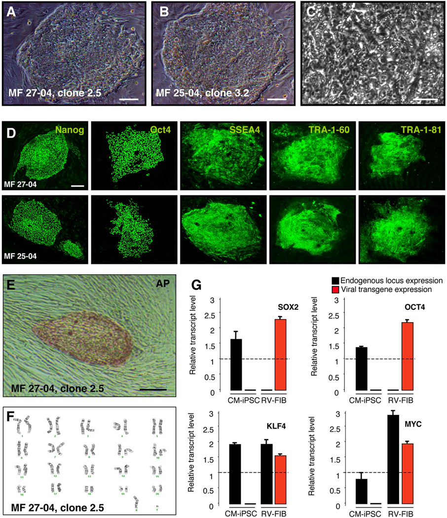
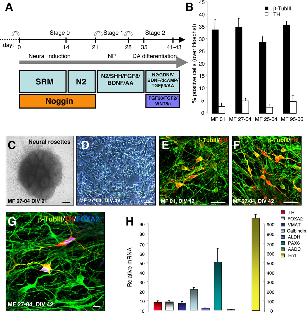
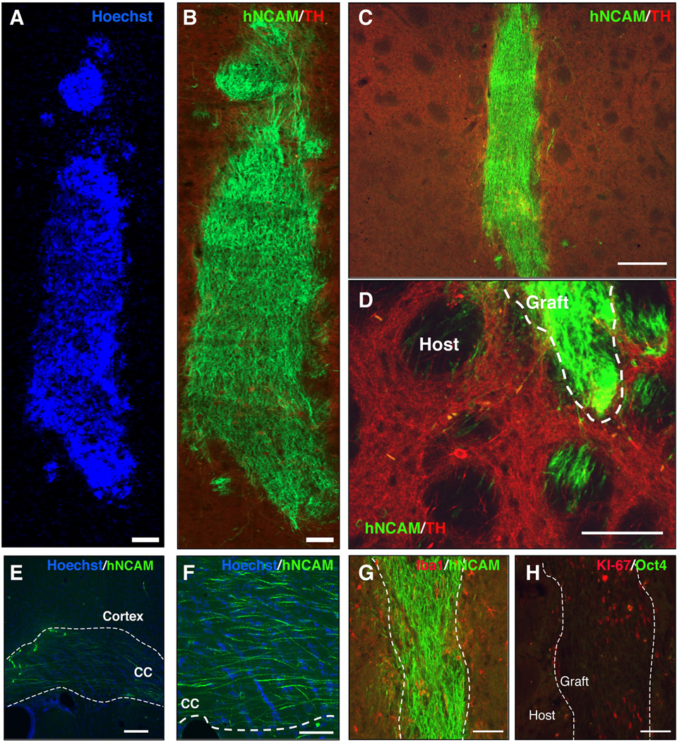
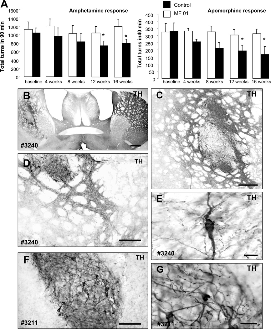
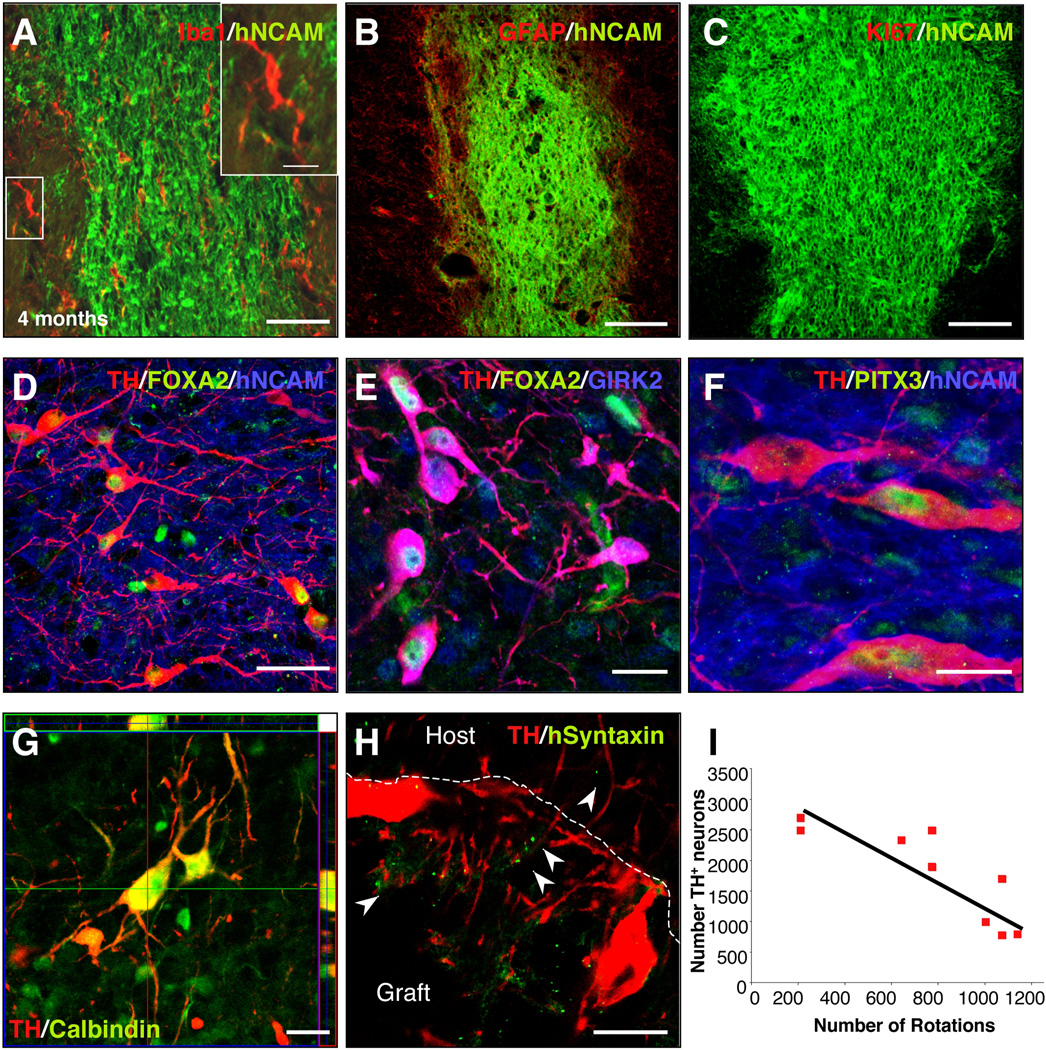
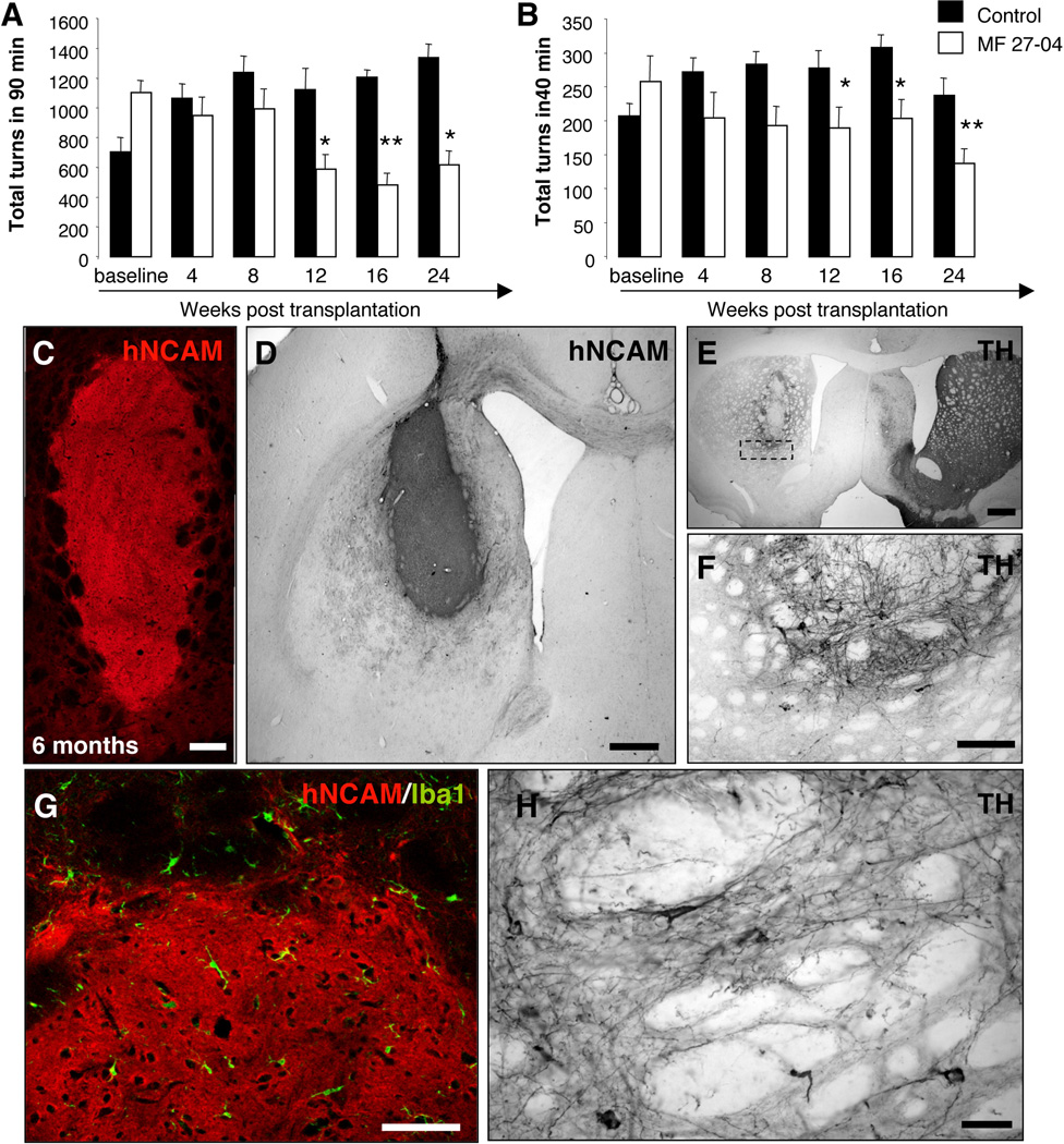
Immune rejection and risk of tumor formation are perhaps the greatest hurdles in the field of stem cell transplantation. Here, we report the generation of several lines of induced pluripotent stem cells (iPSCs) from cynomolgus macaque (CM) skin fibroblasts carrying specific major histocompatibility complex (MHC) haplotypes. To develop a collection of MHC-matched iPSCs, we genotyped the MHC locus of 25 CMs by microsatellite polymerase chain reaction analysis. Using retroviral infection of dermal skin fibroblasts, we generated several CM-iPSC lines carrying different haplotypes. We characterized the immunological properties of CM-iPSCs and demonstrated that CM-iPSCs can be induced to differentiate in vitro along specific neuronal populations, such as midbrain dopaminergic (DA) neurons. Midbrain-like DA neurons generated from CM-iPSCs integrated into the striatum of a rodent model of Parkinson's disease and promoted behavioral recovery. Importantly, neither tumor formation nor inflammatory reactions were observed in the transplanted animals up to 6 months after transplantation. We believe that the generation and characterization of such histocompatible iPSCs will allow the preclinical validation of safety and efficacy of iPSCs for neurodegenerative diseases and several other human conditions in the field of regenerative medicine.


Conflict of interest statement

Disclosure of Potential Conflicts of Interest: the authors indicate no potential conflicts of interest.

Figures

Figure 1
Figure 1. Generation of Cynomolgus Monkey iPSC
(A–B) Phase contrast images showing ESC-like morphology of CM-iPSCs on MEF. (C) High-magnification image of CM-iPSCs showing the high nucleus/cytoplasm ratio and prominent nucleoli. (D) Immunofluorescence staining for the pluripotency markers Nanog, Oct4, SSEA-4, TRA-1–60, and TRA-1–81. (E) CM-iPSC colonies stained positive for alkaline phosphatase (AP). (F) Karyotype analysis of CM-iPSCs (line 27-04, passage 10). CM-iPSCs maintained a normal 42, XY karyotype after expansion.(G) Quantitative RT-PCR showing induction of endogenous transcripts of Sox2, Oct4, Klf4, and c-Myc. Primers specific for endogenously (black) or viral exogenous (red) encoded transcripts of the four reprogramming factors. Monkey dermal fibroblasts, four days after transduction with the four retroviruses (RV-FIB), were used as a positive control for expression of the viral transgenes. Values were normalized by the averaged value of β-actin. Values are expressed as averages + SEM of 3 independent experiments. The value of hESCs (H9) was set to 1 in each experiment. Scale bar, 200 µm (A–B, D–E); 20 µm (C).
Figure 2
Figure 2. Generation of DA neurons from CM-iPSCs
(A) Schematic representation of the in vitro differentiation protocol. (B) Graphs indicate the percentage of cells that stained positive for β-TubIII and TH relative to nuclear Hoechst staining. Values are expressed as averages + SEM of 3 independent experiments. (C) Neural rosettes derived from CM-iPSCs (DIV 21). (D) Differentiated neurons derived from CM-iPSCs (DIV 42). (E–F) Immunofluorescence staining of neuronal cultures derived from CM-iPSCs (lines MF 01, MF 27-04) for β-TubIII (green) and TH (red). (G) Confocal images of neuronal cultures stained for β-TubIII (green), TH (red) and Foxa2 (blue). (H) Quantitative RT-PCR analysis of midbrain transcription factors, DA neurotransmission markers (TH, Foxa2, VMAT, ALDH, Nurr1, En1, AADC, Pax6) and Calbindin in differentiated iPSCs. cDNA was isolated from differentiated iPSCs, and values were normalized to the level of β-actin. Values represent mean + SEM. Three independent experiments were performed in triplicate. Scale bar: 50 µm (C–D); 20 µm (E–G).
Figure 3
Figure 3. Transplantation of CM-iPSC-derived neurons into the adult striatum of naïve rats
CM-iPSCs (line MF 01) were differentiated into DA neurons and 200,000 cells were transplanted into the right striatum of unlesioned rats under immunosuppression. Grafts were analyzed 4 weeks after the transplantation. (A–B) Confocal reconstruction of a representative graft stained for the human/primate-specific neural cell adhesion molecule (NCAM) (green) and TH (red). Nuclei are counterstained with Hoechst. (C) Lower magnification of the graft. (D–F) Photomicrographs of NCAM-stained brain sections 4 weeks after transplantation showing axonal outgrowth of NCAM+ neurons from the graft into the host striatum, along the white matter tracts (corpus callosum) and cerebral cortex. (G) Immunostaining for hNCAM (green) and the microglial marker Iba1 (red) revealed the absence of microglial reaction. (H) Immunostaining for the proliferative marker Ki-67 (red) and Oct4 (green) showing the presence of a low percentage of Ki-67+/Oct4- cells within the graft. Scale bar: 100 µm (A–B,D and F-H), 200 µm (C,E).
Figure 4
Figure 4. CM-iPSC-derived neurons improve behavioral deficits of parkinsonian rats
(A) CM-iPSCs (line MF-01) were differentiated into DA neurons and 400,000 cells were transplanted into the right striatum of 6OHDA-lesioned rats under immunosuppression. Amphetamine and apomorphine response were examined before and at 4, 8, 12 and 16 weeks post-transplantation. Animals showed a progressive reduction in the response compared to pre-transplantation scores. (Transplanted animals n=9, lesion only animals n=4; *, p <0.05 ANOVA repeated-measures over time). (B–G) Tyrosine hydroxylase (TH) immunoreactivity in representative grafts (rat #3211, #3240) derived from differentiated primate iPSCs showing TH + neurons within the graft and areas of dense TH neuritic arborization. Scale bar: 200 µm (B–D); 50 µm (F), 20 µm (E–G).
Figure 5
Figure 5. Analysis of neuronal grafts
Confocal analysis of CM-iPSC-derived grafts, 16 weeks post-transplantation. (A) Staining for the microglial marker Iba1 showed the absence of activated microglial cells around the grafts. The insert shows high magnification of microglial cells with a resting phenotype. (B) Staining for the astrocytic marker GFAP revealed the absence of astrogliosis around the grafts. (C) Staining for the proliferation marker Ki-67 showing absence of proliferating cells 16 weeks after transplantation. (D–F) Confocal analysis of iPSC grafts showed that most grafts contained midbrain-like DA neurons. The grafted TH+ cells (red) were colabeled with antibodies against human NCAM (blue) (D–F), FOXA2 (green) (D–E), GIRK2 (blue) (E), Pitx3 (green) (F). (G) Confocal images showing TH+ neurons (red) in a representative graft co-expressing the calcium-binding protein calbindin (red). (H) Confocal images showing the localization of human syntaxin within the graft and in the host striatum. (I) Correlation between number of TH+ neurons and number of rotations (n=9; simple regression, r=0.885, r2=0.784, P=0.01). Scale bar: 100 µm (A–D), 50 µm (A–E), 20 µm (F–I).
Figure 6
Figure 6. Long-term analysis of neuronal grafts
CM-iPSCs (line MF 27-04) were differentiated into DA neurons and 400,000 cells were transplanted into the right striatum of 6-OHDA-lesioned rats. (A–B) Amphetamine and apomorphine response were examined before and at 4, 8, 12, 16, and 24 weeks post-transplantation. Animals showed a progressive reduction in the response compared to pre-transplantation scores. (Transplanted animals n=11, lesion only animals n=6; *, p <0.05; **, p <0.01, ANOVA repeated-measures over time). (C) Confocal reconstruction of a representative graft stained for the hNCAM (red). (D) NCAM immunoreactivity in a representative graft. (E–F,H) TH immunoreactivity in representative grafts showing survival of TH+ neurons and neuritic arborization. (G) Staining for the microglial marker Iba1 showed the absence of activated microglial cells around the grafts. Scale bar: 100 µm (B–C,G), 200 µm (E), 20 µm (F, H).

Similar articles

Cited by

References

    1. Takahashi K, Tanabe K, Ohnuki M, et al. Induction of pluripotent stem cells from adult human fibroblasts by defined factors. Cell. 2007;131:861–872. - PubMed
    1. Yu J, Vodyanik MA, Smuga-Otto K, et al. Induced pluripotent stem cell lines derived from human somatic cells. Science (New York, N.Y. 2007;318:1917–1920. - PubMed
    1. Park IH, Zhao R, West JA, et al. Reprogramming of human somatic cells to pluripotency with defined factors. Nature. 2008;451:141–146. - PubMed
    1. Nakatsuji N, Nakajima F, Tokunaga K. HLA-haplotype banking and iPS cells. Nature biotechnology. 2008;26:739–740. - PubMed
    1. Bontrop RE. Non-human primates: essential partners in biomedical research. Immunological reviews. 2001;183:5–9. - PubMed

Publication types