Review
. 2011 Sep 20;44(9):828-39.
doi: 10.1021/ar200021p. Epub 2011 May 20.

Photoinducible bioorthogonal chemistry: a spatiotemporally controllable tool to visualize and perturb proteins in live cells

Affiliations
Review

Photoinducible bioorthogonal chemistry: a spatiotemporally controllable tool to visualize and perturb proteins in live cells

Reyna K V Lim et al. Acc Chem Res. .

Abstract

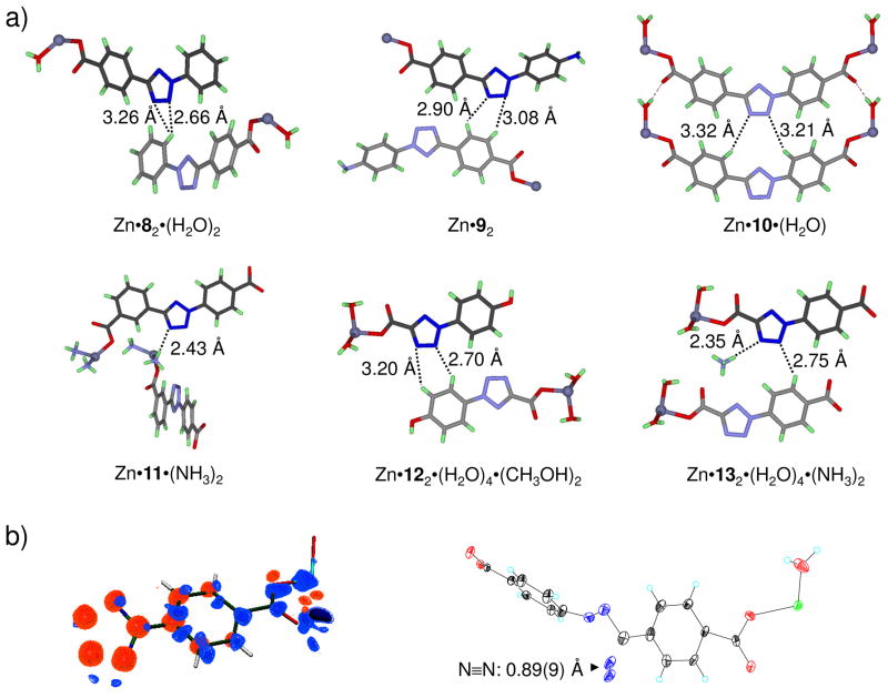
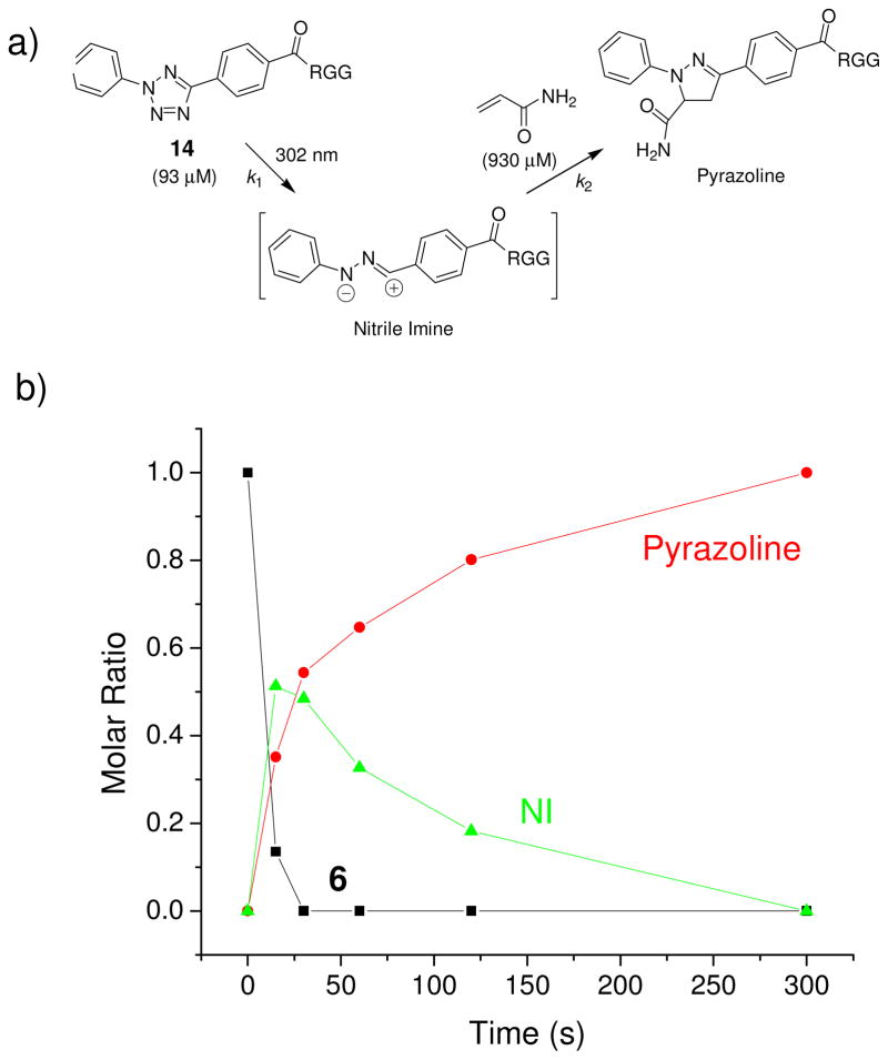
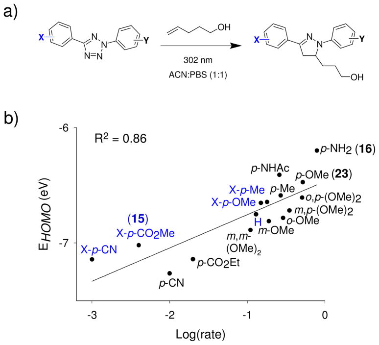
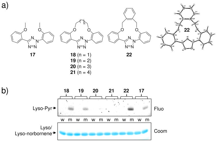
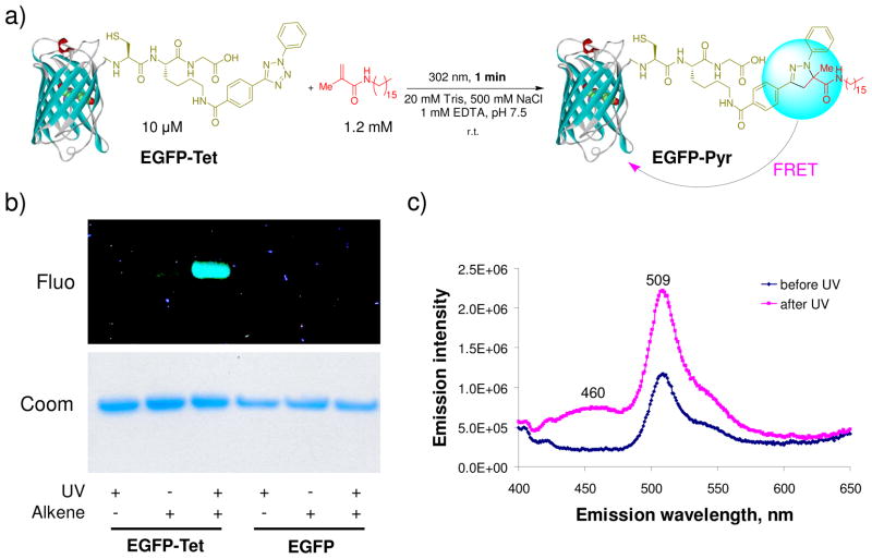
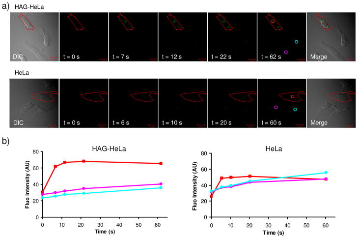
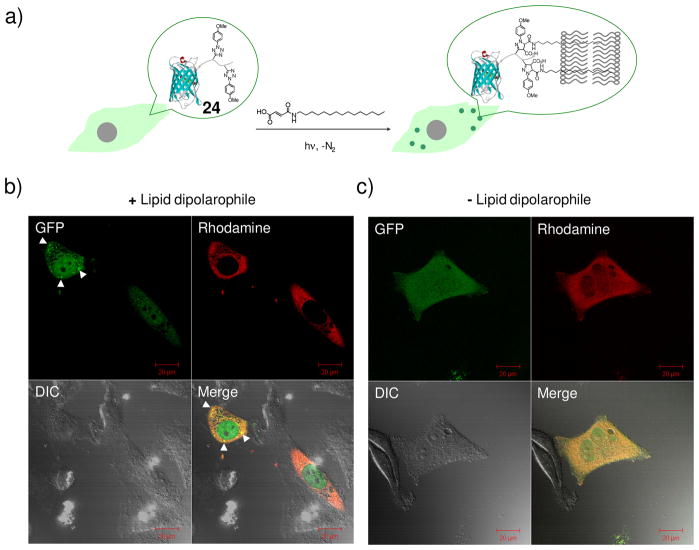
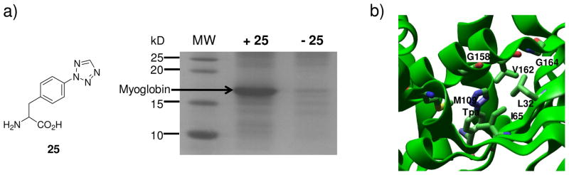
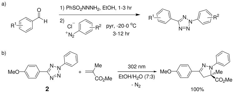
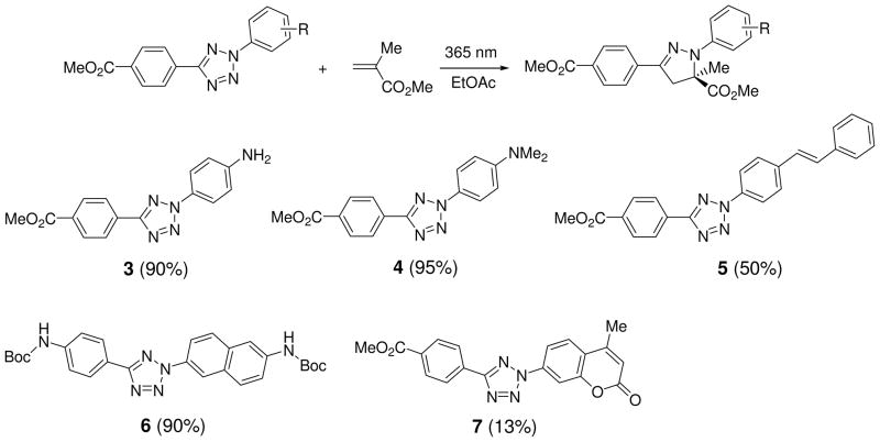
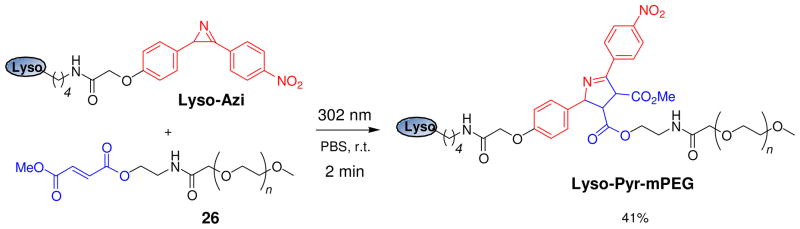
Visualization in biology has been greatly facilitated by the use of fluorescent proteins as in-cell probes. The genes coding for these wavelength-tunable proteins can be readily fused with the DNA coding for a protein of interest, which enables direct monitoring of natural proteins in real time inside living cells. Despite their success, however, fluorescent proteins have limitations that have only begun to be addressed in the past decade through the development of bioorthogonal chemistry. In this approach, a very small bioorthogonal tag is embedded within the basic building blocks of the cell, and then a variety of external molecules can be selectively conjugated to these pretagged biomolecules. The result is a veritable palette of biophysical probes for the researcher to choose from. In this Account, we review our progress in developing a photoinducible, bioorthogonal tetrazole-alkene cycloaddition reaction ("photoclick chemistry") and applying it to probe protein dynamics and function in live cells. The work described here summarizes the synthesis, structure, and reactivity studies of tetrazoles, including their optimization for applications in biology. Building on key insights from earlier reports, our initial studies of the reaction have revealed full water compatibility, high photoactivation quantum yield, tunable photoactivation wavelength, and broad substrate scope; an added benefit is the formation of fluorescent cycloadducts. Subsequent studies have shown fast reaction kinetics (up to 11.0 M(-1) s(-1)), with the rate depending on the HOMO energy of the nitrile imine dipole as well as the LUMO energy of the alkene dipolarophile. Moreover, through the use of photocrystallography, we have observed that the photogenerated nitrile imine adopts a bent geometry in the solid state. This observation has led to the synthesis of reactive, macrocyclic tetrazoles that contain a short "bridge" between two flanking phenyl rings. This photoclick chemistry has been used to label proteins rapidly (within ∼1 min) both in vitro and in E. coli . To create an effective interface with biology, we have identified both a metabolically incorporable alkene amino acid, homoallylglycine, and a genetically encodable tetrazole amino acid, p-(2-tetrazole)phenylalanine. We demonstrate the utility of these two moieties, respectively, in spatiotemporally controlled imaging of newly synthesized proteins and in site-specific labeling of proteins. Additionally, we demonstrate the use of the photoclick chemistry to perturb the localization of a fluorescent protein in mammalian cells.


Figures

Figure 1
Figure 1
Observation of a bent nitrile imine structure in the solid state: (a) Crystal structures of Zn-tetrazole complexes; Zn is shown in silver. The distances between N3-N4 and the nearest surrounding atoms are marked on the structures; (b) Photodifference map (left) of the Zn•10 complex after photoirradiation based on Fo (after) - Fo (before). Blue, 2.0; light blue, 1.0; orange, −1.0; red, −2.0 e/Å3. Only one half of the map is shown because of twofold symmetry; ORTEP representation (right) of the geometry-refined nitrile imine structure and the escaping N2.
Figure 2
Figure 2
Kinetic analysis of the photoinduced tetrazole-alkene cycloaddition reaction in PBS buffer at room temperature: (a) Reaction scheme showing the cycloaddition of a tetrazole-modified Arg-Gly-Gly (RGG) tripeptide 14 to acrylamide; (b) Reaction time course showing the molar ratio changes of starting material 14, nitrile imine (NI), and product over a period of 300 seconds.
Figure 3
Figure 3
The cycloaddition rates are dependent on the nitrile imine HOMO energies: (a) Scheme of the model cycloaddition reactions between substituted 2,5-diphenyltetrazoles and 4-penten-1-ol; (b) Plot of EHOMO vs. Log(rate). The HOMO energies of the nitrile imines were calculated using the Hartree-Fock 3–21G model with the AM1 optimized molecular geometries. The substituents on the X-side were colored in blue while the substituents on the Y-side were colored in black.
Figure 4
Figure 4
Design and evaluation of macrocyclic tetrazoles: (a) Structures of macrocyclic tetrazoles. The X-ray structure of tetrazole 22 shows co-planarity of the aryl rings; (b) Photoinduced cycloaddition reaction of macrocyclic tetrazoles with norbornene-modified lysozyme (m) or wild-type lysozyme (w) in PBS buffer at 302 nm. Top panel, inverted in-gel fluorescence with λex = 365 nm; bottom panel, Coomassie blue staining.
Figure 5
Figure 5
Photoinduced lipidation of EGFP carrying a tetrazole motif at its C-terminus: (a) Scheme for a photoinduced lipidation by a lipid dipolarophile; (b) Fluorescent imaging (top panel, λex = 365 nm) and Coomassie blue staining (bottom panel) of EGFP-Tet and EGFP upon photoirradiation in the presence or absence of the lipid dipolarophile. Duration of 1-min 302-nm UV irradiation was applied to the samples; (c) Fluorescence spectra of EGFP-Tet before and after UV irradiation, λex = 370 nm.
Figure 6
Figure 6
Visualization of a genetically encoded O-allyltyrosine-containing Z-domain protein in E. coli via the photoinduced tetrazole-alkene cycloaddition reaction: (a) Reaction scheme; (b) CFP (cyan fluorescent protein) channel (top panels) and DIC (Differential Interference Contrast) channel (bottom panels) images of bacteria expressing either alkene-Z (left) or wt-Z (right) proteins after treatment with 100 μM of tetrazole 15; scale bar = 10 μm.
Figure 7
Figure 7
Spatiotemporally controlled imaging of HAG-labeled proteins in live HeLa cells: (a) DIC (panel 1), time-lapsed fluorescence (panels 2–6), and merged DIC/fluorescence (panel 7) images of HAG-encoded HeLa cells (top row) and normal HeLa cells (bottom row) upon two-photon illumination. All cells were treated with 200 μM of tetrazole 16. A 5-sec two-photon 700 nm laser was applied to the red encircled area in panels 1 sketched using the LSM-510 software. Scale bar = 20 μm. (b) Time courses of fluorescence development in cytosolic regions in select HeLa cells as indicated in panels 6: red square denotes the subcellular regions that were subjected to two-photon activation; cyan and purple circles denote the regions in the surrounding cells that were not subjected to two-photon activation.
Figure 8
Figure 8
Bioorthogonal chemical control of subcellular localization of 24 in live HeLa cells: (a) Scheme showing the experimental set-up; (b) Confocal micrographs of HeLa cells after the photoinduced reaction with the lipid dipolarophile showing the localization change of 24; (c) Confocal micrographs of HeLa cells after photo illumination in the absence of lipid dipolarophile. HeLa cells were microinjected with 25 μM 24 together with 2 μM Dextran-tetramethylrhodamine (MW ~70 KDa), a red fluorescent cytosol marker. The images were acquired in separate GFP (green), DIC (Differential Interference Contrast), and rhodamine (red) channels following 1-minute 302-nm UV irradiation. The merged cell images were shown at the bottom-right panels; scale bar = 20 μm.
Figure 9
Figure 9
Genetic incorporation of tetrazole amino acid 25 into myoglobin in E. coli: (a) Coomassie blue stained SDS-PAGE gel showing the expression of TAG-mutant myoglobin in the presence or absence of 1 mM p-Tpa (25); (b) A close-up view of the p-Tpa binding pocket with the six mutated residues of MjTyrRS rendered in tube models. The complex structure has been deposited into the Protein Data Bank with access code of 3N2Y.
Scheme 1
Scheme 1
Scheme 2
Scheme 2
Scheme 3
Scheme 3
Scheme 4
Scheme 4
Selective PEGylation of an azirine-containing lysozyme by mPEG-fumarate 26 via a photoinduced azirine-alkene cycloaddition reaction.

References

    1. Tsien RY. Constructing and exploiting the fluorescent protein paintbox (Nobel Lecture) Angew Chem Int Ed. 2009;48:5612–5626. - PubMed
    1. Moritz OL, Tam BM, Papermaster DS, Nakayama T. A Functional Rhodopsin-Green Fluorescent Protein Fusion Protein Localizes Correctly in Transgenic Xenopus laevis Retinal Rods and Is Expressed in a Time-dependent Pattern. J Biol Chem. 2001;276:28242–28251. - PubMed
    1. Marsh DR, Holmes KD, Dekaban GA, Weaver LC. Distribution of an NMDA receptor:GFP fusion protein in sensory neurons is altered by a C-terminal construct. J Neurochem. 2001;77:23–33. - PubMed
    1. Sletten EM, Bertozzi CR. Bioorthogonal chemistry: fishing for selectivity in a sea of functionality. Angew Chem Int Ed Engl. 2009;48:6974–6998. - PMC - PubMed
    1. Lim RK, Lin Q. Bioorthogonal chemistry: recent progress and future directions. Chem Commun. 2010;46:1589–1600. - PMC - PubMed

Publication types