. 2011 May 27;18(5):642-654.
doi: 10.1016/j.chembiol.2011.03.007.

A photoreactive small-molecule probe for 2-oxoglutarate oxygenases

Affiliations

A photoreactive small-molecule probe for 2-oxoglutarate oxygenases

Dante Rotili et al. Chem Biol. .

Abstract

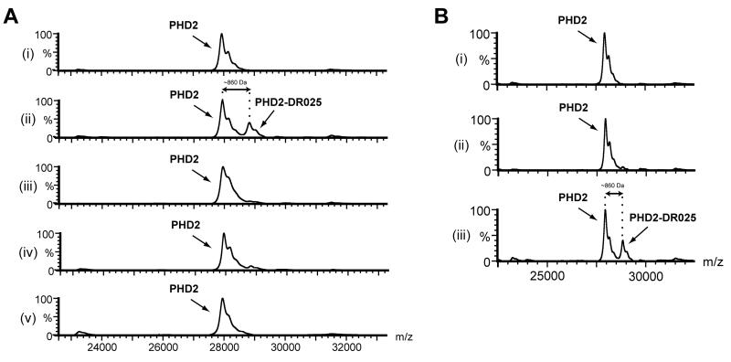
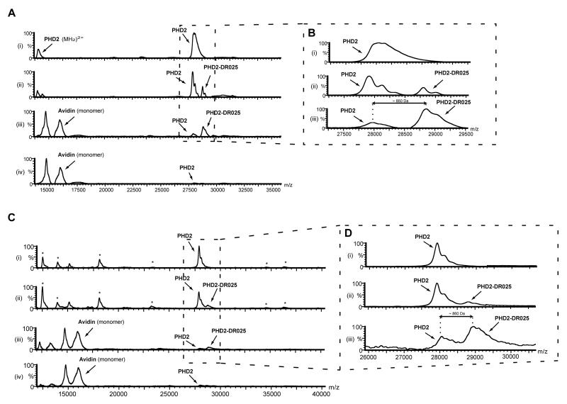
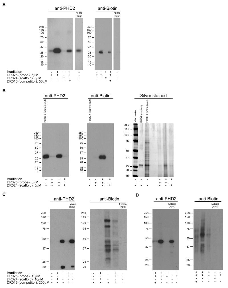
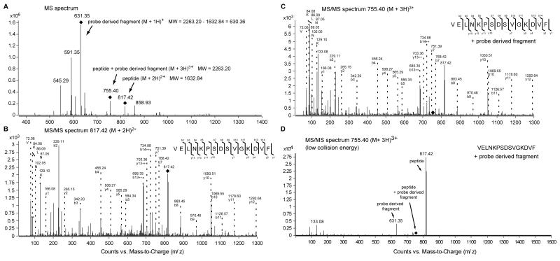
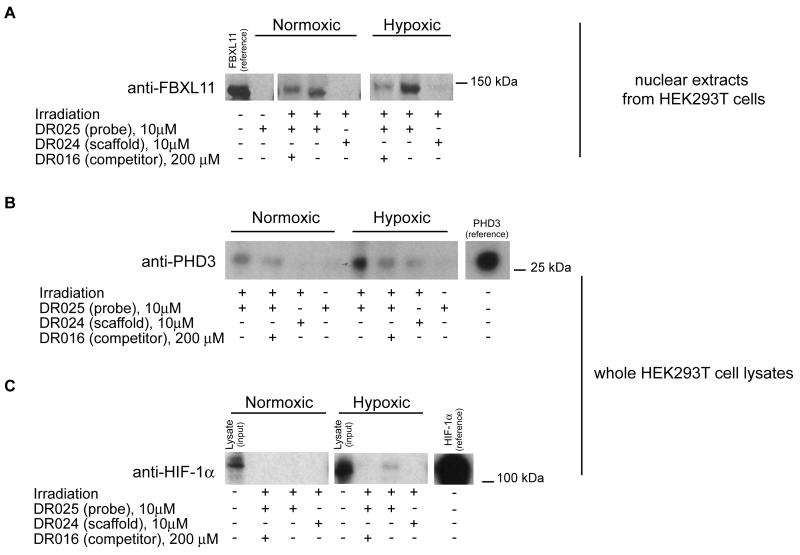
2-oxoglutarate (2-OG)-dependent oxygenases have diverse roles in human biology. The inhibition of several 2-OG oxygenases is being targeted for therapeutic intervention, including for cancer, anemia, and ischemic diseases. We report a small-molecule probe for 2-OG oxygenases that employs a hydroxyquinoline template coupled to a photoactivable crosslinking group and an affinity-purification tag. Following studies with recombinant proteins, the probe was shown to crosslink to 2-OG oxygenases in human crude cell extracts, including to proteins at endogenous levels. This approach is useful for inhibitor profiling, as demonstrated by crosslinking to the histone demethylase FBXL11 (KDM2A) in HEK293T nuclear extracts. The results also suggest that small-molecule probes may be suitable for substrate identification studies.


Figures

Figure 1
Figure 1. Cross-Linking between DR025 and PHD2 is Dependent on Irradiation and Mn(II)
(A) MALDI-MS spectra for photolabelling of PHD2181-426 by DR025. PHD2181-426 (5 μM) and DR025 (25 μM) were incubated (45 min, r.t.) in Tris buffer in the presence of Mn(II) (5 μM). After irradiation on ice (20 min, 365 nm) the resulting solutions were analyzed by MS. (i) and (ii) show results before and after irradiation; (iii) after irradiation in the presence of an inhibitor [(1-chloro-4-hydroxyisoquinoline-3-carbonyl)-amino]-acetic acid (Warshakoon et al., 2006) (25 μM) in a 1:1 concentration ratio with DR025; (iv) competition experiment with DR016 (250 μM) in a 10:1 molar ratio with DR025; (v) control with DR024 (25 μM); for selectivity and efficiency of PHD2 capture by DR025 see Figure S2. (B) Effect of Mn(II) on the efficiency of the PHD2181-426 capture by DR025. (i) and (iii) are relative to a mixture in Tris buffer of PHD2 (5 μM), DR025 (25 μM) and Mn(II) (5 μM) before and after irradiation, respectively. (ii) shows the effects of the irradiation on the same mixture without Mn(II).
Figure 2
Figure 2. Photo-Affinity Labelling and Enrichment of Purified PHD2 by DR025
(A) MALDI spectra for the cross-linking of purified PHD2181-426 by DR025. PHD2181-426 (5 μM) and DR025 (15 μM) were incubated (45 min, r.t.) in Tris buffer in the presence of Mn(II) (5 μM). After irradiation on ice (20 min, 365 nm) the solutions were divided, with one part being analyzed by MALDI and the other incubated (30 min, r.t.) with avidin-coated agarose beads. After washing (see Experimental Procedures), the beads were mixed with the MALDI matrix and analysed. (i) before irradiation in the presence of DR025; (ii) after irradiation in the presence of DR025; (iii) after irradiation and avidin-bead mediated purification; (iv) after the irradiation in the presence of DR024 at the same concentration (15 μM) as DR025 and affinity purification. For the identification of the PHD2 region covalently cross-linked by DR025 see Figure 5 and the text. (B) Magnification of the PHD2181-426 region of spectra in (A). (C) MALDI spectra of the cross-linking of PHD2181-426 by DR025 in the presence of HEK293T cell lysates. PHD2181-426 (5 μM, 5.2 μg), Mn(II) (5 μM) and DR025 (15 μM) were incubated (45 min, r.t.) in Tris buffer in the presence of HEK293T cell lysates (~ 50 μg total protein). After UV treatment on ice (20 min, 365 nm) the resulting solutions were divided, with one part being analyzed by MALDI, and the other incubated (30 min, r.t.) with avidin-coated agarose beads. After washing, the beads were mixed with the MALDI matrix and spotted: (i) Before irradiation in the presence of DR025; (ii) after the irradiation in the presence of DR025; (iii) after irradiation and affinity purification in the presence of DR025; (iv), same as (iii) but in the presence of DR024 at the same concentration (15 μM) as DR025. (D) Magnification of the PHD2181-426 region of the spectra in (C).
Figure 3
Figure 3. Photo-Affinity Labelling and Enrichment of Purified and Endogenous Human PHD2 by DR025
(A) Western blot analysis for the capture of purified PHD2181-426 by DR025. PHD2181-426 (5 μM, 5.2 μg) and DR025 (5 μM) were incubated in the presence of Mn(II) (5 μM) at r.t.. After irradiation and purification by streptavidin-coated beads, proteins were separated by SDS-PAGE and analyzed by anti-PHD2 (polyclonal antibody from rabbit, left panel) and anti-biotin (right panel) immunoblotting. The input purified PHD2181-426 was ~ 4% of the protein corresponding to the other lanes. For an analogous experiment with JMJD2E see Figure S3A. (B) Immunoblotting and silver staining analysis showing capture of purified PHD2181-426 by DR025 in HEK293T cell lysates. The anti-PHD2 (polyclonal antibody from rabbit, left panel), the anti-biotin (central panel) and the silver stained SDS-PAGE gel (right panel) are relative to an experiment carried out under the same conditions as in (A) but in the presence of the HEK293T cell lysates (~ 43 μg total proteins). The input lysate supplemented with recombinant PHD2181-426 was ~ 4% of the total protein corresponding to the other lanes; the purified PHD2181-426 as standard was ~ 4% of the protein corresponding to the other lanes. For an analogous experiment with JMJD2E see Figure S3B. (C) Cross-linking with full length PHD2 in HEK293T cell lysates over-expressing full length PHD2. DR025 (10 μM) was incubated (45 min, r.t.) with an HEK293T cell lysate over-expressing human full length PHD2 (~ 40 μg total protein). After UV treatment (20 min, 365 nm) and purification by mean of avidin-coated agarose beads, proteins released from the beads were analyzed by SDS-PAGE and Western blots using anti-PHD2 (monoclonal antibody from mouse, left panel) and anti-biotin (right panel) antibodies. The input lysate over-expressing full length PHD2 was ~ 2% of the total protein corresponding to the other lanes. (D) Cross-linking of endogenous full length PHD2 in HEK293T cell lysates (i.e. not over-expressing PHD2). Conditions are as in (C) but employing HEK293T cell lysates (~ 200 μg total protein) not over-expressing PHD2. The input lysate was ~ 3% of the total protein corresponding to the other lanes.
Figure 4
Figure 4. Identification by MS of the PHD2 Peptide Sequence Covalently Cross-linked by the Probe
(A) MS spectrum of PHD2181-426 covalently cross-linked by DR025. PHD2181-426 (5 μM), Mn(II) (5 μM) and DR025 (25 μM) were incubated (45 min, r.t.) in Tris buffer. After irradiation (20 min, 365 nm) and purification by the means of streptavidin beads, photo cross-linked PHD2181-426 was digested on the beads by trypsin and analysed by LC-MS/MS. (B) MS/MS spectrum of the doubly charged precursor ion at m/z = 817.42 Da. The doubly charged precursor ion at m/z = 817.42 Da was identified as the peptide VELNKPSDSVGKDVF which represents the C-terminal region of PHD2181-426. The b- and y-fragment ion series, and the detected immoium ions are shown. (C) MS/MS spectrum of the triply charged precursor ion at m/z = 755.40 Da. The triply charged precursor ion at m/z = 755.40 Da exhibits the same fragmentation pattern as the co-eluting precursor at m/z = 817.42 Da with additional ions in the low molecular mass range. The b- and y-fragment ion series, and the detected immoium ions are shown as in (B). (D) MS/MS spectrum of the triply charged precursor ion at m/z = 755.40 Da with reduced collision energy. Under this conditions the triply charged precursor loses a singly charged fragment at m/z = 631.35 Da which generates the doubly charged precursor at m/z = 817.42 Da (MW = 1632.84 Da). For the MS/MS spectra in the low molecular range of the precursor ion masses associated to the cross-linked PHD2 see Figure S4.
Figure 5
Figure 5. Photo-Affinity Cross-Linking Experiments in Lysates from HEK293T Cells Grown under Normoxic and Hypoxic Conditions
(A) Cross-linking of endogenous FBXL11 (KDM2A) in nuclear protein extracts of HEK293T cells. DR025 (10 μM) was incubated (45 min, r.t.) with nuclear extracts (~ 200 μg total protein) of HEK293T cells grown under normoxic (left) and hypoxic (right) conditions. After irradiation (20 min, 365 nm) and purification by streptavidin beads, the proteins released were analyzed by SDS-PAGE and subsequent anti-FBXL11 (polyclonal antibody from rabbit) immunoblotting. The reference protein was a nuclear protein extract of HEK293T cells over-expressing FBXL11 (KDM2A). For the entire immunoblot see Figure S5A. (B) Cross-linking of PHD3 at endogenous levels in HEK293T cell lysates. DR025 (10 μM) was incubated (45 min, r.t.) with cell lysate (~ 200 μg total protein) of HEK293T cells grown under normoxic (left side) and hypoxic (right side) conditions. After irradiation (20 min, 365 nm) and purification by streptavidin beads, released proteins were analyzed by SDS-PAGE and anti-PHD3 (monoclonal antibody from mouse) immunoblotting. The protein reference was a cell lysate (RCC4) over-expressing PHD3. For the entire immunoblot see Figure S5B. (C) Identification of endogenous HIF-1α in HEK293T cell lysates. DR025 (10 μM) was incubated (45 min, r.t.) with a whole lysate (~ 200 μg total protein) of HEK293T cells grown under normoxic (left) and hypoxic (1% O2) (right) conditions. After irradiation (20 min, 365 nm) and purification by streptavidin beads, released proteins were analyzed by SDS-PAGE and anti-HIF-1α (monoclonal antibody from mouse) immunoblotting. The input HEK293T lysate was ~ 3% of the total protein corresponding to the other lanes; the protein reference was a cell lysate (RCC4) over-expressing HIF-1α. For the entire immunoblot see Figure S5C.

Comment in

References

    1. Appelhoff RJ, Tian YM, Raval RR, Turley H, Harris AL, Pugh CW, Ratcliffe PJ, Gleadle JM. Differential function of the prolyl hydroxylases PHD1, PHD2, and PHD3 in the regulation of hypoxia-inducible factor. J. Biol. Chem. 2004;279:38458–38465. - PubMed
    1. Chowdhury R, McDonough MA, Mecinovic J, Loenarz C, Flashman E, Hewitson KS, Domene C, Schofield CJ. Structural basis for binding of hypoxia-inducible factor to the oxygen-sensing prolyl hydroxylases. Structure. 2009;17:981–989. - PubMed
    1. Epstein ACR, Gleadle JM, McNeill LA, Hewitson KS, O’Rourke J, Mole DR, Mukherji M, Metzen E, Wilson MI, Dhanda A, et al. C. elegans EGL-9 and Mammalian Homologs Define a Family of Dioxygenases that Regulate HIF by Prolyl Hydroxylation. Cell. 2001;107:43–54. - PubMed
    1. Evans MJ, Cravatt BF. Mechanism-based profiling of enzyme families. Chem. Rev. 2006;106:3279–3301. - PubMed
    1. Fisher JJ, Graebner(nee’ Baessler) OY, Dalhoff C, Michaelis S, Schrey AK, Ungewiss J, Andrich K, Jeske D, Kroll F, Glinski M, et al. Comprehensive Identification of Staurosporine-Binding Kinases in the Hepatocyte Cell Line HepG2 using Capture Compound Mass Spectrometry (CCMS) J. Proteome Res. 2010;9:806–817. - PubMed

Publication types

MeSH terms