. 2011 Sep 1;39(16):7209-22.
doi: 10.1093/nar/gkr383. Epub 2011 May 23.

Depurination of Brome mosaic virus RNA3 inhibits its packaging into virus particles

Affiliations

Depurination of Brome mosaic virus RNA3 inhibits its packaging into virus particles

Rajita A Karran et al. Nucleic Acids Res. .

Abstract

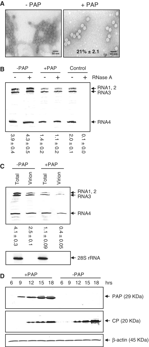
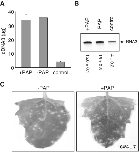
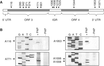
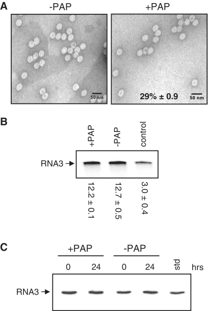
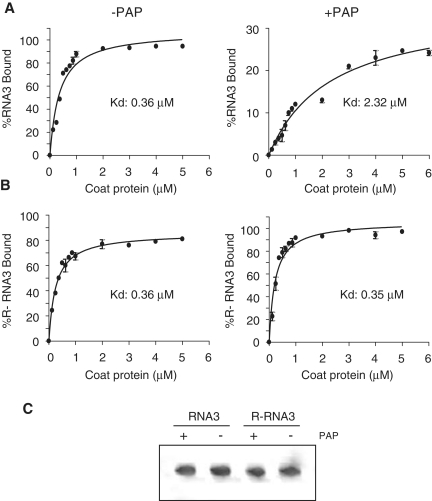
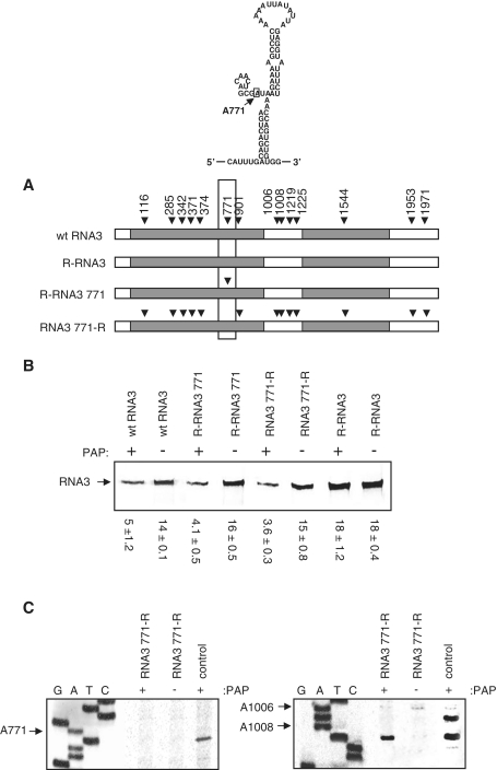
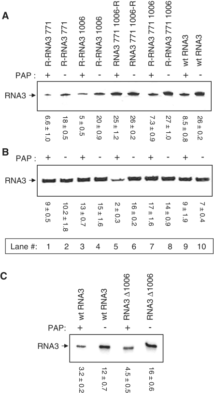
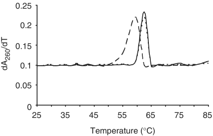
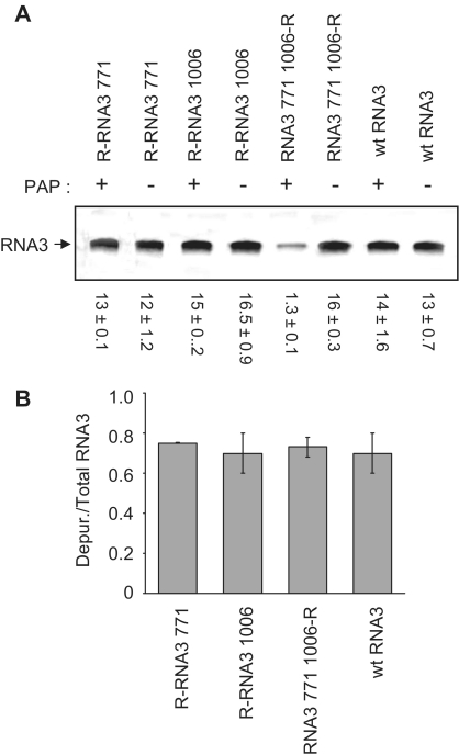
Packaging of the segmented RNA genome of Brome mosaic virus (BMV) into discrete particles is an essential step in the virus life cycle; however, questions remain regarding the mechanism of RNA packaging and the degree to which the viral coat protein controls the process. In this study, we used a plant-derived glycosidase, Pokeweed antiviral protein, to remove 14 specific bases from BMV RNA3 to examine the effect of depurination on virus assembly. Depurination of A771 within ORF3 and A1006 in the intergenic region inhibited coat protein binding and prevented RNA3 incorporation into particles. The disruption of interaction was not based on sequence identity, as mutation of these two purines to pyrimidines did not decrease coat protein-binding affinity. Rather, we suggest that base removal results in decreased thermodynamic stability of local RNA structures required for packaging, and that this instability is detected by coat protein. These results describe a new level of discrimination by coat protein, whereby it recognizes damage to specific viral RNA elements in the form of base removal and selects against incorporating the RNA into particles.


Figures

Figure 1.
Figure 1.
PAP decreases the production of virus from protoplasts. Barley protoplasts were transfected with a plasmid encoding the PAP gene (+PAP) or empty plasmid (−PAP), plus BMV RNA1, 2 and 3 in vitro transcripts and incubated for 18 h. (A) Electron micrographs of virions isolated from PAP-expressing (right) and wild-type protoplasts (left). Viral particles were visualized by staining with uranyl acetate and viewing at 100 000×. The scale bar represents 50 nm. Percentage value indicates the mean number of particles from PAP-expressing cells relative to wild-type cells ± SE from five fields of view. (B) Viral RNAs were purified from equal volume (20 µl) of the isolated viral particles and analyzed by northern blot for the presence of positive-strand BMV RNAs. To ensure that no unincorporated BMV RNAs were included, a portion of the pelleted virion fraction was resuspended and treated with RNase A or left untreated. Control indicates BMV RNAs isolated from virions treated or untreated with RNase A. Values are means of intensities for BMV RNAs ± SE for three separate experiments. (C) Northern blot analysis indicating the levels of total BMV RNAs from protoplast cell lysates and from virus particles isolated from PAP-expressing or wild-type cells, 18 h after transfection. Total RNA from cell lysates was also probed for 28S rRNA as a loading control. Values are means of intensities for BMV RNAs ± SE for three separate experiments. (D) Immunoblot analysis of total cell lysate from protoplasts transfected with a PAP cDNA and in vitro transcripts of BMV RNA1, 2 and 3 over time. Zero hours represent the time at transfection and 18 h represent the time of cell harvest. Equal amounts of total protein (10 µg) were separated by 12% SDS–PAGE, transferred to nitrocellulose and probed with an antibody to PAP (1:5000), BMV CP (1:5000) or β-actin (1:5000).
Figure 2.
Figure 2.
PAP does not alter the quality of virus from protoplasts. Barley protoplasts were transfected with a plasmid encoding the PAP gene (+PAP) or empty plasmid (−PAP), plus BMV RNA1, 2 and 3 in vitro transcripts and incubated for 18 h. Virion particles were isolated from cells and BMV RNAs associated with particles were separated through 7 M urea/4.5% acrylamide gel. Gel purified RNA3 (0.5 µg) was reverse transcribed (RT) and used for quantitative PCR (A) and radioactive PCR (B). Quantitative PCR results are plotted as microgram amount of DNA product. The control lane represents in vitro PAP-treated RNA3 used for RT–PCR. Values of radioactive PCR are means of intensities for RNA3 ± SE for three separate experiments. (C) Virion particles were isolated from PAP-expressing and wild-type protoplasts and equal amounts (15 µg) were used to infect Chenopodium quinoa plants. Infectivity of viral particles was assessed by counting the number of lesions, 9 days post infection. Percentage value indicates the mean number of lesions on leaves of plants infected with particles taken from PAP-expressing barley protoplasts (+PAP), relative to particles isolated from wild-type protoplasts (–PAP) ± SE from five leaves.
Figure 3.
Figure 3.
PAP depurinates BMV RNA3 in vivo. (A) Schematic of BMV RNA3 illustrating the sites of depurination by their nucleotide number. The 5′ untranslated region (5′-UTR; 1–91 nt), the open reading frame of RNA3 (ORF3; 92–1003 nt), the intergenic region (IGR; 1004–1246 nt), the open reading frame for RNA4 (ORF4; 1247–1813 nt) and the 3′ untranslated region (3′-UTR; 1814–2113 nt) are also indicated. (B) Representative depurination sites as determined by primer extension. Barley protoplasts were transfected with a plasmid encoding the PAP gene (+PAP) or an empty plasmid (−PAP) along with wild-type BMV RNA1, 2 and 3 in vitro transcripts. Following incubation for 18 h, RNA3 was gel purified from total protoplast RNA and 1.0 µg was extended with reverse transcriptase using radiolabeled cDNA primers distributed along the length of RNA3. cDNA products were separated in a 7 M urea/6% acrylamide gel and bands were visualized using a phosphorimager. The bases depurinated by PAP are indicated with arrows and nucleotide numbers. Deoxynucleotide sequencing of BMV DNA3 was conducted using the same primers to identify the depurination sites.
Figure 4.
Figure 4.
Depurination inhibits packaging of RNA3 in vitro. (A) Electron micrographs of particles assembled in vitro from isolated CP with either PAP-treated (right) or untreated RNA3 (left). Particles were visualized by staining with uranyl acetate and viewing at 100 000×. The scale bar represents 50 nm. Percentage value indicates the mean number of particles from PAP-treated RNA3 samples relative to untreated samples ± SE from five fields of view. (B) Viral RNAs were isolated from in vitro assembled particles and gel purified RNA3 (0.5 µg) was used for reverse transcription and radioactive PCR. The control lane represents in vitro PAP-treated RNA3 used for RT–PCR. Values are means of intensities for RNA3 ± SE for three separate experiments. (C) Northern blot analysis of RNA3 treated with PAP (+PAP) or buffer alone (−PAP), before its assembly (0 h) and isolated from in vitro particles 24 h following assembly. In vitro transcript of RNA3 was loaded into one lane to serve as a size marker (std).
Figure 5.
Figure 5.
Depurination inhibits binding of CP to RNA3. (A) Binding curves of CP with PAP-treated and untreated radiolabeled wild-type BMV RNA3 and (B) PAP-resistant RNA3 (R-RNA3). Transcripts were incubated with increasing concentrations of BMV CP before passing through a nitrocellulose membrane. The amount of retained RNA3 was quantified by scintillation counting and corrected by subtracting background counts in the absence of CP. Points are means ± SE for three separate experiments. The Kd values were obtained after results were fitted to the one site binding equation using Graphpad Prism 4. (C) Radiolabeled wild-type RNA3 (RNA3) and RNA3 resistant to depurination (R-RNA3) were treated with PAP (+PAP) or buffer alone (−PAP). The RNAs were separated through 7 M urea/6% acrylamide gel and visualized with a phosphorimager prior to incubation with CP.
Figure 6.
Figure 6.
Depurination of A771 inhibits packaging of RNA3. RNA3 constructs were created by site-directed mutagenesis: one that cannot be depurinated by PAP (R-RNA3), one that can be depurinated by PAP only at nucleotide A771 (R-RNA3 771), and RNA3 that cannot be depurinated at A771 only but can be depurinated at other sites (RNA3 771-R). (A) Schematics indicating the sites of depurination for wild-type RNA3, R-RNA3, R-RNA3 771 and RNA3 771-R. An ∼200-nt region of RNA3 is boxed and the modeled secondary structure of a portion containing A771 is shown above. (B) Wt RNA3, R-RNA3 771, RNA3 771-R and R-RNA3 were treated with either PAP or buffer only and used for in vitro particle assembly assays with isolated CP. Viral RNA3 was isolated from equal volume of assembled particles and analyzed by northern blot for the presence of positive-strand BMV RNA3. Values are means of intensities for RNA3 ± SE for three separate experiments. (C) Primer extension analysis of RNA3 771-R isolated from virions. cDNA primers distributed over the length of the viral RNA3 were annealed and extended with reverse transcriptase. Radiolabeled cDNA products were separated in a 7 M urea/6% acrylamide gel and visualized with a phosphorimager. The control lane on the right panel represents PAP-treated RNA3 771-R transcript prior to virion assembly, and on the left panel control represents wild-type RNA3 treated with PAP, to illustrate the location of A771 depurination. The bases depurinated by PAP are indicated with arrows and nucleotide numbers. Deoxynucleotide sequencing of BMV DNA3 was conducted using the same primers to identify the depurination sites.
Figure 7.
Figure 7.
Effect of depurination and deletion of A1006 on packaging efficiency. RNA3 constructs, R-RNA3 771, R-RNA3 1006, RNA3 771 1006-R and R-RNA3 771 1006 were created by site directed mutagenesis. Transcripts of these four RNA3 mutants and wild-type RNA3 were treated with PAP or buffer only. PAP-treated and untreated RNA3 were used to assemble particles in vitro with isolated CP. (A) RNA was isolated from equal volume of in vitro assembled particles and analyzed by northern blot for the presence of positive-strand BMV RNA3. Values are means of intensities for RNA3 ± SE for three separate experiments. (B) Equal amounts of gel purified RNA3 (0.5 µg) from in vitro assembled particles were used for reverse transcription and radioactive PCR. Values are means of intensities for RNA3 ± SE for three separate experiments. (C) The deletion construct RNA3 Δ1006, missing A1006 from RNA3, was created by site-directed mutagenesis and its in vitro transcript was treated with PAP or buffer alone. PAP-treated and untreated RNA3 were used to assemble particles in vitro with isolated CP. RNA was isolated from equal volume of assembled particles and analyzed by northern blot for the presence of positive-strand BMV RNA3. Values are means of intensities for RNA3 ± SE for three separate experiments.
Figure 8.
Figure 8.
CP interaction with RNA3 fragments depurinated at A771 and A1006. (A) Schematics showing regions of RNA3 (R-RNA3 771 1006, depurinated only at A771 and A1006) tested for their binding affinity to isolated CP by filter binding assay and EMSA. RNA3 fragments were treated with PAP or buffer only and incubated with increasing concentration of BMV CP before passing through a nitrocellulose membrane. The amount of retained RNA was quantified by scintillation counting and corrected by subtracting background counts in the absence of CP. The Kd values were obtained after results were fitted to the one site binding equation using Graphpad Prism 4, and appear in the table to the right. (B) Radiolabeled RNA3 fragments were incubated with PAP or buffer only, followed by incubation with BMV CP. Samples were separated through a 6% acrylamide non-denaturing gel and the bands were visualized using a phosphorimager.
Figure 9.
Figure 9.
Thermal stability of RNA3 fragment depurinated at A771. The 162-nt cDNA constructs of RNA3 containing either A771 (wt cDNA3 fragment) or this nucleotide mutated to T771 (R-cDNA3 fragment) were produced by PCR. In vitro transcripts of these cDNAs were treated with PAP or buffer alone. The melting temperatures of the RNA fragments were measured by UV spectrophotometry. The first derivative of the absorbance at 260 nm was plotted as a function of temperature. Wild-type RNA3 fragment is represented by a continuous line, PAP-treated wild-type RNA3 fragment by long broken lines and R-RNA3 fragment by short broken lines. The absorbance of each fragment was measured independently and values are means of each ± SE for three separate experiments.
Figure 10.
Figure 10.
Effect of depurination at A771 and A1006 on the quality of RNA3 packaged in vivo. In vitro transcripts of RNA1, 2 and either wild-type or mutant RNA3 were transfected into barley protoplasts along with a plasmid encoding the PAP gene (+PAP) or empty plasmid (−PAP) and allowed to replicate for 18 h before viral particles were isolated. (A) RNAs were isolated from viral particles and equal amounts of gel purified RNA3 (0.5 µg) were used for reverse transcription and radioactive PCR. Values are means of intensities for RNA3 ± SE for three separate experiments. (B) Total RNA was isolated from protoplasts, RNA3 was gel purified and reverse transcribed in the presence of [α33P]-dATP. The intensities of truncated bands, due to depurination, were plotted relative to total RNA3. Bars represent mean percent depurination ± SE for three separate experiments of wtRNA3 and each mutant.

Similar articles

Cited by

References

    1. Schmitz I, Rao AL. Molecular studies on bromovirus capsid protein. I. Characterization of cell-to-cell movement-defective RNA3 variants of brome mosaic virus. Virology. 1996;226:281–293. - PubMed
    1. Goldbach R, LeGall O, Wellink J. Alpha-like viruses in plants. Semin. Virol. 1991;2:19–25.
    1. Ahlquist P. Bromovirus RNA replication and transcription. Curr. Opin. Genet. Dev. 1992;2:71–76. - PubMed
    1. Ahola T, den Boon JA, Ahlquist P. Helicase and capping enzyme active site mutations in brome mosaic virus protein 1a cause defects in template recruitment, negative-strand RNA synthesis, and viral RNA capping. J. Virol. 2000;74:8803–8811. - PMC - PubMed
    1. Kao CC, Quadt R, Hershberger RP, Ahlquist P. Brome mosaic virus RNA replication proteins 1a and 2a from a complex in vitro. J. Virol. 1992;66:6322–6329. - PMC - PubMed

Publication types