

Case Reports
doi: 10.1503/cmaj.091521.
Epub 2011 May 24.
Tinnitus: identifying the ominous causes
Affiliations
- PMID: 21609995
- PMCID: PMC3255117
- DOI: 10.1503/cmaj.091521
Item in Clipboard
Case Reports
Tinnitus: identifying the ominous causes
CMAJ.
.
No abstract available
Figures
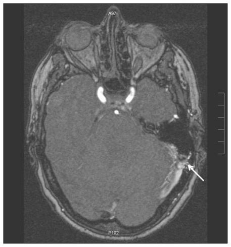
Magnetic resonance image of the head of a 67-year-old woman with pulsatile tinnitus. A dural arteriovenous fistula (arrow) and dural enhancement consistent with venous hypertension can be seen on the patient’s left side.
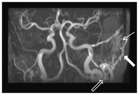
Magnetic resonance angiogram of the patient’s cranial circulation. A dural arteriovenous fistula with prominent blood supply from the left occipital artery can be seen (thick white arrow). An enlarged external carotid artery (black arrow) and signal within the transverse sinus at the level of the fistula (thin white arrow) are also evident.
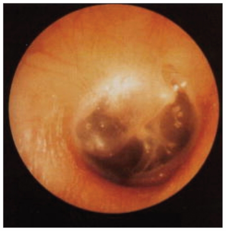
A high-riding jugular bulb, appearing during otoscopy as a purplish mass behind the intact tympanic membrane, in the right ear of a 12-year-old boy who presented with pulsatile tinnitus. Reprinted with permission from Laryngoscope 1997;107:321–7. Copyright © 1997 John Wiley & Sons, Inc.

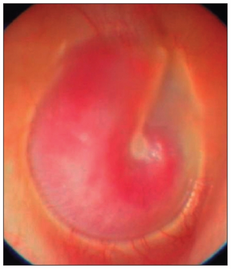
An aberrant carotid artery appearing during otoscopy as a curved reddish formation behind an intact and mobile tympanic membrane, in the right ear of a 28-year-old woman. Reprinted with permission from Arch Otolaryngol Head Neck Surg 2004;130:1120. Copyright © 2004 American Medical Association. All rights reserved.
A glomus tumour. A reddish retrotympanic mass can be seen filling the middle ear space of the right ear. Reprinted from Acta Otorrinolaringol Esp 2007;58:426–33. Copyright © 2007 Elsevier Limited.
References
-
- Liyanage SH, Singh A, Savundra P, et al. Pulsatile tinnitus. J Laryngol Otol 2006;120:93–7 - PubMed
-
- Crummer RW, Hassan GA. Diagnostic approach to tinnitus. Am Fam Physician 2004;69:120–6 - PubMed
-
- Sismanis A. Pulsatile tinnitus. Otolaryngol Clin North Am 2003; 36:389–402 - PubMed
-
- Biesinger E, Del Bo L, De Ridder D, et al. Algorithm for the diagnostic and therapeutic management of tinnitus. Tinnitus Clinic Network, Tinnitus Research Initiative; Available: www.tinnitusresearch.org/en/documents/downloads/TRI_Tinnitus_Flowchart.pdf (accessed 2010 Aug. 9).
-
- Lewis JE, Stephens SD, McKenna L. Tinnitus and suicide. Clin Otolaryngol Allied Sci 1994;19:50–4 - PubMed