. 2011 Jun;138(12):2555-65.
doi: 10.1242/dev.056804.

The MADS box transcription factor MEF2C regulates melanocyte development and is a direct transcriptional target and partner of SOX10

Affiliations

The MADS box transcription factor MEF2C regulates melanocyte development and is a direct transcriptional target and partner of SOX10

Pooja Agarwal et al. Development. 2011 Jun.

Abstract

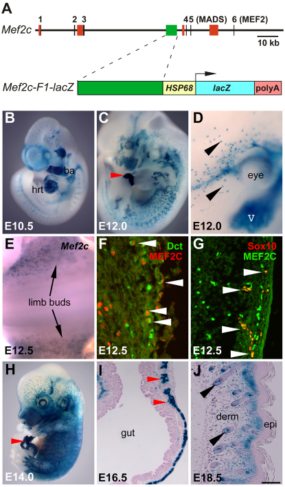
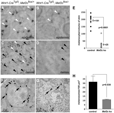
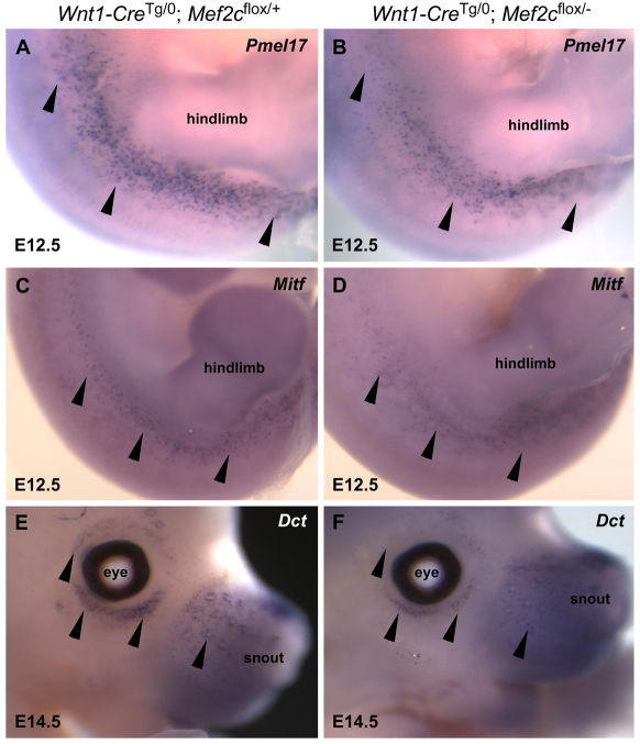
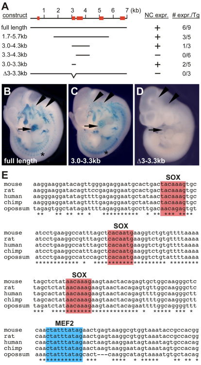
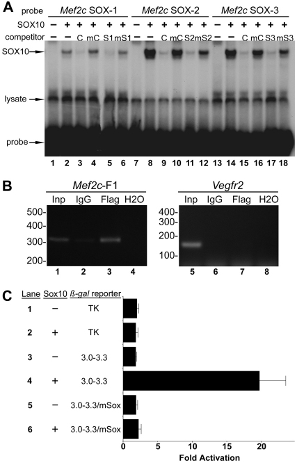
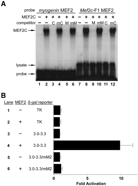
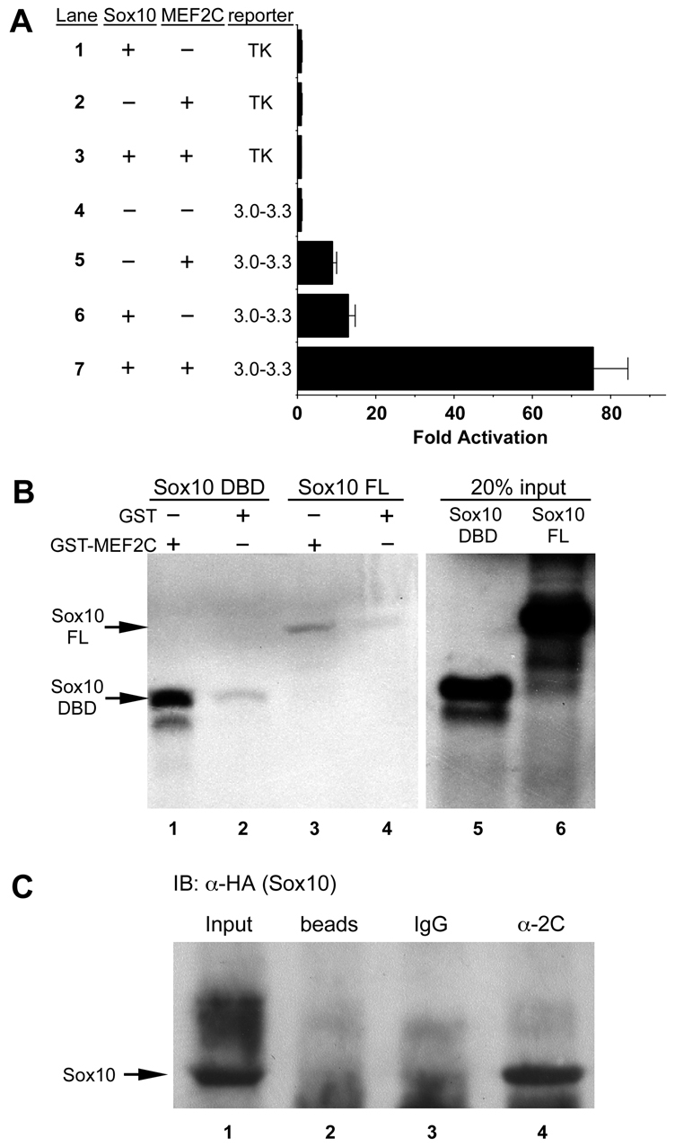
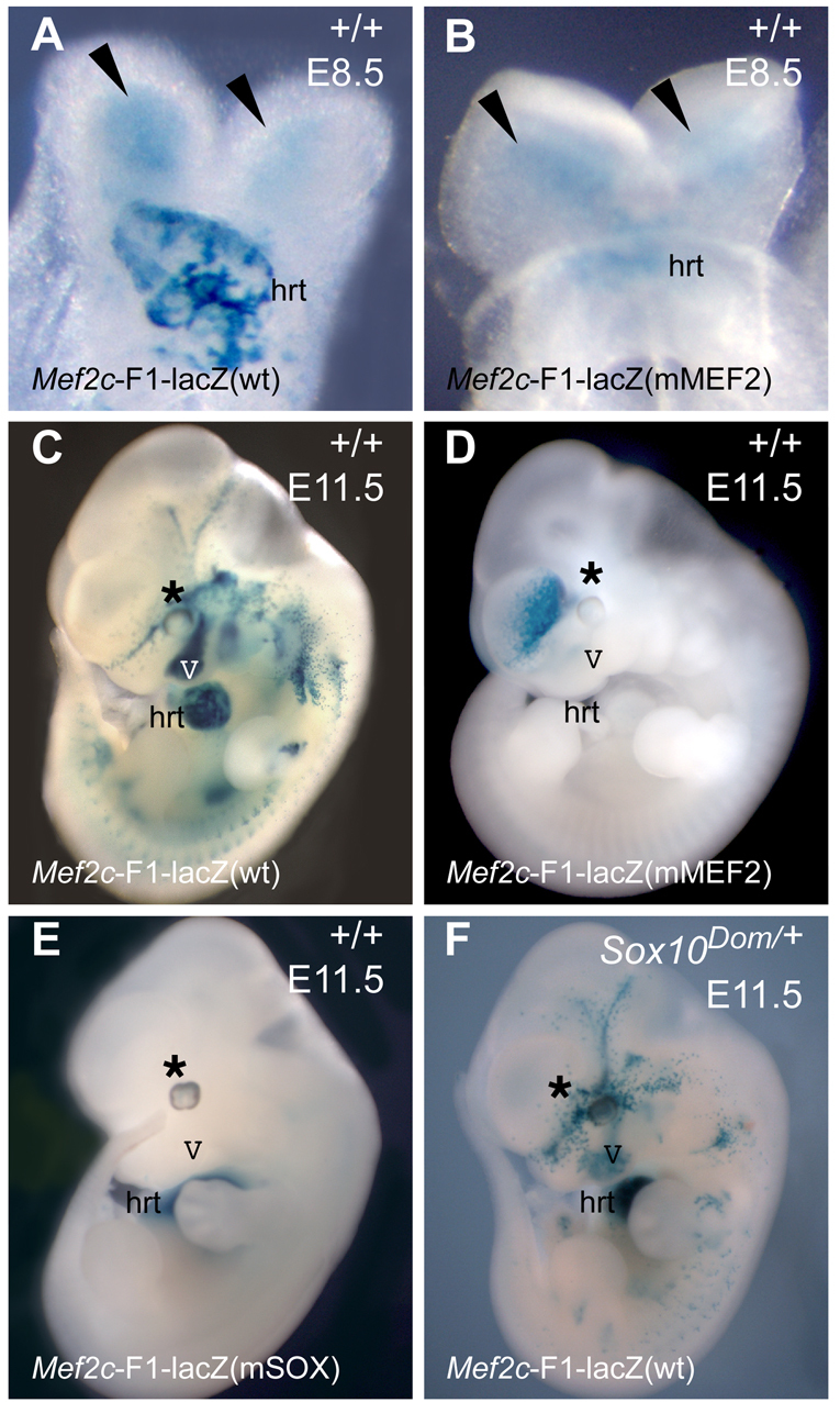
Waardenburg syndromes are characterized by pigmentation and autosensory hearing defects, and mutations in genes encoding transcription factors that control neural crest specification and differentiation are often associated with Waardenburg and related disorders. For example, mutations in SOX10 result in a severe form of Waardenburg syndrome, Type IV, also known as Waardenburg-Hirschsprung disease, characterized by pigmentation and other neural crest defects, including defective innervation of the gut. SOX10 controls neural crest development through interactions with other transcription factors. The MADS box transcription factor MEF2C is an important regulator of brain, skeleton, lymphocyte and cardiovascular development and is required in the neural crest for craniofacial development. Here, we establish a novel role for MEF2C in melanocyte development. Inactivation of Mef2c in the neural crest of mice results in reduced expression of melanocyte genes during development and a significant loss of pigmentation at birth due to defective differentiation and reduced abundance of melanocytes. We identify a transcriptional enhancer of Mef2c that directs expression to the neural crest and its derivatives, including melanocytes, in transgenic mouse embryos. This novel Mef2c neural crest enhancer contains three functional SOX binding sites and a single essential MEF2 site. We demonstrate that Mef2c is a direct transcriptional target of SOX10 and MEF2 via this evolutionarily conserved enhancer. Furthermore, we show that SOX10 and MEF2C physically interact and function cooperatively to activate the Mef2c gene in a feed-forward transcriptional circuit, suggesting that MEF2C might serve as a potentiator of the transcriptional pathways affected in Waardenburg syndromes.


Figures

Fig. 1.
Fig. 1.
Identification of Mef2c-F1, a neural crest enhancer from Mef2c. (A) The top line represents the 5′ end of the mouse Mef2c locus. Vertical black lines depict Mef2c exons. Colored rectangles represent known enhancers. A schematic of the Mef2c-F1-lacZ transgene is shown below. (B-J) Whole-mount (B-E,H) or transverse sections (F,G,I,J) of X-gal-stained Mef2c-F1-lacZ transgenic (B-D,H-J) or wild-type embryos examined by in situ hybridization for Mef2c (E) or by immunofluorescence using antibodies against MEF2C (red in F, green in G), DCT (green in F) and SOX10 (red in G). Black or white arrowheads mark developing melanocytes. Red arrowheads mark peripheral nerves in the gut. V, fifth cranial nerve; ba, branchial arch; derm, dermis; epi, epidermis; hrt, heart. Scale bar: 100 μm.
Fig. 2.
Fig. 2.
Mef2c is required for melanocyte development in mice. (A-D) Whole-mount DOPA-stained epidermis (A,B) or dermis (C,D) from Wnt1-CreTg/0; Mef2cflox/+ control (A,C) and Wnt1-CreTg/0; Mef2cflox/− neural crest knockout (NC KO; B,D) neonatal mice showed many fewer stained follicular (white arrowheads) and interfollicular (black arrowheads) melanocytes in Mef2c NC KO skin compared with controls. (E) Quantification of DOPA-stained melanocytes from Wnt1-CreTg/0; Mef2cflox/+ control and Wnt1-CreTg/0; Mef2cflox/− NC KO epidermis. Filled circles represent individual control mice (191±16 DOPA-stained melanocytes per sample, n=7) and open circles represent individual Mef2c NC KO mice (25±10 DOPA-stained melanocytes per sample, n=9), P<0.0001. (F,G) Electron micrographs of DOPA-stained melanocytes from Wnt1-CreTg/0; Mef2cflox/+ control mice (F) and Wnt1-CreTg/0; Mef2cflox/− NC KO mice (G) showed that Mef2c NC KO mice had fewer melanosomes per melanocyte (black arrows). (H) Quantification of melanosomes in individual DOPA-stained control and Mef2c NC KO neonates indicated the mean number of melanosomes in 100 μm2 for control skin was 31.7±6.8; n=3 mice. The mean number of melanosomes in 100 μm2 within stained melanocytes in Mef2c NC KO neonates was 11.0±0.6; n=3 mice (P=0.038). An average of six randomly selected melanocytes from each neonatal mouse was scored. Data are expressed as the mean number of melanosomes in 100 μm2 + s.e.m.
Fig. 3.
Fig. 3.
Mef2c is required for normal expression of melanocyte marker genes during development. (A-F) Whole-mount in situ hybridization for Pmel17 (A,B), Mitf (C,D) and Dct (E,F) transcripts from Wnt1-CreTg/0; Mef2cflox/− conditional knockout embryos (B,D,F) showed reduced expression of each of the melanocyte markers during embryonic development compared with expression in Wnt1-CreTg/0; Mef2cflox/+ control embryos (A,C,E). Black arrowheads point to melanocytes in the trunk region at E12.5 and in the sub- and supra-optic region at E14.5.
Fig. 4.
Fig. 4.
A minimal fragment of the Mef2c-F1 neural crest enhancer is necessary and sufficient for enhancer activity and contains consensus binding sites for MEF2 and SOX10. (A) Schematic of the Mef2c-F1 enhancer region and the deletion constructs tested for enhancer activity in transgenic mice. Red rectangles depict regions of high conservation between the marsupial opossum and the mouse genomic sequences. The column on the left lists the regions of the Mef2c-F1 fragment tested in deletion analyses. The columns on the right denote the activity and the fraction of independently derived transgenic (Tg) embryos that displayed activity in neural crest (NC) derivatives. (B-D) Representative transgenic embryos for the indicated constructs. Asterisks denote transgene expression in neural crest-derivatives in the trunk. Arrows denote the location of the heart. Arrowheads indicate regions of melanocyte progenitors. (E) ClustalW analysis of Mef2c gene sequences in the core region of the 300-bp necessary and sufficient region of the Mef2c-F1 enhancer. Asterisks denote conserved nucleotides.
Fig. 5.
Fig. 5.
SOX10 binds and activates the Mef2c-F1 enhancer in a SOX site-dependent manner. (A) In vitro-translated SOX10 was incubated with the following Mef2c-F1 radiolabeled probes: SOX-1 (lanes 1-6), SOX-2 (lanes 7-12) and SOX-3 (lanes 13-18). SOX10 efficiently retarded the mobility of each of the sites (lanes 2, 8, 14). Binding was competed by excess unlabeled control probe (C; lanes 3, 9, 15) but not by a mutant version (mC; lanes 4, 10, 16). Binding was also competed by excess unlabeled self probe (S1, lane 5; S2, lane 11; S3, lane 17) but not by excess mutant versions (mS1, lane 6; mS2, lane 12; mS3, lane 18). Unprogrammed reticulocyte lysates are shown in lanes 1, 7 and 13. Lysate-derived, non-specific bands and the free, unbound probe are indicated. (B) Sox10[Flag]-transfected B16F10 mouse melanoma cells were subjected to ChIP to detect SOX10 bound to the endogenous Mef2c-F1 enhancer using an anti-FLAG antibody. Following ChIP, the region of the Mef2c gene surrounding the SOX sites in the Mef2c-F1 enhancer (lanes 1-4), or a region of the Vegfr2 enhancer (lanes 5-8) as a non-specific control, was amplified by PCR and analyzed by agarose gel electrophoresis. Lanes 1 and 5: amplification prior to immunoprecipitation; lanes 2 and 6: amplification following non-specific isotype-matched IgG ChIP; lanes 3 and 7: amplification following ChIP with anti-FLAG to detect SOX10-bound DNA; lanes 5 and 8: amplification without added template (H20). Sizes in bp are shown. (C) Co-transfection of the Mef2c[3-3.3]-F1-TK-lacZ reporter plasmid with pRK5-Sox10 expression plasmid (+) or with pRK5 (−) resulted in strong activation in a SOX10-dependent manner (lanes 3, 4). Mutation of all three SOX sites abolished the ability of SOX10 to transactivate the Mef2c-F1 enhancer (lane 6). SOX10 did not transactivate the parental TK-lacZ reporter (lane 2). Data are expressed as the mean fold activation + s.e.m. from three independent transfections and analyses.
Fig. 6.
Fig. 6.
MEF2C binds the MEF2 site in the Mef2c-F1 enhancer. (A) In vitro translated MEF2C was incubated with radiolabeled probes corresponding to a consensus bona fide MEF2 site from the myogenin promoter (lanes 1-6) or from the Mef2c-F1 enhancer (lanes 7-12). MEF2C bound the Mef2c-F1 MEF2 (lane 8) and myogenin control MEF2 (lane 2) sites with similar affinity. Excess unlabeled Mef2c-F1 MEF2 site (M; lanes 5, 9) and myogenin control (C; lanes 3, 11) probes each competed efficiently for binding. Mutant versions of the control probe (mC) or the Mef2c-F1 probe (mM) failed to compete for binding to either labeled probe (lanes 4, 6, 10, 12). Unprogrammed reticulocyte lysate was used in EMSA with each probe (lanes 1, 7) and no specific binding was observed. Lysate-derived, non-specific binding and the free, unbound probe are indicated. (B) Co-transfection of the Mef2c[3-3.3]-F1-TK-lacZ reporter plasmid with a MEF2C-VP16 activator plasmid (+) or with pRK5 (−) demonstrates that MEF2 can activate the Mef2c-F1 enhancer (lanes 3, 4). Mutation of the MEF2 site in Mef2c-F1 completely abolished activation by MEF2C-VP16 (lane 6). No activation of the parental minimal promoter-containing reporter plasmid TK-lacZ by MEF2C-VP16 was observed (lane 2). Data are expressed as the mean fold activation + s.e.m. from three independent transfections and analyses.
Fig. 7.
Fig. 7.
MEF2C and SOX10 physically interact and synergistically activate the Mef2c-F1 enhancer. (A) Co-transfection of expression plasmids for MEF2C-VP16 and SOX10 with the Mef2c[3-3.3]-F1-TK-lacZ reporter plasmid resulted in synergistic activation of the Mef2c-F1 enhancer (lane 7) compared with transfection of the reporter with MEF2C-VP16 (lane 5) or SOX10 (lane 6) alone. No activation of the reporter with empty expression vectors or of the parental TK-lacZ reporter was observed (lanes 1-4). Data are expressed as the mean fold activation + s.e.m. from three independent transfections and analyses. (B) Agarose conjugated to either GST-MEF2C (lanes 1, 3) or GST alone (lanes 2, 4) were used as bait in pull-down assays with radiolabeled DNA binding domain (DBD) of SOX10 (lanes 1, 2) or with radiolabeled full length (FL) SOX10 (lanes 3, 4). Twenty percent of the input amount of radiolabeled SOX10 DBD and SOX10 FL are included in lanes 5 and 6, respectively. (C) MEF2C and SOX10 interact in vivo in B16F10 melanoma cells. MEF2C- and SOX10[HA]-transfected cells were subjected to immunoprecipitation with anti-MEF2C followed by western blot analysis with anti-HA (SOX10). SOX10 was only detected following precipitation with anti-MEF2C (α-2C, lane 4) but not with isotype-matched IgG (lane 3) or with beads alone (lane 2). Lane 1 contains input sample only for western blot (no prior immunoprecipitation) as a positive control for SOX10[HA] detection.
Fig. 8.
Fig. 8.
The Mef2c-F1 neural crest enhancer is regulated by MEF2 and SOX10 in vivo. (A,C) The wild-type Mef2c-F1 transgene in an otherwise wild-type background (+/+) directs expression to the nascent neural crest adjacent to the neural folds at E8.5 (A) and in neural crest derivatives, including melanocytes, at E11.5 (C). The same pattern was observed in six out of nine independent transgenic lines examined at both E8.5 and E11.5. (B,D) Mutation of the MEF2 site in Mef2c-F1 completely abolished transgene expression in the neural crest and its derivatives at E11.5 (D) but not at E8.5 (B). Note that the expression in the forebrain region in D was not reproducible and represents ectopic transgene expression. Similar activity of the MEF2 mutant transgene was observed in each of three independent transgenic lines examined at E8.5 and E11.5 and an additional three F0 transient transgenic embryos examined at E11.5 only. (E) Mutation of all three SOX sites in the Mef2c-F1 enhancer abolished transgene expression in neural crest derivatives, but not in the heart (hrt), in each of five independent mSOX transgenic lines examined. (F) Crossing the wild-type Mef2c-F1-lacZ transgene into a Sox10 heterozygous (Sox10Dom/+) background resulted in a diminution of transgene expression in neural crest derivatives at E11.5 but did not affect transgene expression in the heart. Arrowheads indicate neural crest cells. Asterisks indicate melanocytes in the supraocular region at E11.5. hrt, heart; V, fifth cranial nerve.

References

    1. Amin N. M., Shi H., Liu J. (2010). The FoxF/FoxC factor LET-381 directly regulates both cell fate specification and cell differentiation in C. elegans mesoderm development. Development 137, 1451-1460. - PMC - PubMed
    1. Baker C. V., Bronner-Fraser M. (1997). The origins of the neural crest. Part I: embryonic induction. Mech. Dev. 69, 3-11. - PubMed
    1. Baxter L. L., Hou L., Loftus S. K., Pavan W. J. (2004). Spotlight on spotted mice: a review of white spotting mouse mutants and associated human pigmentation disorders. Pigment Cell Res. 17, 215-224. - PubMed
    1. Bi W., Drake C. J., Schwarz J. J. (1999). The transcription factor MEF2C-null mouse exhibits complex vascular malformations and reduced cardiac expression of angiopoietin 1 and VEGF. Dev. Biol. 211, 255-267. - PubMed
    1. Black B. L., Cripps R. M. (2010). Myocyte enhancer factor 2 transcription factors in heart development and disease. In Heart Development and Regeneration, Vol. 2 (ed. Rosenthal N., Harvey R. P.), pp. 673-699. Oxford: Academic Press.

Publication types

Supplementary concepts