. 2011 Jul 22;286(29):26258-66.
doi: 10.1074/jbc.M111.256305. Epub 2011 May 24.

An interaction between the Walker A and D-loop motifs is critical to ATP hydrolysis and cooperativity in bacteriophage T4 Rad50

Affiliations

An interaction between the Walker A and D-loop motifs is critical to ATP hydrolysis and cooperativity in bacteriophage T4 Rad50

Metzere Bierlein De la Rosa et al. J Biol Chem. .

Abstract

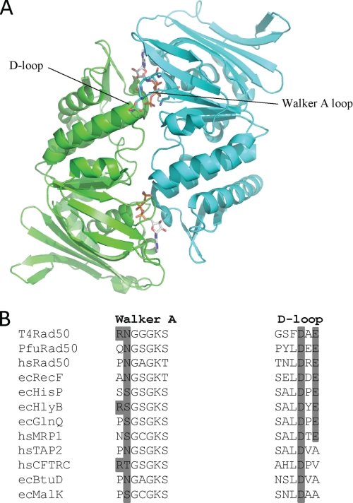
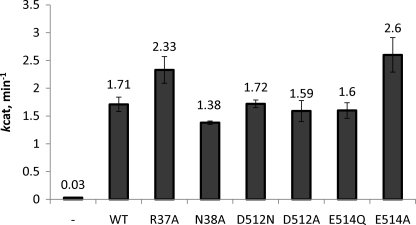
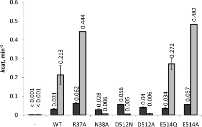
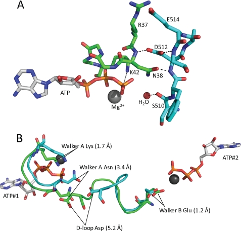
The ATP binding cassette (ABC) proteins make up a large superfamily with members coming from all kingdoms. The functional form of the ABC protein nucleotide binding domain (NBD) is dimeric with ATP binding sites shared between subunits. The NBD is defined by six motifs: the Walker A, Q-loop, Signature, Walker-B, D-loop, and H-loop. The D-loop contains a conserved aspartate whose function is not clear but has been proposed to be involved in cross-talk between ATP binding sites. Structures of various ABC proteins suggest an interaction between the D-loop aspartate and an asparagine residue located in Walker A loop of the opposing subunit. Here, we evaluate the functional role of the D-loop using a bacteriophage T4 ABC protein, Rad50 (gp46). Mutation of either the D-loop aspartate or the Walker A asparagine results in dramatic reductions in ATP affinity, hydrolysis rate, and cooperativity. The mutant proteins bind Mre11 (gp47) and DNA normally, but no longer support the ATP-dependent nuclease activities of Mre11. We propose that the D-loop aspartate functions to stabilize the Walker A asparagine in a position favorable for catalysis. We find that the asparagine is crucially important to the mechanism of ATP hydrolysis by increasing the affinity for ATP and positioning the γ-phosphate of ATP for catalysis. Additionally, we propose that the asparagine acts as a γ-phosphate sensor and, through its interaction with the conserved D-loop aspartate, transmits conformational changes across the dimer interface to the second ATP binding site.


Figures

FIGURE 1.
FIGURE 1.
A, ribbon structure of the dimeric PfuRad50 nucleotide binding domain. The subunits of the dimer (PDB code 1F2U) are colored green and cyan with the ATP molecules located at the dimer interface. The Walker A and D-loop motifs from opposing subunits that form a single ATP active site are indicated. B, multiple amino acid sequence alignment of the Walker A and D-loop motifs from selected Rad50 and ABC proteins. The protein names are preceded by the organism abbreviations, which are as follows: T4, bacteriophage T4; Pfu, P. furiosus; hs, Homo sapiens; and ec, E. coli. The dark gray shading indicates the conservation of the residues mutated in this study.
FIGURE 2.
FIGURE 2.
WT and mutant steady-state exonuclease activity on the 2nd position 2-AP DNA substrate. Exonuclease activity was measured by the release of the fluorescent nucleotide analog, 2-AP, as a function of time. The assay consisted of Rad50 (50 or 100 nm), Mre11 (53 or 105 nm), and 1.3 μm DNA using buffer conditions described under “Experimental Procedures.” The dash indicates the nuclease activity of Mre11 in the absence of Rad50. The 2-AP probe is located at the 2nd position relative to the 3′ end of the DNA substrate. The value given above the bars is the specific activity estimate averaged from four independent measurements (two measurements at each protein concentration). Error bars represent the S.D. of the averaged values.
FIGURE 3.
FIGURE 3.
WT and mutant steady-state exonuclease activity on the 17th position 2-AP DNA substrate. Exonuclease activity was measured by the release of the fluorescent nucleotide analog, 2-AP, as a function of time. The assay consisted of 400 nm Rad50, 420 nm Mre11, and 1.3 μm DNA substrate using buffer conditions described under “Experimental Procedures.” The dash indicates the nuclease activity of Mre11 in the absence of Rad50. A nuclease rate of 0.001 min−1 represents the limit of detection. The 2-AP probe is located at the 17th position relative to the 3′ end of the DNA substrate. The light and dark gray bars represent the average specific activity estimates in the presence and absence of ATP, respectively. The concentration of ATP, when included, was held at a concentration of 2 mm. The value given above the bars is the specific activity estimate averaged from three independent measurements and the error bars represent the S.D. of the averaged values.
FIGURE 4.
FIGURE 4.
A, a close up of the Walker A and D-loop residues from the T4 homology model generated using the Swiss model server and based on PDB code 1F2U. ATP, Mg2+, the putative catalytic water, and the residues relevant to this study are noted. The coloring is the same as shown in Fig. 1. Dashes represent potential hydrogen bonds. B, a structural alignment of the Walker A, D-loop, and Walker B motifs in the presence (green) and absence (cyan) of ATP. The alignment was generated with the iterative magic fit function of the DeepView software (36) using the apo and ATP-bound x-ray crystal structures of PfuRad50 (PDB codes 1F2T and 1F2U, respectively). The overall root mean square deviation for the alignment of all of the c-α carbons for the apo and ATP-bound structures was 1.2 Å. The positions of the relevant residues are indicated by black lines and the distance each residue moves between the apo and ATP-bound forms of the enzyme is indicated in the parentheses.

References

    1. Connelly J. C., Leach D. R. (2002) Trends Biochem. Sci. 27, 410–418 - PubMed
    1. Aguilera A., Gómez-González B. (2008) Nat. Rev. Genet. 9, 204–217 - PubMed
    1. Hoeijmakers J. H. J. (2007) Mech. Ageing Dev. 128, 460–462 - PubMed
    1. Borde V. (2007) Chromosome Res. 15, 551–563 - PubMed
    1. Soulas-Sprauel P., Rivera-Munoz P., Malivert L., Le Guyader G., Abramowski V., Revy P., de Villartay J. (2007) Oncogene 26, 7780–7791 - PubMed

Publication types

MeSH terms