. 2011 Jul;152(7):2870-82.
doi: 10.1210/en.2011-0219. Epub 2011 May 24.

Steroidogenic factor-1 (SF-1)-driven differentiation of murine embryonic stem (ES) cells into a gonadal lineage

Affiliations

Steroidogenic factor-1 (SF-1)-driven differentiation of murine embryonic stem (ES) cells into a gonadal lineage

Unmesh Jadhav et al. Endocrinology. 2011 Jul.

Abstract

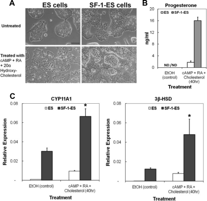
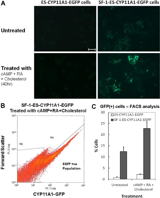
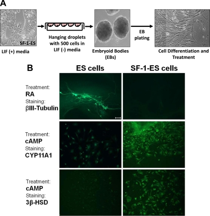
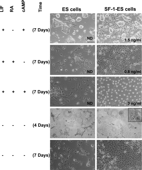
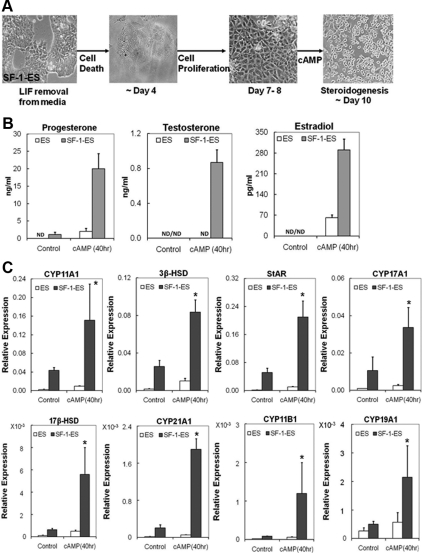
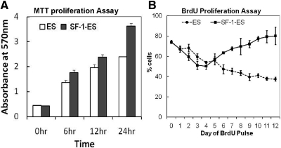
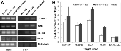
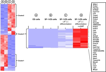
Steroidogenic factor 1 (SF-1) is essential for the development and function of steroidogenic tissues. Stable incorporation of SF-1 into embryonic stem cells (SF-1-ES cells) has been shown to prime the cells for steroidogenesis. When provided with exogenous cholesterol substrate, and after treatment with retinoic acid and cAMP, SF-1-ES cells produce progesterone but do not produce other steroids such as cortisol, estradiol, or testosterone. In this study, we explored culture conditions that optimize SF-1-mediated differentiation of ES cells into defined steroidogenic lineages. When embryoid body formation was used to facilitate cell lineage differentiation, SF-1-ES cells were found to be restricted in their differentiation, with fewer cells entering neuronal pathways and a larger fraction entering the steroidogenic lineage. Among the differentiation protocols tested, leukemia inhibitory factor (LIF) removal, followed by prolonged cAMP treatment was most efficacious for inducing steroidogenesis in SF-1-ES cells. In this protocol, a subset of SF-1-ES cells survives after LIF withdrawal, undergoes morphologic differentiation, and recovers proliferative capacity. These cells are characterized by induction of steroidogenic enzyme genes, use of de novo cholesterol, and production of multiple steroids including estradiol and testosterone. Microarray studies identified additional pathways associated with SF-1 mediated differentiation. Using biotinylated SF-1 in chromatin immunoprecipitation assays, SF-1 was shown to bind directly to multiple target genes, with induction of binding to some targets after steroidogenic treatment. These studies indicate that SF-1 expression, followed by LIF removal and treatment with cAMP drives ES cells into a steroidogenic pathway characteristic of gonadal steroid-producing cells.


Figures

Fig. 1.
Fig. 1.
Characterization of SF-1-ES cells. A, Morphological changes seen in the ES cells after stable transfection with SF-1 to produce SF-1-ES cells and after treatment for steroidogenic induction. B, Progesterone production by ES cells and SF-1-ES cells after induction of steroidogenesis for 40 h. The results represent averages of three or more independent experiments performed at least in triplicate. Similar results were obtained when these experiments were repeated with three different SF-1-ES cell clones. C, Real-time RT-PCR of CYP11A1 and 3β-HSD mRNAs. Relative mRNA expression levels were calibrated to 18S RNA. Values are means ± sd (n = 3). *, P ≤ 0.05. ND, Not detectable.
Fig. 2.
Fig. 2.
Quantitation of SF-1-ES cells expressing CYP11A1. A, EGFP fluorescence reflects CYP11A1 promoter activity in SF-1-ES cells before and after treatment with cAMP, RA, and cholesterol. B, FACS sorting of SF-1-ES-CYP11A1-EGFP (+) cells after treatment with cAMP, RA, and cholesterol. C, Quantitation of FACS analysis of SF-1-ES-CYP11A1-EGFP (+) cells. Bar, 100 μm.
Fig. 3.
Fig. 3.
Differentiation of SF-1-ES cells after the formation of EBs. A, Schematic illustration of the protocol for differentiation of cells through EB formation. B, ES cells and SF-1-ES cells were differentiated as EBs and then treated with RA for neuronal differentiation or with cAMP and stained with neuronal (βIII-tubulin) or with steroidogenic markers (CYP11A1; 3β-HSD). Bar, 100 μm.
Fig. 4.
Fig. 4.
Effects of different treatment protocols on the differentiation of SF-1-ES cells. ES cells and SF-1-ES cells were differentiated in modified culture media as indicated at the left side of the panel. Progesterone production was measured in case of the cells differentiated in presence of LIF and absence of an external source of cholesterol. The amount of progesterone produced is indicated in the bottom right of the corresponding figures. Progesterone production in the absence of LIF is described in Fig. 5. ND, Not detected.
Fig. 5.
Fig. 5.
Expression of steroidogenic pathway genes and steroids in SF-1-ES cells differentiated in the absence of LIF. In presence of SF-1, ES cells differentiate in LIF(−) media to form a proliferating cell population capable of effective steroidogenesis upon cAMP induction. A, Schematic illustration of the protocol for differentiation of SF-1-ES cells into a steroidogenic cell population. B, Steroidogenesis in the differentiated SF-1-ES cells after cAMP treatment as assessed by progesterone, testosterone, and estradiol production. C, Real-time RT-PCR analysis characterizing steroidogenic pathway gene induction in differentiated SF-1-ES cells after cAMP treatment. Relative mRNA expression levels were calibrated to 18S RNA. Values are means ± sd (n = 3). *, P ≤ 0.05. ND, Not detectable.
Fig. 6.
Fig. 6.
Effect of SF-1 expression on proliferation of ES cells in the presence and absence of LIF. A, Proliferation of ES cells and SF-1-ES cells grown in media containing LIF, as analyzed by the MTT assay. B, Proliferation of native ES cells and SF-1-ES cells during differentiation in LIF(−) culture media as analyzed by BrdU incorporation.
Fig. 7.
Fig. 7.
SF-1 binds the control regions of steroidogenic target genes in ES cells. A, ChIP-PCR analysis of known SF-1 target regions shows selective enrichment of SF-1 bound DNA elements in SF-1-ES cells compared with the ES cells. B, Quantitation of SF-1 binding to target sequences after treatment to induce steroidogenesis in SF-1-ES cells.
Fig. 8.
Fig. 8.
Microarray gene expression profiles in SF-1-ES cells and differentiated SF-1-ES cells. The figure shows heat map for differentially expressed genes among the samples: ES cells, SF-1-ES cells, SF-1-ES cells differentiated in LIF(−) medium and differentiated SF-1-ES cells + cAMP. With the microarray probe signals, principal component analysis was performed to assess sample variability. The search for genes varying among the conditions was performed with the bioconductor package limma to construct an F-like test. The moderated F-statistic P values derived above were further adjusted for multiple testing by Benjamini and Hochberg's method to control false discovery rate (FDR). The FDR cutoff of less than 0.1% was used to obtain the list of DEG among the conditions. Hierarchical clustering analysis and a heat map were generated after the signals of the DEG were standardized with mean = 0 and sd = 1. Cluster 1: SF-1-ES cells compared with native ES cells; cluster 2: genes up-regulated in SF-1-ES cells after differentiation; cluster 3: genes up-regulated only in differentiated SF-1-ES cells treated with cAMP (steroidogenic cell population). The heat diagram represents expression of individual genes relative to the mean of the four different cell/treatment samples. Blue indicates down-regulation and red indicates up-regulation of a gene.

References

    1. Ikeda Y, Lala DS, Luo X, Kim E, Moisan MP, Parker KL. 1993. Characterization of the mouse FTZ-F1 gene, which encodes a key regulator of steroid hydroxylase gene expression. Mol Endocrinol 7:852–860 - PubMed
    1. Morohashi K, Honda S, Inomata Y, Handa H, Omura T. 1992. A common trans-acting factor, Ad4-binding protein, to the promoters of steroidogenic P-450s. J Biol Chem 267:17913–17919 - PubMed
    1. Morohashi K, Zanger UM, Honda S, Hara M, Waterman MR, Omura T. 1993. Activation of CYP11A and CYP11B gene promoters by the steroidogenic cell-specific transcription factor, Ad4BP. Mol Endocrinol 7:1196–1204 - PubMed
    1. Ikeda Y, Luo X, Abbud R, Nilson JH, Parker KL. 1995. The nuclear receptor steroidogenic factor 1 is essential for the formation of the ventromedial hypothalamic nucleus. Mol Endocrinol 9:478–486 - PubMed
    1. Sadovsky Y, Crawford PA, Woodson KG, Polish JA, Clements MA, Tourtellotte LM, Simburger K, Milbrandt J. 1995. Mice deficient in the orphan receptor steroidogenic factor 1 lack adrenal glands and gonads but express P450 side-chain-cleavage enzyme in the placenta and have normal embryonic serum levels of corticosteroids. Proc Natl Acad Sci USA 92:10939–10943 - PMC - PubMed

Publication types

MeSH terms