. 2011 May;5(5):e1028.
doi: 10.1371/journal.pntd.0001028. Epub 2011 May 17.

Orientia tsutsugamushi stimulates an original gene expression program in monocytes: relationship with gene expression in patients with scrub typhus

Affiliations

Orientia tsutsugamushi stimulates an original gene expression program in monocytes: relationship with gene expression in patients with scrub typhus

Wiwit Tantibhedhyangkul et al. PLoS Negl Trop Dis. 2011 May.

Expression of concern in

Abstract

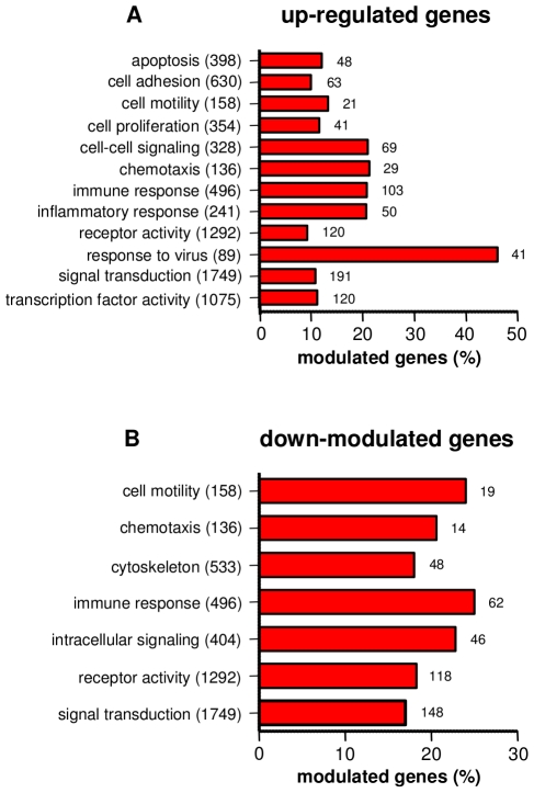
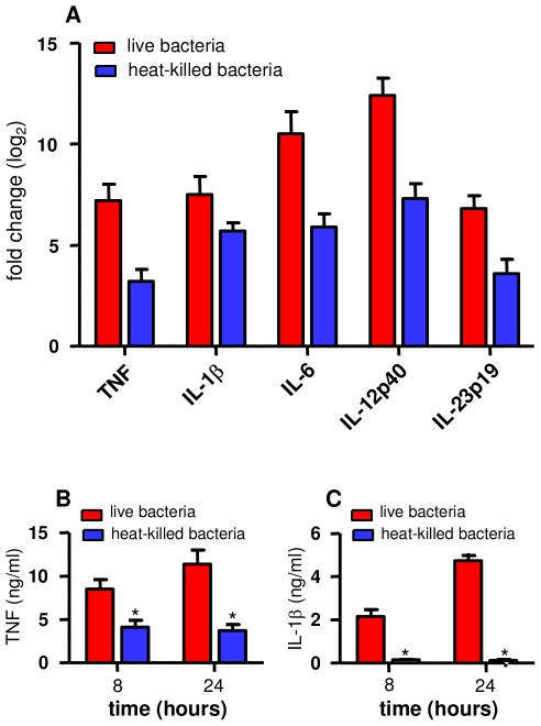
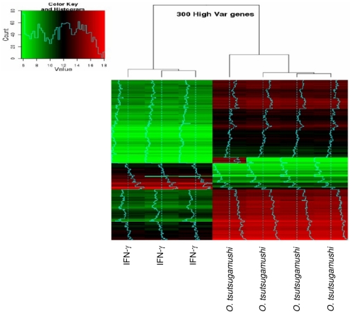
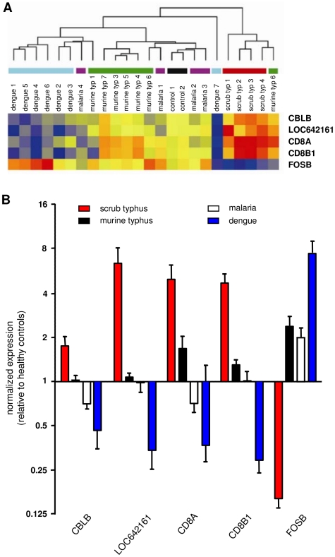
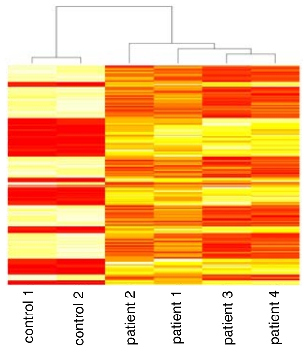
Orientia tsutsugamushi is the causal agent of scrub typhus, a public health problem in the Asia-Pacific region and a life-threatening disease. O. tsutsugamushi is an obligate intracellular bacterium that mainly infects endothelial cells. We demonstrated here that O. tsutsugamushi also replicated in monocytes isolated from healthy donors. In addition, O. tsutsugamushi altered the expression of more than 4,500 genes, as demonstrated by microarray analysis. The expression of type I interferon, interferon-stimulated genes and genes associated with the M1 polarization of macrophages was significantly upregulated. O. tsutsugamushi also induced the expression of apoptosis-related genes and promoted cell death in a small percentage of monocytes. Live organisms were indispensable to the type I interferon response and apoptosis and enhanced the expression of M1-associated cytokines. These data were related to the transcriptional changes detected in mononuclear cells isolated from patients with scrub typhus. Here, the microarray analyses revealed the upregulation of 613 genes, which included interferon-related genes, and some features of M1 polarization were observed in these patients, similar to what was observed in O. tsutsugamushi-stimulated monocytes in vitro. This is the first report demonstrating that monocytes are clearly polarized in vitro and ex vivo following exposure to O. tsutsugamushi. These results would improve our understanding of the pathogenesis of scrub typhus, during which interferon-mediated activation of monocytes and their subsequent polarization into an M1 phenotype appear critical. This study may give us a clue of new tools for the diagnosis of patients with scrub typhus.


Conflict of interest statement

The authors have declared that no competing interests exist.

Figures

Figure 1
Figure 1. O. tsutsugamushi replication within monocytes.
Monocytes and L929 cells were infected with O. tsutsugamushi (two viable bacteria per cell) for different periods of time. A. The number of bacterial DNA copies was determined using qRT-PCR. The data are expressed as the mean ± SD of two independent experiments performed in triplicate. B. Monocytes were infected with O. tsutsugamushi, and the bacteria were detected using indirect immunofluorescence. C. Monocytes infected with O. tsutsugamushi for 5 days were labeled with bodipy phallacidin to detect filamentous actin and bacteria were detected using indirect immunofluorescence (in red). One representative micrograph performed in confocal microscopy is shown.
Figure 2
Figure 2. GO analysis of differentially expressed genes.
Monocytes were stimulated with O. tsutsugamushi or mock stimulated for 8 hours, and the modulation of genes was analyzed using microarrays and GO term tools. The upregulated (A) and downregulated (B) genes were classified based on the major biological processes in which they are involved. The total number of genes present in each biological process and the number of differentially expressed genes are indicated. The results are expressed as the percentage of the upregulated or downregulated genes.
Figure 3
Figure 3. Quantitative RT-PCR of selected genes in stimulated monocytes.
Monocytes were stimulated with or without O. tsutsugamushi for 8 (A) and 24 (B) hours. RNA was extracted, and qRT-PCR was performed on 16 genes involved in the immune response that were differentially expressing in the microarray experiments. The results, expressed as the log2 ratio of fold changes, are presented as the mean ± SEM of three experiments performed in triplicate.
Figure 4
Figure 4. Bacterial viability and monocyte responses.
Monocytes were stimulated with live or heat-killed O. tsutsugamushi for 8 (A) or 24 (B, C) hours. A. RNA was extracted, and qRT-PCR was performed to detect several genes that were differentially expressed in the microarray experiments. The results, expressed as the log2 ratio of fold changes, are presented as the mean ± SEM of two experiments performed in triplicate. B and C. Culture supernatants were analyzed for the presence of TNF (B) and IL-1β (C) using ELISAs. The results are expressed in ng/ml and are presented as the mean ± SD of two experiments performed in duplicate. *p<0.05.
Figure 5
Figure 5. Hierarchical clustering of differentially expressed genes in stimulated monocytes.
Monocytes were stimulated with O. tsutsugamushi or IFN-γ (500 UI/ml) for 8 hours, and genome-wide expression studies were performed using microarrays from Agilent Technologies. A hierarchical clustering consisting of 300 highly altered genes is shown.
Figure 6
Figure 6. Hierarchical clustering in patients with scrub typhus.
RNA was isolated from PBMCs from healthy controls and patients with different infectious diseases, and microarray studies were performed using Illumina Human-6 v2 BeadChips. The unsupervised hierarchical clustering of 22 patients and 2 RNA pools from healthy controls was performed based on the expression of 65 genes specific to scrub typhus (typ). The normalized expression level in each sample was baseline-adjusted to the mean expression level of the healthy control group and color-scaled, with red indicating increased expression and blue indicating decreased expression.
Figure 7
Figure 7. Reduced transcriptional signature of scrub typhus.
The microarrays performed on RNA isolated from healthy controls or patients with infectious diseases suggested that five genes can be used as a specific signature of scrub typhus. A. The unsupervised hierarchical clustering of 22 patients and 2 pools of RNA from healthy controls was performed based on the expression of these five genes. The normalized expression level in each sample was baseline-adjusted to the mean expression level of the healthy control group and color-scaled, with red indicating increased expression and blue indicating decreased expression. B. The expression of the five genes was assessed using qRT-PCR. The results, expressed as the log2 ratio of fold changes, are presented as the mean ± SEM of all the patients in each group performed in duplicate.
Figure 8
Figure 8. Comparison between patient blood samples and in vitro-infected monocytes.
Monocytes from healthy donors were stimulated with O. tsutsugamushi for 8 hours, and Agilent microarrays were used to detect the differential expression of 2,015 genes that corresponded to 1,606 genes in the Illumina microarrays. Among these genes, 184 (250 probes) were altered in patients with scrub typhus with a p value less than 0.01. The hierarchical clustering of these genes demonstrates that the resulting transcriptional signature was specific to scrub typhus.

References

    1. Seong SY, Choi MS, Kim IS. Orientia tsutsugamushi infection: overview and immune responses. Microbes Infect. 2001;3:11–21. - PubMed
    1. Watt G, Parola P. Scrub typhus and tropical rickettsioses. Curr Opin Infect Dis. 2003;16:429–436. - PubMed
    1. Chattopadhyay S, Richards AL. Scrub typhus vaccines: past history and recent developments. Hum Vaccin. 2007;3:73–80. - PubMed
    1. Tamura A, Ohashi N, Urakami U, Miyamura S. Classification of Rickettsia tsutsugamushi in a new genus, Orientia gen. nov., as Orientia tsutsugamushi comb. nov. Int J Syst Bacteriol. 1995;45:589–591. - PubMed
    1. Cho NH, Kim HR, Lee JH, Kim SY, Kim J, et al. The Orientia tsutsugamushi genome reveals massive proliferation of conjugative type IV secretion system and host-cell interaction genes. Proc Natl Acad Sci U S A. 2007;104:7981–7986. - PMC - PubMed

Publication types