

Case Reports
doi: 10.2349/biij.6.1.e6.
Epub 2010 Jan 1.
Leigh syndrome: MRI findings in two children
Affiliations
- PMID: 21611066
- PMCID: PMC3097793
- DOI: 10.2349/biij.6.1.e6
Item in Clipboard
Case Reports
Leigh syndrome: MRI findings in two children
Biomed Imaging Interv J.
2010 Jan-Mar.
Abstract
Leigh syndrome is a progressive neurodegenerative disorder of childhood. The symmetrical necrotic lesions in the basal ganglia and/or brainstem which appear as hyperintense lesions on T2-weighted MRI is characteristic and one of the essential diagnostic criteria. Recognising this MR imaging pattern in a child with neurological problems should prompt the clinician to investigate for Leigh syndrome. We present here two cases of Leigh syndrome due to different biochemical/genetic defects, and discuss the subtle differences in their MR neuroimaging features.
Keywords: Leigh syndrome; MRI; SURF1; mitochondrial.
Figures
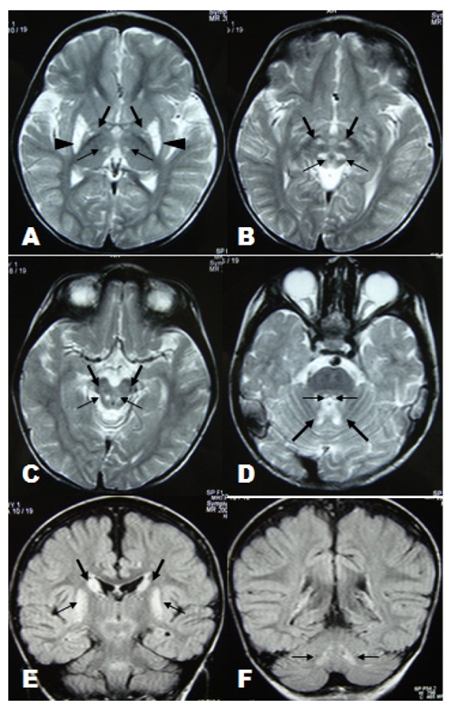
(A): Axial T2-weighted image through the basal ganglia shows symmetrical hyperintense lesions involved thalamic posteromedial ventral nuclei (thin arrow), globus pallidi (thick arrow) and putamina (arrowhead). (B-D): Axial T2-weighted images through the brainstem show symmetrical involvement of reticular formation of midbrain (thin arrow in B), subthalamic nuclei (thick arrow in B), substantia nigra (thick arrow in C), dorsal midbrain (thin arrow in C) central tegmental tracts (thin arrow in D) and cerebellar nuclei region (thick arrow in D). (E-F): Coronal FLAIR images show symmetrical involvement of head of caudate nuclei (thick arrow in E), putamina (thin arrow in E) and dentate nuclei (thin arrow in F).
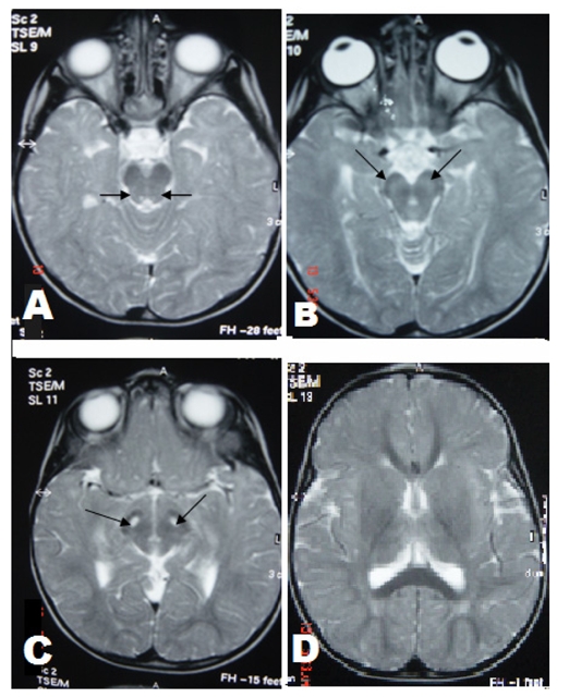
(A-C): Axial T2-weighted images through the brainstem show symmetrical hyperintense lesions involving central tegmental tracts (thin arrow in A), substantia nigra (thin arrow in B and C). (D): Axial T2-weighted image through basal ganglia shows putamen, globus pallidus and thalamus were not involved.
References
-
- Rahman S, Blok RB, Dahl HH, et al. Leigh syndrome: clinical features and biochemical and DNA abnormalities. Ann Neurol. 1996;139(3):343–51. - PubMed
-
- Finsterer J. Leigh and Leigh-like syndrome in children and adults. Pediatr Neurol. 2008;39(4):223–35. - PubMed
-
- Savoiardo M, Zeviani M, Uziel G, et al. MRI in Leigh syndrome with SURF1 gene mutation. Ann Neurol. 2002;51(1):138–9. - PubMed
Publication types
LinkOut - more resources
Full Text Sources