. 2011;6(5):e19716.
doi: 10.1371/journal.pone.0019716. Epub 2011 May 18.

Identification of small-molecule inhibitors of Yersinia pestis Type III secretion system YscN ATPase

Affiliations

Identification of small-molecule inhibitors of Yersinia pestis Type III secretion system YscN ATPase

Wieslaw Swietnicki et al. PLoS One. 2011.

Abstract

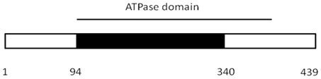
Yersinia pestis is a gram negative zoonotic pathogen responsible for causing bubonic and pneumonic plague in humans. The pathogen uses a type III secretion system (T3SS) to deliver virulence factors directly from bacterium into host mammalian cells. The system contains a single ATPase, YscN, necessary for delivery of virulence factors. In this work, we show that deletion of the catalytic domain of the yscN gene in Y. pestis CO92 attenuated the strain over three million-fold in the Swiss-Webster mouse model of bubonic plague. The result validates the YscN protein as a therapeutic target for plague. The catalytic domain of the YscN protein was made using recombinant methods and its ATPase activity was characterized in vitro. To identify candidate therapeutics, we tested computationally selected small molecules for inhibition of YscN ATPase activity. The best inhibitors had measured IC(50) values below 20 µM in an in vitro ATPase assay and were also found to inhibit the homologous BsaS protein from Burkholderia mallei animal-like T3SS at similar concentrations. Moreover, the compounds fully inhibited YopE secretion by attenuated Y. pestis in a bacterial cell culture and mammalian cells at µM concentrations. The data demonstrate the feasibility of targeting and inhibiting a critical protein transport ATPase of a bacterial virulence system. It is likely the same strategy could be applied to many other common human pathogens using type III secretion system, including enteropathogenic E. coli, Shigella flexneri, Salmonella typhimurium, and Burkholderia mallei/pseudomallei species.


Conflict of interest statement

Competing Interests: The authors have declared that no competing interests exist.

Figures

Figure 1
Figure 1. yscN gene deletion in Y.pestis CO92.
The yscN gene fragment (black box) was deleted in the pCD1 plasmid as described under Materials and Methods. The approximate position of ATPase domain is marked with the bar on the top of the box. Numbers below the box correspond to approximate positions in the amino acid sequence marking the boundaries of the deleted region.
Figure 2
Figure 2. Deletion of the yscN gene in Y. pestis CO92 strain does not affect the growth of bacteria.
Fresh dilutions of the wild type and ΔyscN strains of bacteria were grown at 28°C as described under Materials and Methods. A graph for one of the representative experiments is presented.
Figure 3
Figure 3. Steady-state kinetics of ATP hydrolysis by the optimized catalytic domain.
(A) The ATP hydrolysis is inhibited above 4 mM ATP concentration. (B) The hydrolysis of ATP by the enzyme shows a positive cooperativity up to 4 mM ATP concentration. The kinetics of hydrolysis was measured by following phosphate release in 10 mM Tris, pH = 7.6, 150 mM NaCl, and 1 mM Mg+2 at 37°C as described under Materials and Methods. Total protein concentration was 9.6 µg. Error bars correspond to standard deviation of triplicate measurements.
Figure 4
Figure 4. Small-molecule inhibitors are capable of fully blocking the optimized catalytic domain ATPase activity.
The small molecules derived from computational screen of ZINC database were incubated with the enzyme at 37°C and ATP hydrolysis was determined by measuring ADP release as described under Materials and Methods. Inhibitor concentrations were 100 µM for all reactions. The numbers on the X axis correspond to in-house IDs for individual compounds. Error bars correspond to standard deviation of triplicate measurements.
Figure 5
Figure 5. Structural model of YscN ATPase with compounds docked into the active site.
(A) Protein fold of the YscN model (colored yellow) built by comparative modeling methods and illustrated with the docked compound ID number 7812 (depicted by atomic spheres). (B) Molecular surface of the YscN model showing the active-site region (colored gold) bound by compound ID 7812 in its conformational pose (stick representation, where in general the color cyan are carbon atoms, nitrogen atoms are blue, oxygen are red, and sulfur are yellow). (C) Modeled YscN complex with compound ID 7146. (D) Modeled YscN complex with compound ID 7086.
Figure 6
Figure 6. Inhibition of YopE secretion by selected small molecules.
The secretion of YopE was measured in the supernatants of bacterial cell culture by ELISA using anti-YopE antibody as described under Materials and Methods. The data were collected 1 hr post-induction with EGTA. Inhibitor concentrations were 100 µM. Positive and negative controls correspond to the results obtained for co-solvent (DMSO) without and with EGTA, respectively. Error bars correspond to standard deviation of five measurements.
Figure 7
Figure 7. Inhibition of Y. pestis KIM growth by selected small molecules.
Attenuated Y. pestis KIM19 strain was grown in the presence of 100 mM inhibitor in HBI medium at 30°C as described under Materials and Methods. Control experiment was performed with 2% DMSO. Data were normalized to the starting OD620 = 0.15 at 0 hr time. For a reference, the secretion of YopE protein (Fig. 6) was measured at t = 3 hrs.

References

    1. Butler T. Plague into the 21st century. Clin Infect Dis. 2009;49:736–742. - PubMed
    1. Veenendaal AK, Sundin C, Blocker AJ. Small-molecule type III secretion system inhibitors block assembly of the Shigella type III secreton. J Bacteriol. 2009;191:563–570. - PMC - PubMed
    1. Felise HB, Nguyen HV, Pfuetzner RA, Barry KC, Jackson SR, et al. An inhibitor of gram-negative bacterial virulence protein secretion. Cell Host Microbe. 2008;4:325–336. - PMC - PubMed
    1. Kline T, Felise HB, Barry KC, Jackson SR, Nguyen HV, et al. Substituted 2-imino-5-arylidenethiazolidin-4-one inhibitors of bacterial type III secretion. J Med Chem. 2008;51:7065–7074. - PMC - PubMed
    1. Tree JJ, Wang D, McInally C, Mahajan A, Layton A, et al. Characterization of the effects of salicylidene acylhydrazide compounds on type III secretion in Escherichia coli O157:H7. Infect Immun. 2009;77:4209–4220. - PMC - PubMed

Publication types

MeSH terms