. 2011;6(5):e19809.
doi: 10.1371/journal.pone.0019809. Epub 2011 May 18.

Wnt3a induces myofibroblast differentiation by upregulating TGF-β signaling through SMAD2 in a β-catenin-dependent manner

Affiliations

Wnt3a induces myofibroblast differentiation by upregulating TGF-β signaling through SMAD2 in a β-catenin-dependent manner

Jon M Carthy et al. PLoS One. 2011.

Abstract

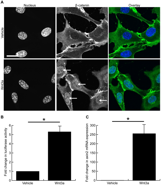
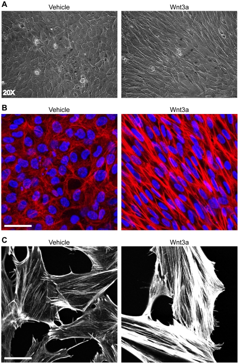
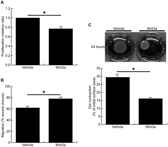
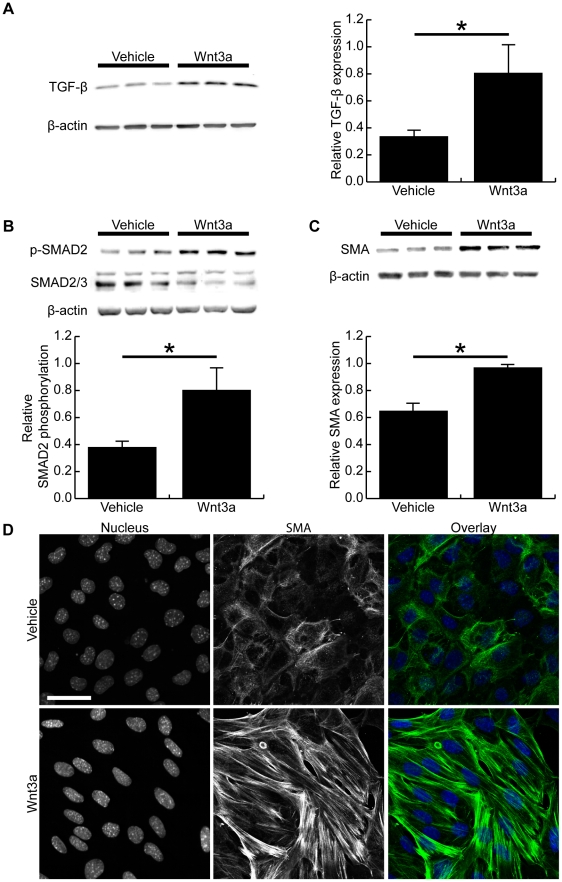
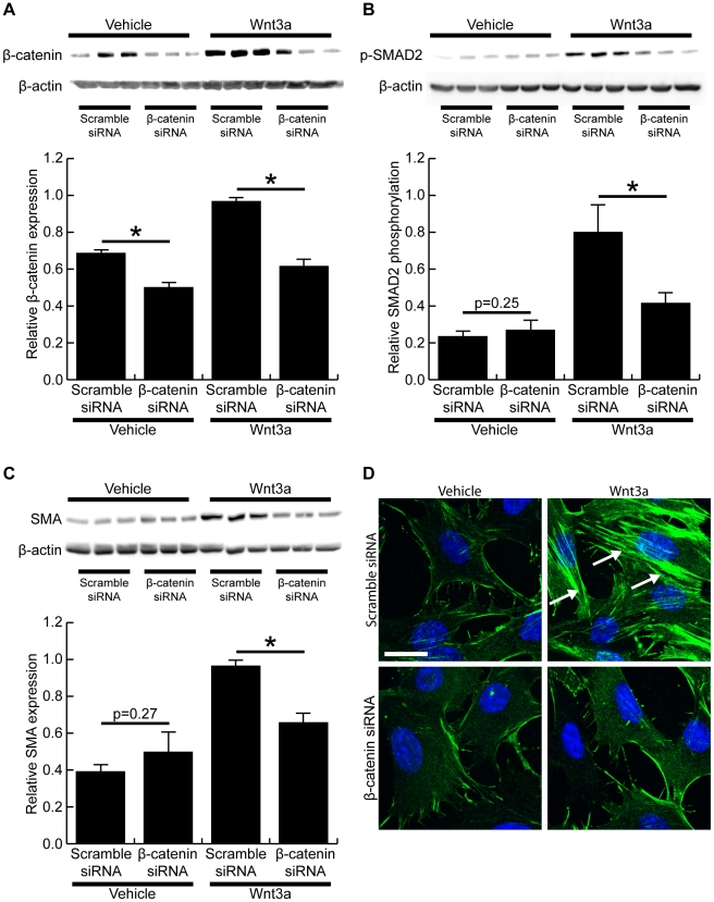
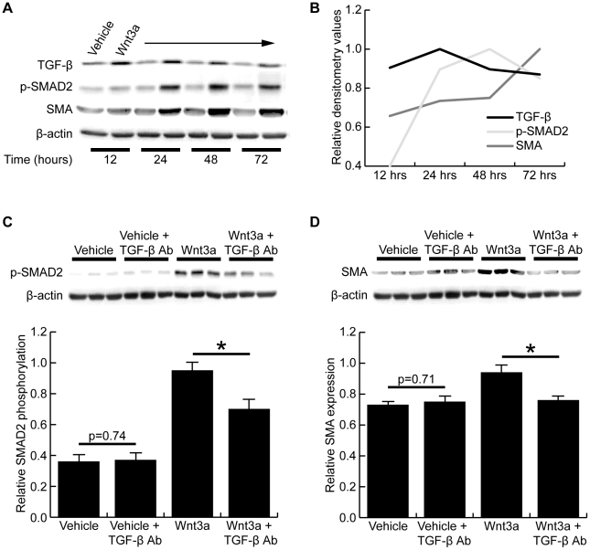
Growing evidence suggests the Wnt family of secreted glycoproteins and their associated signaling pathways, linked to development, are recapitulated during wound repair and regeneration events. However, the role of the Wnt pathway in such settings remains unclear. In the current study, we treated mouse fibroblasts with 250 ng/mL of recombinant Wnt3a for 72 hours and examined its affect on cell morphology and function. Wnt3a induced a spindle-like morphology in fibroblasts characterized by the increased formation of stress fibres. Wnt3a decreased the proliferation of fibroblasts, but significantly increased cell migration as well as fibroblast-mediated contraction of a collagen lattice. Wnt3a significantly increased the expression of TGF-β and its associated signaling through SMAD2. Consistent with this, we observed significantly increased smooth muscle α-actin expression and incorporation of this contractile protein into stress fibres following Wnt3a treatment. Knockdown of β-catenin using siRNA reversed the Wnt3a-induced smooth muscle α-actin expression, suggesting these changes were dependent on canonical Wnt signaling through β-catenin. Neutralization of TGF-β with a blocking antibody significantly inhibited the Wnt3a-induced smooth muscle α-actin expression, indicating these changes were dependent on the increased TGF-β signaling. Collectively, this data strongly suggests Wnt3a promotes the formation of a myofibroblast-like phenotype in cultured fibroblasts, in part, by upregulating TGF-β signaling through SMAD2 in a β-catenin-dependent mechanism. As myofibroblasts are critical regulators of wound healing responses, these findings may have important implications for our understanding of normal and aberrant injury and repair events.


Conflict of interest statement

Competing Interests: The authors have declared that no competing interests exist.

Figures

Figure 1
Figure 1. Wnt3a induces canonical Wnt signaling in mouse fibroblasts.
Confocal images of fibroblasts treated for 24 hours with vehicle (top panels) or 250 ng/mL Wnt3a (bottom panels) and immunonstained for β-catenin (green) and nuclei (blue). Wnt3a treatment induced clear nuclear accumulation of β-catenin in murine fibroblasts (arrows). (B) TOPFlash reporter assay demonstrated Wnt3a significantly increased luciferase activity 5.3±1.6 fold after a 24 hour treatment (p<0.05). (C) Wnt3a treatment induced a 255±71 fold increase in the mRNA expression of axin2, a target of classical Wnt signaling (p<0.05). (Scale bar = 23.00 µm in A, * denotes p<0.05)
Figure 2
Figure 2. Wnt3a induces a spindle-like morphology with increased stress fibre formation after 72 hours of treatment.
(A) Light microscope images of mouse fibroblasts that had been treated for 72 hours with vehicle (left panel) or 250 ng/mL Wnt3a (right panel). Wnt3a treatment induced a spindle-like morphology in fibroblasts. (B) Confocal images of vehicle-treated (left panel) or Wnt3a-treated (right panel) fibroblasts immunostained for f-actin (red) and nuclei (blue) showing the increased formation and parallel organization of stress fibres following 72 hours Wnt3a treatment. (C) Low density culture of vehicle-treated (left panel) or Wnt3a-treated (right panel) fibroblasts highlights the increased formation of stress fibres seen after Wnt3a treatment. (Scale bars = 47.00 µm in B, 23.00 µm in C)
Figure 3
Figure 3. Wnt3a inhibits cell proliferation, but increases cell migration and contraction after 72 hour treatment.
(A) Cell proliferation was measured after 72 hours of treatment with Wnt3a or vehicle. Wnt-treated cells grew at 77.4±4.5% the rate of vehicle treated cells (p<0.05). (B) Cells were treated for 72 hours with Wnt3a or vehicle, and then a scratch wound assay was performed to measure cell migration. Wnt-treated cells closed the scratch wound at a significantly faster rate than vehicle-treated cells, as measured 48 hours after injury (78.1±2.1% vs 61.9±3.8%, p<0.05). (C) Cells were treated for 72 hours with Wnt3a or vehicle and then a fibroblast-populated collagen lattice contraction assay was performed. Images of contracted gels taken at 24 hours are shown along with the quantified surface areas of contracted gels. Wnt3a treatment significantly increased the fibroblast-mediated contraction of collagen gels (16.1±0.6% vs 29.4±1.3% of initial surface area, p<0.05). (* denotes p<0.05)
Figure 4
Figure 4. Wnt3a increases TGF-β expression, SMAD2 phosphorylation and smooth muscle α-actin expression.
(A) Representative Western blot of TGF-β expression in vehicle-treated and Wnt3a-treated fibroblasts after 72 hours. Densitometry showed TGF-β expression to be significantly increased after Wnt3a treatment (p<0.05). (B) Western blot of SMAD2 phosphorylation after 72 hours of vehicle or Wnt3a treatment. Densitometry showed Wnt3a significantly increased SMAD2 phosphorylation at 72 hours. (C) Western blot of smooth muscle α-actin expression in vehicle-treated or Wnt3a-treated cells. Wnt3a-treatment significantly increased the expression of smooth muscle α-actin expression in mouse fibroblasts, as measured by densitometry (p<0.05). (D) Confocal images of fibroblasts immunostained for smooth muscle α-actin (green) and nuclei (blue). Wnt3a-treated fibroblasts had clearly visible smooth muscle α-actin positive stress fibres while the vehicle-treated cells did not display expression of smooth muscle α-actin in their stress fibres. (Scale bar = 47.00 µm in D, * denotes p<0.05)
Figure 5
Figure 5. Wnt3a-induced change in cell phenotype is dependent on β-catenin.
(A) Western blot demonstrated β-catenin siRNA significantly decreased β-catenin expression in vehicle- and Wnt-treated fibroblasts when compared to a scrambled siRNA. (B) Western blot showed knock down of β-catenin expression significantly inhibited the Wnt3a-induced SMAD2 phosporylation (p<0.05). No difference in SMAD2 phosporylation was detected in vehicle treated cells (p = 0.25). (C) Western blot of smooth muscle α-actin expression demonstrated that β-catenin siRNA significantly decreased smooth muscle α-actin expression in Wnt3a-treated fibroblasts (p<0.05). No significant difference was seen in the vehicle-treated cells (p = 0.27). (D) Immunohistochemistry showed Wnt3a promoted smooth muscle α-actin stress fibre formation in control siRNA transfected cells (green, arrows), but β-catenin siRNA completely inhibited the Wnt3a-induced smooth muscle α-actin expression. Cell nuclei are stained blue with DAPI. (Scale bar = 23.00 µm in D, * denotes p<0.05)
Figure 6
Figure 6. Wnt3a-induced change in cell phenotype is dependent on TGF-β expression.
(A) Representative Western blots of vehicle- and Wnt3a-treated fibroblasts showing TGF-β expression, SMAD2 phosphorylation, and smooth muscle α-actin expression at 12, 24, 48, and 72 hours of treatment. (B) Graphical representation of the densitometry results for the blots in A shows, in a sequential manner, that TGF-β expression peaks between 12 and 24 hours, followed by SMAD2 phosphorylation peaking between 24 and 48 hours, which is then followed by smooth muscle α-actin expression peaking after 72 hours of treatment. (C) Western blot of SMAD2 phosphorylation in fibroblasts treated with or without Wnt3a and a TGF-β neutralizing antibody. Densitometry demonstrated the TGF-β neutralizing antibody significantly inhibited Wnt3a-induced SMAD2 phosphorylation (p<0.05). No change was seen in the vehicle-treated cells (p = 0.74). (D) Western blot of smooth muscle α-actin expression in fibroblasts treated with or without Wnt3a and the TGF-β neutralizing antibody. Densitometry confirmed TGF-β neutralization significantly inhibited the Wnt3a-induced smooth muscle α-actin expression (p<0.05). No change was seen in vehicle-treated cells (p = 0.71). (* denotes p<0.05)

References

    1. Martin P. Wound healing-aiming for perfect skin regeneration. Science. 1997;276:75–81. - PubMed
    1. Barrientos S, Stojadinovic O, Golinko MS, Brem H, Tomic-Canic M. Growth factors and cytokines in wound healing. Wound Repair Regen. 2008;16:585–601. - PubMed
    1. Zhao J, Kim KA, Abo A. Tipping the balance: modulating the Wnt pathway for tissue repair. Trends Biotechnol. 2009;27:131–136. - PubMed
    1. Zhang DL, Gu LJ, Liu L, Wang CY, Sun BS, et al. Effect of Wnt signaling pathway on wound healing. Biochem Biophys Res Commun. 2009;378:149–151. - PubMed
    1. Logan CY, Nusse R. The Wnt signaling pathway in development and disease. Annu Rev Cell Dev Biol. 2004;20:781–810. - PubMed

Publication types

MeSH terms