

Theranostic nanoshells: from probe design to imaging and treatment of cancer
- PMID: 21612199
- PMCID: PMC3888233
- DOI: 10.1021/ar200023x
Theranostic nanoshells: from probe design to imaging and treatment of cancer
Abstract
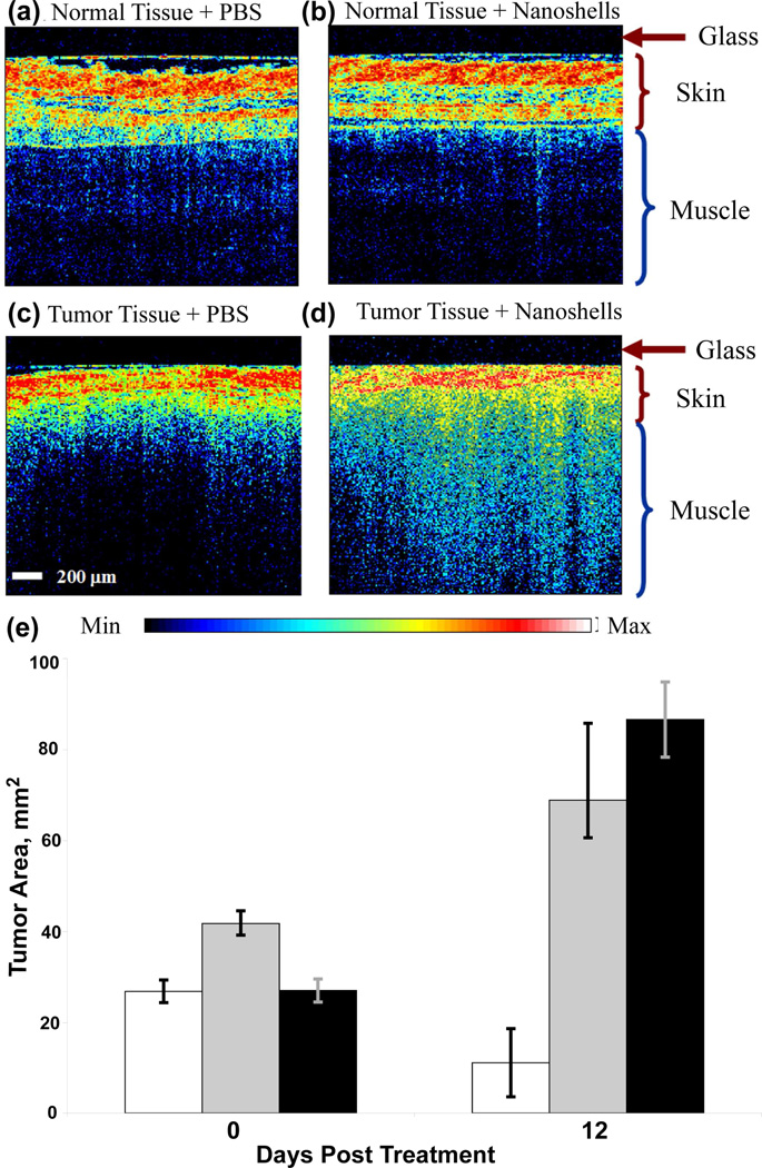
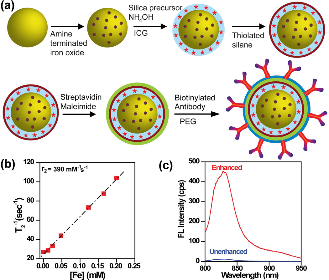
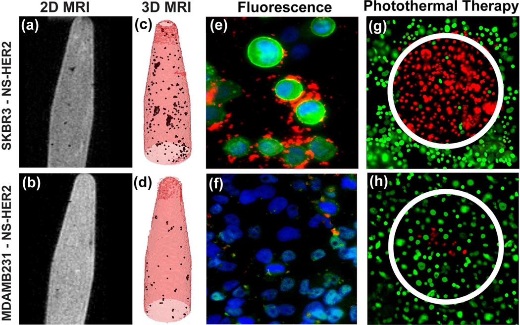
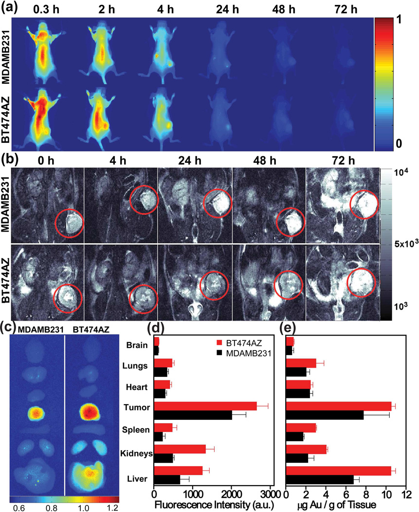
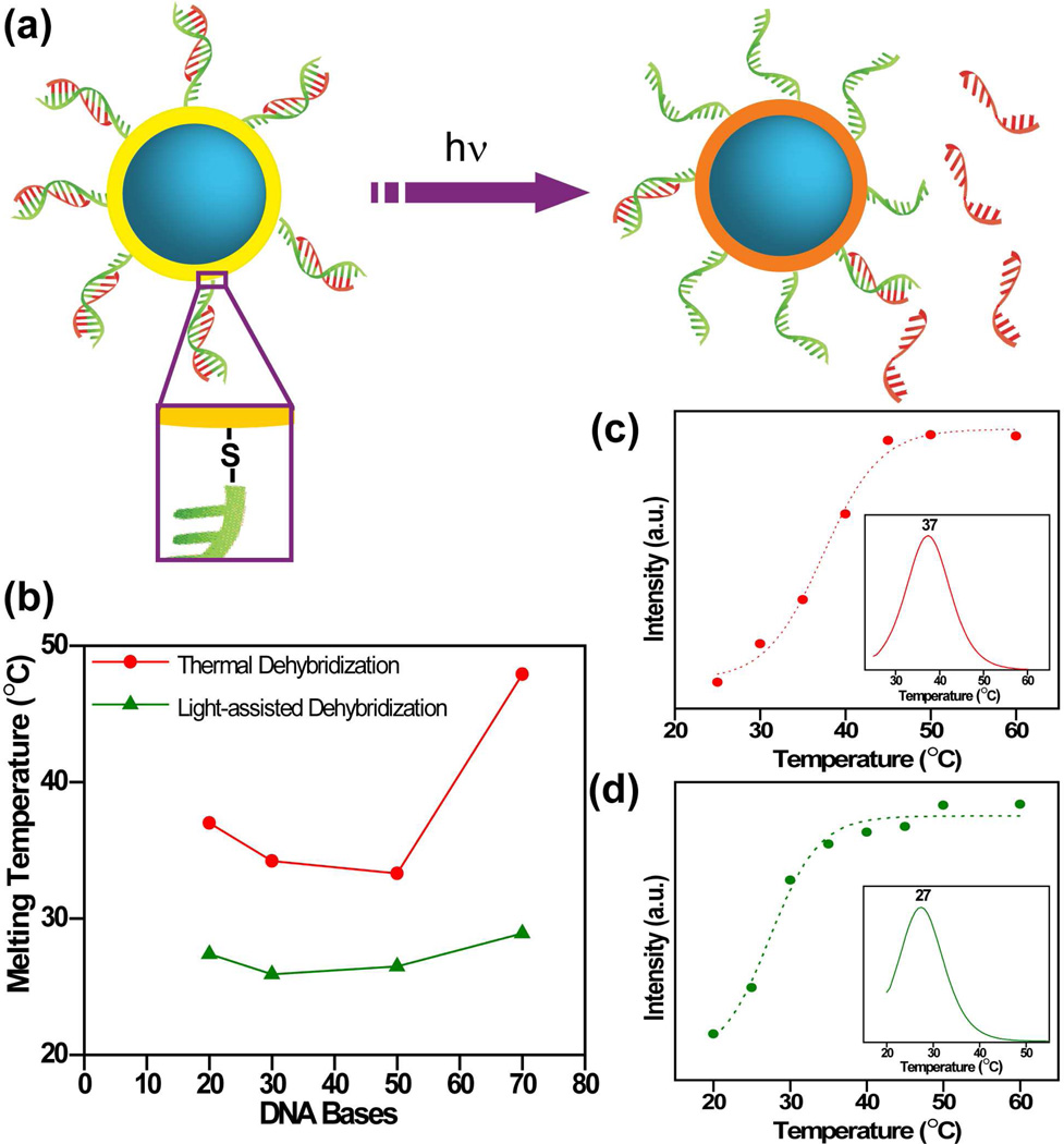
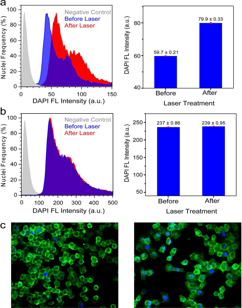
Recent advances in nanoscience and biomedicine have expanded our ability to design and construct multifunctional nanoparticles that combine targeting, therapeutic, and diagnostic functions within a single nanoscale complex. The theranostic capabilities of gold nanoshells, spherical nanoparticles with silica cores and gold shells, have attracted tremendous attention over the past decade as nanoshells have emerged as a promising tool for cancer therapy and bioimaging enhancement. This Account examines the design and synthesis of nanoshell-based theranostic agents, their plasmon-derived optical properties, and their corresponding applications. We discuss the design and preparation of nanoshell complexes and their ability to enhance the photoluminescence of fluorophores while maintaining their properties as MR contrast agents. In this Account, we discuss the underlying physical principles that contribute to the photothermal response of nanoshells. We then elucidate the photophysical processes that induce nanoshells to enhance the fluorescence of weak near-infrared fluorophores. Nanoshells illuminated with resonant light are either strong optical absorbers or scatterers, properties that give rise to their unique capabilities. These physical processes have been harnessed to visualize and eliminate cancer cells. We describe the application of nanoshells as a contrast agent for optical coherence tomography of breast carcinoma cells in vivo. Our recent studies examine nanoshells as a multimodal theranostic probe, using these nanoparticles for near-infrared fluorescence and magnetic resonance imaging (MRI) and for the photothermal ablation of cancer cells. Multimodal nanoshells show theranostic potential for imaging subcutaneous breast cancer tumors in animal models and the distribution of tumors in various tissues. Nanoshells also show promise as light-triggered gene therapy vectors, adding temporal control to the spatial control characteristic of nanoparticle-based gene therapy approaches. We describe the fabrication of DNA-conjugated nanoshell complexes and compare the efficiency of light-induced and thermally-induced release of DNA. Double-stranded DNA nanoshells also provide a way to deliver small molecules into cells: we describe the delivery and light-triggered release of DAPI (4',6-diamidino-2-phenylindole), a dye molecule used to stain DNA in the nuclei of cells.
Figures
References
-
- ACS, Cancer Facts & Figures. American Cancer Society. 2010.
-
- Grobner T, Prischl FC. Gadolinium and nephrogenic systemic fibrosis. Kidney Inter. 2007;72:260–264. - PubMed
-
- Wang J, Short D, Sebire NJ, Lindsay I, Newlands ES, Schmid P, Savage PM, Seckl MJ. Salvage Chemotherapy of Relapsed or High-risk Gestational Trophoblastic Neoplasia (GTN) with Paclitaxel/cisplatin Alternating with Paclitaxel/etoposide (TP/TE) Ann. Oncol. 2008;19:1578–1583. - PubMed
-
- Davis ME, Chen ZG, Shin DM. Nanoparticle therapeutics: an emerging treatment modality for cancer. Nat. Rev. Drug Discovery. 2008;7:771–782. - PubMed
-
- Peer D, Karp JM, Hong S, Farokhzad OC, Margalit R, Langer R. Nanocarriers as an emerging platform for cancer therapy. Nat. Nanotech. 2007;2:751–760. - PubMed
Publication types
MeSH terms
Substances
Grants and funding
LinkOut - more resources
Full Text Sources
Other Literature Sources
Miscellaneous
