Review
. 2011 Oct;82(3):117-26.
doi: 10.1016/j.diff.2011.04.008. Epub 2011 May 25.

Normal and abnormal epithelial differentiation in the female reproductive tract

Affiliations
Review

Normal and abnormal epithelial differentiation in the female reproductive tract

Takeshi Kurita. Differentiation. 2011 Oct.

Abstract

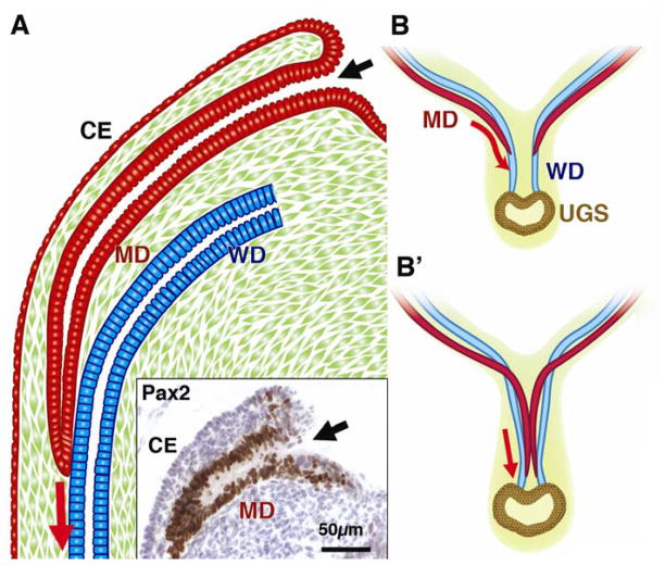
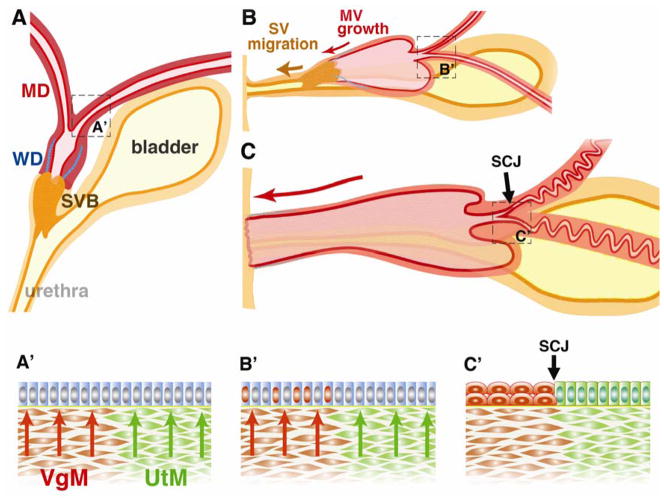
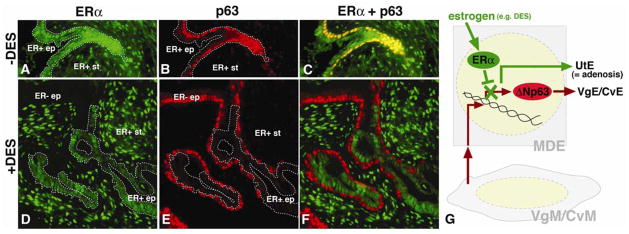
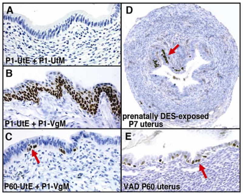
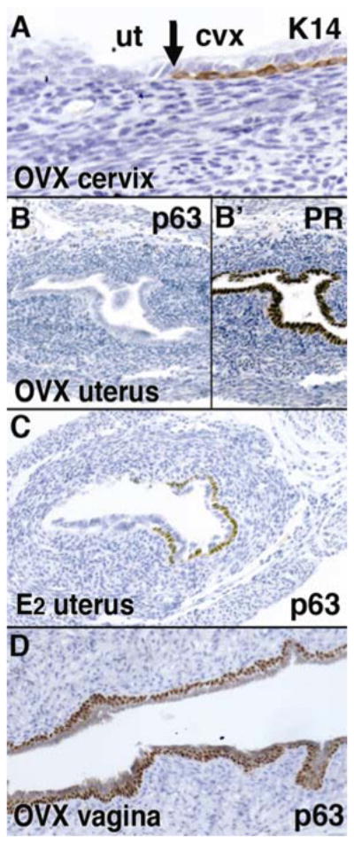
In mammals, the female reproductive tract (FRT) develops from a pair of paramesonephric or Müllerian ducts (MDs), which arise from coelomic epithelial cells of mesodermal origin. During development, the MDs undergo a dynamic morphogenetic transformation from simple tubes consisting of homogeneous epithelium and surrounding mesenchyme into several distinct organs namely the oviduct, uterus, cervix and vagina. Following the formation of anatomically distinctive organs, the uniform MD epithelium (MDE) differentiates into diverse epithelial cell types with unique morphology and functions in each organ. Classic tissue recombination studies, in which the epithelium and mesenchyme isolated from the newborn mouse FRT were recombined, have established that the organ specific epithelial cell fate of MDE is dictated by the underlying mesenchyme. The tissue recombination studies have also demonstrated that there is a narrow developmental window for the epithelial cell fate determination in MD-derived organs. Accordingly, the developmental plasticity of epithelial cells is mostly lost in mature FRT. If the signaling that controls epithelial differentiation is disrupted at the critical developmental stage, the cell fate of MD-derived epithelial tissues will be permanently altered and can result in epithelial lesions in adult life. A disruption of signaling that maintains epithelial cell fate can also cause epithelial lesions in the FRT. In this review, the pathogenesis of cervical/vaginal adenoses and uterine squamous metaplasia is discussed as examples of such incidences.


Figures

Figure 1
Figure 1. Development of the MD
CE; coelomic epithelium, MD; Müllerian duct, WD; Wolffian duct, UGS; urogenital sinus A; Formation of MD The MD arises as an invagination of CE at the cranial end of the urogenital ridge. The insert demonstrates Pax2 immunohistochemistry on the E13.5 female mouse embryo. The Pax2 is essential for the development of the MD (Torres et al., 1995) and its expression differentiates the MDE (brown cells in the insert) from the CE. The site of infolding remains open throughout development (black arrows). The MD grows caudally through the urogenital ridge mesenchyme and the tip comes into contact with the WD within a common basement membrane. Afterwards, the tip of the growing MD maintains close contact with WD while the cranial portion is separated from the WD by intervening mesenchyme. B; Caudal growth and fusion of the MDs The MDs remain in contact with the WDs and use them as a guide during their caudal growth. As the MDs grow caudally, they cross over the WDs and meet in the midline to fuse with each other (B′). The caudal tips of the MDs remain separated to keep contact with the WDs (Hashimoto, 2003). Right before the MD tips reach the urogenital sinus, they finally become united and fuse with the UGS.
Figure 2
Figure 2. Formation of mouse cervix and vagina
MD; Müllerian duct, WD; Wolffian duct, UGS; urogenital sinus, SVB; sinovaginal bulb. SV; sinus vagina, MV; Müllerian vagina, SJC; squamocolumnar junction, VgM; vaginal mesenchyme, UtM; uterine mesenchyme. A. Late embryonic state (~E16). The MD, WD and UGS are present. The cranial portion of WD is regressed by this stage. At this stage, the MDE is uniformly undifferentiated (A′). B. Perinatal stage. The SV moves caudally as the MV elongates caudally. During this caudal migration, the SV maintains its connection to the urethra. Around this time, ΔNp63 expression is induced in epithelial cells (red nuclei in B′) of the cervix and vagina in response to the mesenchymal induction (red arrows). By this time, the majority of the WD (blue lines) is regressed. C. Pubertal stage. The MV reaches the posterior body wall in the neonatal stage. At puberty, the solid epithelial cord of the SV is canalized and the vaginal orifice is formed. The entire vagina is lined by epithelial cells derived from MDE. In the cervix, the SCJ is formed as a result of mesenchymal induction (C′). The residual segments of WDs may still be present in the vaginal stroma, and the amount of WDE remnants varies among adult female mice.
Figure 3
Figure 3. Cell-autonomous inhibition of p63 expression by DES/ERα in MDE
ERα; estrogen receptor α, UtE; uterine epithelium, VgE; vaginal epithelium, CvE; cervical epithelium, VgM; vaginal mesenchyme, CvM; cervical mesenchyme, ep; epithelium, st; stroma, ER+; ERα positive ER−; ERα negative. Dotted line indicates ER+ epithelial cells. The uterine epithelial cells from P1 ERα null and wild-type mice were mixed, combined with P1 rat VgM and grafted under the subrenal capsule of female host nude mice with/without subcutaneous implantation of a 25μg DES pellet (Kurita et al., 2004). In the absence of DES, p63 was induced in the entire epithelium, which contained both ERα positive and negative epithelial cells. The panels A –C show the area with double positive epithelial cells for ERα and p63. In contrast, in the presence of DES, p63 was induced only in the ERα negative cells. The panels D– F show exclusive expression of ERα and p63 in the epithelium in the DES-treated host. These data confirm the conclusion of our previous study that DES inhibits expression of p63 through ERα in the epithelial cells. ERα in the VgM/CvM does not inhibit induction of p63 in the MDE. Furthermore, co-localization of ER+/p63-negative and ER−/p63-positive epithelial cells indicates that the inhibitory effect of DES/ERα on p63 expression is cell-autonomous, as illustrated in G.
Figure 4
Figure 4. Uterine squamous metaplasia
UtE; uterine epithelium, VgE; vaginal epithelium, UtM; uterine mesenchyme, VgM; vaginal mesenchyme A– C; Tissue recombinants composed with P1 UtE + P1 UtM (A), P1 UtE + P1 VgM (B), and P60 UtE + P1 VgM (C). When the UtE from the P1 mouse is associated with a P1 UtM, it develops into simple columnar UtE, which is negative for p63 (A). The same P1 UtE can be induced to be stratified squamous VgE by P1 VgM (B). In contrast, UtE from mature mice have limited potential to transdifferentiate into VgE. UtE from two month old virgin mice remains mostly simple columnar when it is combined with P1 VgM (C). However, the bipotency of UtE is not completely lost even in the adult mice, and a small number of UtE cells can be induced to express p63 in response to the P1 VgM (C, red arrow). These developmentally plastic cells are likely to be the target of VAD-induced squamous metaplasia (E). D. Expression of p63 in the uterus of prenatally (gestational day 14 – 17) DES-exposed mouse. The ectopic expression of p63 in the uterus was detected at P7 (red arrow). These cells are believed to develop into squamous metaplasia in a mature animal. E. Vitamin A deficiency induces expression of p63 in the adult uterus. When female mice were fed a vitamin A deficient diet from P21, p63 positive cells were detected in the UtE by four-month-old.
Figure 5
Figure 5
Uterine and vaginal epithelial differentiation phenotypes of Wnt7a null mutant, postaxial hemimelia (px) mice. Female px mice were ovariectomized (OVX) at P35 and the expressions of K14 (A), PR (B′) and p63 (B, C and D) were analyzed at P60. The SCJ was normally formed at cervix (A). UtE was columnar, positive for PR (B′) and negative for p63 and K14 (B), whereas CvE/VgE were stratified squamous with the expression of p63 (D). However, IP injection of 125ng 17β-estradiol (E2)/day for 3 days from P60 induced p63 expression in the uterus (C). Therefore, Wnt7a is required for stabilization of the epithelial cell fate in the uterus, but is dispensable for induction of the uterine/vaginal epithelial cell fate.

References

    1. Accetta SG, Rivoire WA, Monego HI, Vettori DV, De Oliveira Freitas DM, Edelweiss MI, Capp E. Vaginal adenosis in a non-diethylstilbestrol-exposed 6-year-old patient. Gynecologic and obstetric investigation. 2001;51:271–273. - PubMed
    1. Acién P. Embryological observations on the female genital tract. Human reproduction (Oxford, England) 1992;7:437–445. - PubMed
    1. Arey LB. Developmental anatomy: a textbook and laboratory manual of embryology. W.B. Saunders; Philadelphia: 1954.
    1. Bloomfield A, Frazer JE. The Development of the Lower End of the Vagina. Journal of anatomy. 1927;62:9–32. - PMC - PubMed
    1. Boutin EL, Battle E, Cunha GR. The response of female urogenital tract epithelia to mesenchymal inductors is restricted by the germ layer origin of the epithelium: prostatic inductions. Differentiation; research in biological diversity. 1991a;48:99–105. - PubMed

Publication types

MeSH terms

Substances