

Characterization of chemical, radiochemical and optical properties of a dual-labeled MMP-9 targeting peptide
- PMID: 21612930
- PMCID: PMC3148023
- DOI: 10.1016/j.bmc.2011.04.054
Characterization of chemical, radiochemical and optical properties of a dual-labeled MMP-9 targeting peptide
Abstract
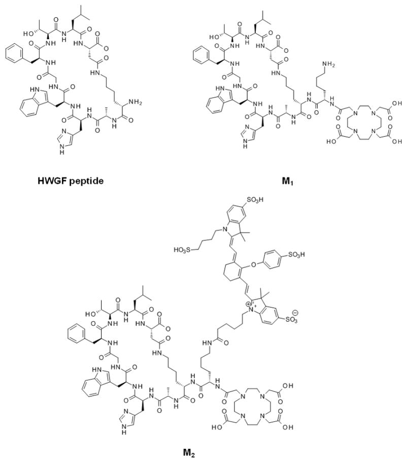
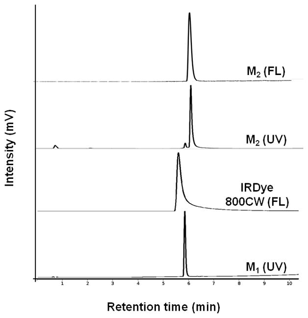
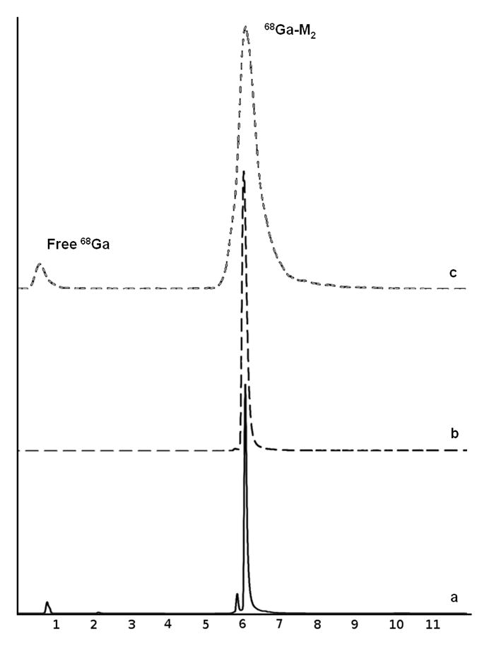
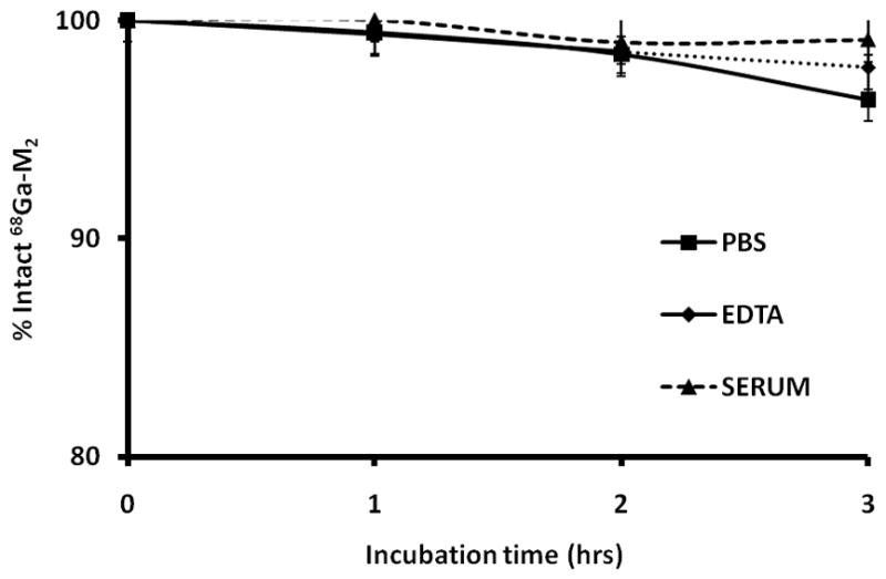
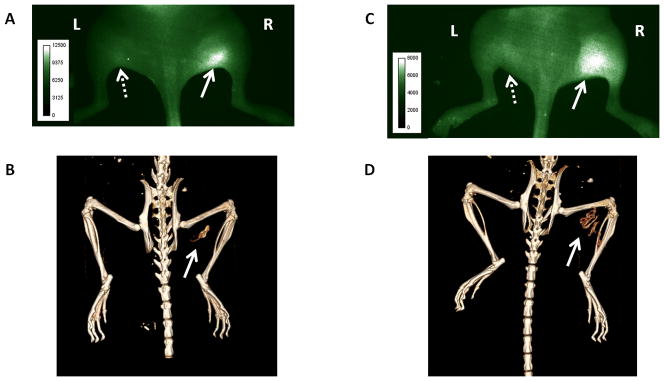
Optical imaging possesses similar sensitivity to nuclear imaging and has led to the emergence of multimodal approaches with dual-labeled nuclear/near-infrared (NIR) agents. The growing impact of (68)Ga (t(1/2)=68 min) labeled peptides on preclinical and clinical research offers a promising opportunity to merge the high spatial resolution of NIR imaging with the clinically-accepted positron emission tomography (PET). Previously, dual-labeled agents have been prepared with longer-lived radiometals and showed no detrimental effects on optical properties as a result of radiolabeling. In this study, we selected a peptide (M(2)) that targets MMP-2/9 and is dual-labeled with IRDye 800 CW and (68)Ga. Since (68)Ga chelation typically requires low pH (3.5-4) and elevated heating temperatures (95 °C), we sought to evaluate the impact of (68)Ga labeling on the optical properties of M(2). An efficient method for preparation of (68)Ga-M(2) was developed and reaction conditions were optimized. Stability studies in PBS, DTPA, and serum were performed and high levels of intact agent were evident under each condition. The addition of multiple reporters to a targeting agent adds further complexity to the characterization and validation and thus requires not only testing to ensure the agent is stable chemically and radiochemically, but also optically. Therefore, fluorescence properties were evaluated using a spectrofluorometer as well as by fluorescence detection via HPLC. It was determined that (68)Ga-labeling conditions did not impair the fluorescent properties of the agent. The agent was then used for in vivo imaging in a mouse model of heterotopic ossification (HO) with activated MMP-9 expression as an early biomarker which precedes mineralization. Although (68)Ga-complexation greatly reduced binding affinity of the peptide and negated tracer uptake on PET, NIR imaging showed consistent fluorescent signal that correlated to MMP-9 expression. This attests to the feasibility of using (68)Ga/NIR for dual-labeling of other peptides or small molecules for multimodality molecular imaging.
Copyright © 2011. Published by Elsevier Ltd.
Figures
References
Publication types
MeSH terms
Substances
Grants and funding
LinkOut - more resources
Full Text Sources
Other Literature Sources
Miscellaneous
