. 2011 Aug;85(15):7788-96.
doi: 10.1128/JVI.00555-11. Epub 2011 May 25.

Structure of respiratory syncytial virus fusion glycoprotein in the postfusion conformation reveals preservation of neutralizing epitopes

Affiliations

Structure of respiratory syncytial virus fusion glycoprotein in the postfusion conformation reveals preservation of neutralizing epitopes

Jason S McLellan et al. J Virol. 2011 Aug.

Abstract

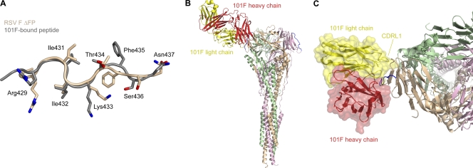
Respiratory syncytial virus (RSV) invades host cells via a type I fusion (F) glycoprotein that undergoes dramatic structural rearrangements during the fusion process. Neutralizing monoclonal antibodies, such as 101F, palivizumab, and motavizumab, target two major antigenic sites on the RSV F glycoprotein. The structures of these sites as peptide complexes with motavizumab and 101F have been previously determined, but a structure for the trimeric RSV F glycoprotein ectodomain has remained elusive. To address this issue, we undertook structural and biophysical studies on stable ectodomain constructs. Here, we present the 2.8-Å crystal structure of the trimeric RSV F ectodomain in its postfusion conformation. The structure revealed that the 101F and motavizumab epitopes are present in the postfusion state and that their conformations are similar to those observed in the antibody-bound peptide structures. Both antibodies bound the postfusion F glycoprotein with high affinity in surface plasmon resonance experiments. Modeling of the antibodies bound to the F glycoprotein predicts that the 101F epitope is larger than the linear peptide and restricted to a single protomer in the trimer, whereas motavizumab likely contacts residues on two protomers, indicating a quaternary epitope. Mechanistically, these results suggest that 101F and motavizumab can bind to multiple conformations of the fusion glycoprotein and can neutralize late in the entry process. The structural preservation of neutralizing epitopes in the postfusion state suggests that this conformation can elicit neutralizing antibodies and serve as a useful vaccine antigen.


Figures

Fig. 1.
Fig. 1.
Structure of RSV F ΔFP in the postfusion state. The RSV F ectodomain with a truncated fusion peptide adopts the postfusion state, as evidenced by the characteristic postfusion six-helix bundle of N- and C-terminal helices. (Left) Ribbon representation of an F2/F1 monomer, colored from blue to red for the N terminus (term) of F2 to the C terminus of F1, respectively. (Middle) Ribbon representation of an F2/F1 monomer colored tan and rotated 90° about the y axis. Stick representations of the N-acetyl-d-glucosamines (NAG) attached to Asn70 and Asn500 are shown, with oxygen atoms colored red and nitrogen atoms colored blue. (Right) Ribbon representation of the trimeric F glycoprotein, with each F2/F1 monomer a different color. The location of the fused membrane is represented by two solid lines.
Fig. 2.
Fig. 2.
Neutralizing epitopes on RSV F ΔFP. Epitopes for motavizumab (antigenic site II), 101F (antigenic site IV), and 131-2a (antigenic site I) are solvent exposed and in conformations compatible with antibody binding. Residues 254 to 277 are colored red (antigenic site II), residues 429 to 437 are colored blue (antigenic site IV), and atoms in Pro389 are shown as spheres (antigenic site I).
Fig. 3.
Fig. 3.
Binding of neutralizing antibodies to RSV F ΔFP as measured by surface plasmon resonance. Antibodies 101F, palivizumab, motavizumab, and 131-2a bind tightly to the postfusion conformation of RSV F. For each antigen-binding fragment, five 2-fold dilutions starting at 25 nM were measured, with the 3.1 nM concentration measured in duplicate. The red lines represent the best fit of the kinetic data to a 1:1 binding model.
Fig. 4.
Fig. 4.
Structural conservation of the 101F epitope. The 101F epitope on the postfusion RSV F ΔFP glycoprotein is in a conformation similar to that of the 101F-bound peptide, and modeling suggests that CDRL1 contacts regions on the F glycoprotein outside the linear epitope. (A) Least-squares superposition of residues 429 to 437 from RSV F ΔFP (tan) and the 101F-bound peptide structure (gray) (28) (PDB ID 3O45), as viewed by the antibody. Side chains of residues that contact 101F in the peptide-bound structure are shown as sticks. Oxygen atoms are colored red; nitrogen atoms are colored blue. (B) Model of 101F bound to RSV F ΔFP based on the alignment in panel A. The 101F heavy chain is colored red, and the light chain is colored yellow. RSV F ΔFP residues 429 to 437 are colored blue. (C) Top view of 101F binding to RSV F ΔFP. A transparent molecular surface of 101F is shown over a ribbon representation of the heavy and light chains. The 101F CDRL1 is labeled.
Fig. 5.
Fig. 5.
Structural conservation of the motavizumab epitope. The motavizumab epitope on the postfusion RSV F ΔFP glycoprotein exists in a conformation that is very similar to that of the motavizumab-bound peptide. A model of motavizumab bound to RSV F ΔFP suggests that motavizumab contacts residues on two protomers in the trimer. (A) Least-squares superposition of residues 255 to 276 from RSV F ΔFP (tan) and the motavizumab-bound peptide structure (gray) (29) (PDB ID 3IXT) as viewed by the antibody. Side chains of residues that contact motavizumab in the peptide-bound structure are shown as sticks. Oxygen atoms are colored red; nitrogen atoms are blue. (B) Model of motavizumab bound to RSV F ΔFP based on the alignment in panel A. The motavizumab heavy chain is colored green, and the light chain is colored blue. RSV F ΔFP residues 254 to 277 are colored red. (C) Close-up of motavizumab binding to RSV F ΔFP. The helix-loop-helix epitope (red) resides in the tan protomer, whereas Asn454 and Lys465 are from the pink protomer.
Fig. 6.
Fig. 6.
Comparison of paramyxovirus F glycoproteins in the postfusion state. Superposition of RSV F ΔFP with uncleaved PIV3 and NDV F0 glycoproteins reveals that the structures are similar, though differences in the region containing the motavizumab epitope exist. RSV F ΔFP is colored tan, PIV3 F0 (43) (PDB ID 1ZTM) is colored magenta, NDV F0 (39) (PDB ID 3MAW) is colored blue, and all structures are shown as Cα ribbons. The boxed images magnify the regions containing the motavizumab epitope.
Fig. 7.
Fig. 7.
Binding of RSV-neutralizing molecules to different F glycoprotein conformations. A schematic of the RSV F glycoprotein in the prefusion, intermediate, and postfusion states is shown, along with a table indicating whether various RSV-neutralizing molecules are expected to bind the different states. The green cylinders represent heptad repeat B (HRB), the red ovals represent domains I and II and the portion of domain III that appears to be conformationally static (as described by Yin et al. [44]), and the blue ovals represent the portion of domain III that does refold to form heptad repeat A (blue cylinders). TMC353121 is a small molecule that inhibits fusion by disrupting formation of the postfusion six-helix bundle (35).

References

    1. Adams P. D., et al. 2002. PHENIX: building new software for automated crystallographic structure determination. Acta Crystallogr. D. Biol. Crystallogr. 58:1948–1954 - PubMed
    1. Anderson L. J., Bingham P., Hierholzer J. C. 1988. Neutralization of respiratory syncytial virus by individual and mixtures of F and G protein monoclonal antibodies. J. Virol. 62:4232–4238 - PMC - PubMed
    1. Arbiza J., et al. 1992. Characterization of two antigenic sites recognized by neutralizing monoclonal antibodies directed against the fusion glycoprotein of human respiratory syncytial virus. J. Gen. Virol. 73:2225–2234 - PubMed
    1. Aricescu A. R., Lu W., Jones E. Y. 2006. A time- and cost-efficient system for high-level protein production in mammalian cells. Acta Crystallogr. D Biol. Crystallogr. 62:1243–1250 - PubMed
    1. Beeler J. A., van Wyke Coelingh K. 1989. Neutralization epitopes of the F glycoprotein of respiratory syncytial virus: effect of mutation upon fusion function. J. Virol. 63:2941–2950 - PMC - PubMed

Publication types

LinkOut - more resources